Live your best life & take care
![]() Collagen is a vital component of the skin's extracellular matrix, providing essential structural support and elasticity. Collagen-stimulating treatments, skincare products, and supplements have gained popularity for their effectiveness in gradual prejuvenation and rejuvenation approaches. These methods can help maintain skin health and combat signs of aging when used appropriately. However, it's important to note that excessive collagen stimulation can potentially lead to adverse effects, including fibrosis and skin stiffness, which may be detrimental to overall skin health and beauty. Therefore, a balanced and informed approach to collagen stimulation is crucial for achieving optimal results while minimizing potential risks. TYPES OF COLLAGEN AND THEIR ROLES 1. Type I collagen: Predominantly found in skin, tendons, and bones, providing tensile strength. 2. Type III collagen: Often found alongside Type I, contributing to skin elasticity and firmness. While these types are beneficial for youthful skin, excessive production can lead to fibrotic tissue formation and stiffness [1]. More about collagen types click here EXCESSIVE COLLAGEN STIMULATION Excessive collagen production, particularly type I collagen, can contribute to fibrosis and scarring in pathological conditions: 1. In hypertrophic scars, there is an overproduction of primarily type III collagen, which is later replaced by type I collagen. These scars contain "an overload of primarily type III collagen oriented parallel to the epidermal surface with multiple nodules containing myofibroblasts, large extracellular collagen filaments and abundant acidic mucopolysaccharides" [2]. 2. Many rejuvenating in-office treatments (for example energy based devices)are based on "controlled damage and repair”, thus wound healing. During wound healing, abnormal extracellular matrix (ECM) reconstruction, particularly abnormal collagen remodelling, leads to the formation of hypertrophic scars. In these scars, "thin collagen fibres with increased synthesis and crosslinks result in raised scars" [2]. 3. The relative ratio of type III to type I collagen is reduced in pathological scars compared to unscarred adult dermis. Additionally, hydroxylation of type I collagen was found to be significantly higher in keloids, leading to excessive collagen cross-linking [3]. IN-OFFICE TREATMENTS AND COLLAGEN STIMULATION These treatments aim to maintain or restore natural collagen production rather than overstimulate it to unnatural levels. Some examples are: 1. Exosomes and Polynucleotides: Aim to stimulate healthy collagen production but require careful application. 2. Radiofrequency and Ultrasound: Use heat to remodel collagen. While generally safe, a study by Zelickson et al. [4] reported that excessive heating during RF treatments could potentially lead to collagen denaturation and subsequent fibrosis if not properly controlled. 3. Microneedling: Promotes collagen production but risks scarring if not performed properly. A review by Iriarte et al. [5] noted that while microneedling is generally safe, excessive or improper use could potentially lead to scarring or hyperpigmentation. 4. Laser treatments: Excessive use of ablative lasers can potentially lead to scarring and fibrosis. A study by Hantash et al. [6] found that ablative fractional resurfacing can induce dermal remodeling and new collagen formation, but also noted that improper use could lead to adverse effects. It's important to emphasize that these potential adverse effects are typically associated with improper use, overtreatment, or individual susceptibility rather than being inherent risks of the treatments themselves when performed correctly. More research is needed to fully understand the long-term effects of repeated collagen stimulation treatments on skin structure and function. POTENTIAL RISKS ▌Excessive collagen production: Can lead to fibrosis, characterized by stiff, non-functional tissue: increased extracellular matrix deposition, with collagen being the main component, leading to a drastic reduction of tissue functionality [7]. In skin, this can result in reduced elasticity and increased stiffness. ▌Imbalance in collagen types: Overproduction of certain collagen types can lead to reduced skin elasticity and increased stiffness. The ratio of type I to type III collagen naturally increases with age, which is associated with changes in skin tension, elasticity, and healing [7]. RECOMMENDATIONS FOR SAFE USE ▌ Prejuvenation: Focus on treatments (performed by a professional) that promote balanced collagen production without overstimulation. The effect of a collagen-stimulating procedure is a gradual process and can take up to 12 weeks or longer before a final result. This gradual improvement is due to the time required for the body to produce new collagen in response to the stimulation. Laser treatments, for example, can trigger collagen synthesis deep within the skin, with effects continuing for several months post-treatment [8]. Leave sufficient time in between procedures. Support your skin with a skincare routine tailored to your skintype, goals and use of daily sunscreen. Be very rigorous when it comes to the use of home devices or treatments. Many of them are not well researched or might cause damage when not properly used or performed. ▌Rejuvenation: Opt for treatments or a combination of treatments that complement each other, working in different layers of the skin in different ways. Don't expect a "one-day transformation". Rebuilding collagen takes time and a consistent approach. The skin is not able to replenish what it lost over a period of many years in just a few days [9]. Support in-office collagen stimulating treatments with a good skincare regimen, daily use of sunscreen, healthy lifestyle and diet or supplementation if necessary [10]11]. The effectiveness of combining different treatments for skin rejuvenation has been demonstrated in clinical studies. For instance, a study published in the Journal of Clinical and Aesthetic Dermatology showed that a combination of microneedling and platelet-rich plasma significantly improved skin texture and collagen production compared to microneedling alone [12]. The importance of a consistent skincare regimen and sun protection in maintaining collagen levels has been well-documented. A review in the Archives of Dermatological Research highlighted that daily use of broad-spectrum sunscreen can prevent collagen degradation caused by UV radiation [13]. ![]() While collagen stimulation is beneficial for skin prejuvenation, "banking" or rejuvenation, it is crucial to balance its production to avoid the formation of fibrotic tissue and maintain healthy skin elasticity. Further research is needed to optimize treatment protocols and minimize risks associated with excessive collagen stimulation. Always consult a qualified healthcare professional to determine the most suitable approach for your skin goals, health, and beauty. Take care Anne-Marie References: [1] Wang Kang , Wen Dongsheng , Xu Xuewen , Zhao Rui , Jiang Feipeng , Yuan Shengqin , Zhang Yifan , Gao Ya , Li Qingfeng Extracellular matrix stiffness—The central cue for skin fibrosis Frontiers in Molecular Biosciences 2023 DOI=10.3389/fmolb.2023.1132353 [2] Meirte J, Moortgat P, Anthonissen M, Maertens K, Lafaire C, De Cuyper L, Hubens G, Van Daele U. Short-term effects of vacuum massage on epidermal and dermal thickness and density in burn scars: an experimental study. Burns Trauma. 2016 Jul 8;4:27. doi: 10.1186/s41038-016-0052-x. PMID: 27574695; PMCID: PMC4964043. [3] Zhou Claire Jing , Guo Yuan Mini review on collagens in normal skin and pathological scars: current understanding and future perspective Frontiers in Medicine 2024 [4] Zelickson, B. D., Kist, D., Bernstein, E., Brown, D. B., Ksenzenko, S., Burns, J., ... & Kilmer, S. (2004). Histological and ultrastructural evaluation of the effects of a radiofrequency‐based nonablative dermal remodeling device: a pilot study. Archives of Dermatology, 140(2), 204-209. [5] Iriarte, C., Awosika, O., Rengifo-Pardo, M., & Ehrlich, A. (2017). Review of applications of microneedling in dermatology. Clinical, Cosmetic and Investigational Dermatology, 10, 289-298. [6] Hantash, B. M., Bedi, V. P., Kapadia, B., Rahman, Z., Jiang, K., Tanner, H., ... & Zachary, C. B. (2007). In vivo histological evaluation of a novel ablative fractional resurfacing device. Lasers in Surgery and Medicine, 39(2), 96-107. [7] Wang, C., Rong, Y., Ning, F., & Zhang, G. (2011). The content and ratio of type I and III collagen in skin differ with age and injury. African Journal of Biotechnology, 10(13), 2524-2529. https://doi.org/10.5897/AJB10.1999 [8] Alam, M., Hughart, R., Champlain, A., Geisler, A., Paghdal, K., Whiting, D., Hammel, J. A., Maisel, A., Rapcan, M. J., West, D. P., & Poon, E. (2018). Effect of Platelet-Rich Plasma Injection for Rejuvenation of Photoaged Facial Skin: A Randomized Clinical Trial. JAMA Dermatology, 154(12), 1447-1452. https://doi.org/10.1001/jamadermatol.2018.3977 [9] Ganceviciene, R., Liakou, A. I., Theodoridis, A., Makrantonaki, E., & Zouboulis, C. C. (2012). Skin anti-aging strategies. Dermato-endocrinology, 4(3), 308-319. https://doi.org/10.4161/derm.22804 [10] Katta, R., & Desai, S. P. (2014). Diet and dermatology: the role of dietary intervention in skin disease. The Journal of clinical and aesthetic dermatology, 7(7), 46-51. [11] Addor, F. A. S. (2017). Antioxidants in dermatology. Anais brasileiros de dermatologia, 92, 356-362. https://doi.org/10.1590/abd1806-4841.20175697 [12] Asif, M., Kanodia, S., & Singh, K. (2016). Combined autologous platelet-rich plasma with microneedling verses microneedling with distilled water in the treatment of atrophic acne scars: a concurrent split-face study. Journal of Cosmetic Dermatology, 15(4), 434-443. https://doi.org/10.1111/jocd.12207 [13] Battie, C., & Verschoore, M. (2012). Cutaneous solar ultraviolet exposure and clinical aspects of photodamage. Indian Journal of Dermatology, Venereology, and Leprology, 78, S9-S14. https://doi.org/10.4103/0378-6323.97351
Comments
12/7/2024 Comments Collagen banking![]() Collagen banking is a proactive skincare strategy falling under the category prejuvenation aimed at preserving and stimulating collagen production to maintain youthful, firm and excellent skin quality over time. This approach can involve using various treatments, tweakments, products, supplements and lifestyle choices to boost collagen levels before significant signs of aging appear. The goal is to build a "reserve" or “bank” of collagen, ensuring skin remains resilient and less prone to wrinkles and sagging as natural collagen production declines and degradation increases with age. To start banking collagen as early as in your twenties makes sense, as the producing cell called the dermal fibroblast is still very healthy and active. Moreover as the loss is not yet so great (just a few percent loss), thus less invasive methods work well to maintain a youthful status quo. I´s never too late to start “banking” collagen, although when you are more mature, the word rejuvenation might be more suitable. There is no direct scientific evidence that collagen stimulation is more effective in your twenties than in your sixties. However, starting collagen stimulation earlier may be beneficial: ▌Collagen production begins to decline around age 25-30, decreasing by about 1% per year. ▌By the 50s and beyond, the collagen loss is greater >30%, becomes more noticeable and it´s always harder to get back what you lost than to maintain what you have. ▌Peak collagen levels occur at twenty years of age, thus maintaining what you have the highest achievable level. ▌Starting collagen stimulation treatments earlier may help prevent further collagen loss and require less invasive and number of treatments. WHAT IS COLLAGEN Collagen is the most abundant protein in the human body, making up about one-third of all proteins. 1. Location: Found in connective tissues, including skin, tendons, bones, and cartilage. 2. Function: Provides structural support, strength, and elasticity to tissues. 3. Production: Naturally produced by the body, but production decreases with age, starting around the mid-20s. Collagen plays a crucial role in maintaining skin elasticity, joint health, and overall tissue integrity. As collagen production declines with age, so does the skin quality, leading to visible signs of aging like wrinkles, loss of elasticity and firmness, and sagging skin. Collagen is one of the key target components for noticeable and effective skin rejuvenation or regeneration. ![]() There are at least 28 types of collagen in the human body, but the most abundant and relevant for skin are: [1] Type I Collagen: ▌Most abundant type in the skin, making up about 80-90% of skin's collagen. ▌Provides tensile strength and structure to the skin. ▌Maintains skin elasticity and firmness. Type III Collagen: ▌Found alongside Type I collagen in the skin, comprising about 8-12% of skin collagen. ▌Contributes to skin firmness and elasticity. ▌Important in early stages of wound healing and fetal development. Type IV Collagen: ▌Found in the basement membrane of the skin. ▌Provides support and filtration in the basement membranes. ▌Crucial for overall skin health and function. Type V and VI Collagen: ▌Present in smaller amounts in the skin. ▌Support skin health and collagen fibril formation. ![]() Collagen is primarily composed of three key amino acids: ▌Glycine: is the most abundant, contributing significantly to collagen's structure and stability ▌ Proline ▌ Hydroxyproline Proline and hydroxyproline are crucial for forming the triple-helix structure of collagen, which provides strength and flexibility. Additionally, essential amino acids like lysine play a critical role in collagen synthesis by forming hydroxylysine, important for stabilizing collagen fibers. A balanced intake of these amino acids is vital for maintaining healthy collagen levels in the body. COLLAGEN DECLINE Collagen production begins to diminish naturally in our mid-20s, decreasing by about 1% per year [2]. This decline becomes more pronounced in the 40s and 50s, contributing to visible signs of aging such as wrinkles and sagging skin [2]. Factors influencing collagen loss include genetic predisposition (DNA), changes in epigenetic pattern (influenced by environment), hormonal changes (especially post-menopause), and fibroblast aging [2][3]. ![]() Environmental factors like UV exposure and pollution, and lifestyle decisions like smoking, and poor diet, poor sleep and stress further accelerate collagen degradation [4]: 1. UV exposure stimulates the production of matrix metalloproteinases (MMPs), enzymes that break down collagen in the skin. 2. Smoking constricts blood vessels in the skin, depriving it of oxygen and nutrients crucial for collagen production. It also increases MMP production and generates free radicals that damage collagen fibers. 3. Poor diet, particularly high sugar consumption, can lead to glycation, a process that makes collagen dry, brittle, and weak. COLLAGEN DEGRADATION Collagen degradation is a complex process involving several key enzymes, primarily from the matrix metalloproteinase (MMP) family, along with other proteases. The degradation of collagen as one of the components of the ECM (extracellular matrix) is a very important process in the development, morphogenesis, tissue remodeling, and repair. ![]() 1. Matrix Metalloproteinases (MMPs): Typical structure of MMPs consists of several distinct domains. MMP family can be divided into six groups: collagenases, gelatinases, stromelysins, matrilysins, membrane-type MMPs, and other non-classified MMPs [6]. ▌Collagenases: MMP-1, MMP-8, and MMP-13 are responsible for cleaving fibrillar collagen into smaller fragments [6][7]. ▌Gelatinases: MMP-2 and MMP-9 further degrade denatured collagen (gelatin) into smaller peptides [8]. ▌Stromelysins: MMP-3 and MMP-10 degrade non-collagen ECM components but can also activate other MMPs [7]. ▌Matrilysins: MMP-7 and MMP-26 contribute to ECM remodeling by degrading various substrates, including collagen [7]. 2. Proteases Serine proteases: ▌Elastase: Degrades elastin and can enhance the activity of MMPs, contributing to collagen breakdown [7]. Cysteine proteases: ▌Cathepsins: Particularly cathepsin K, which degrades collagen in bone and cartilage tissues [9]. Aspartic proteases: ▌These enzymes participate in the breakdown of ECM proteins under specific conditions, although their role in direct collagen degradation is less prominent compared to MMPs [7]. Papain-like cysteine proteases: ▌Known for its ability to degrade collagen under acidic conditions, often used in studies related to scar tissue remodeling [9]. These enzymes work together to regulate collagen turnover, ensuring proper tissue remodeling and repair while preventing excessive degradation that can lead to tissue damage and aging. ![]() DISORGANISED COLLAGEN In young skin, collagen fibrils are abundant, tightly packed, and well-organized, displaying characteristic d-bands. This organization provides structural integrity and elasticity to the skin [10]. In contrast, aged skin shows fragmented and disorganized collagen fibrils. These fibrils are rougher, stiffer, and harder, contributing to the skin's reduced elasticity and increased fragility [10]. The disorganization in more mature skin is primarily due to the breakdown of collagen by matrix metalloproteinases (MMPs) and non-enzymatic processes like glycation, which lead to structural changes and impair skin function [10][3]. ![]() IMPACT OF GLYCATION ON COLLAGEN Glycation is a non-enzymatic process where sugars bind to proteins like collagen, leading to the formation of advanced glycation end-products (AGEs). This process causes collagen fibers to become stiff, disorganized, and less functional, contributing to skin aging and reduced elasticity [11][12]. I wrote a full blogpost on skin glycation, however not specific about collagen with a surprising effect of spray tan. Read more. Prevention and treatment of glycation [13][14][15] 1. Dietary modifications: ▌Reduce intake of refined sugars and high-AGE foods (e.g., fried and processed foods). ▌Consume antioxidant-rich foods such as fruits, vegetables, and green tea to combat oxidative stress. 2. Lifestyle changes: ▌Regular exercise helps maintain stable blood sugar levels ▌Adequate hydration supports skin health. 3. Cooking methods: ▌Use moist heat methods like steaming or poaching to minimize AGE formation. 4. Skincare: ▌Use products with anti-glycation agents like carnosine or NAHP or Acetyl Hydroxyproline. ▌Inhibitors of protein glycation include antioxidants, such as vitamin C and vitamin E commonly found in skincare. ![]() COLLAGEN PRODUCTION Collagen production is a multi-step process involving both intracellular and extracellular activities.
SKINCARE INGREDIENTS THAT STIMULATE COLLAGEN PRODUCTION 1. Vitamin A and derivatives Retinoids (Retinol = cosmetic ingredient, Tretinoin = prescription strenght) Retinoids increase collagen production by promoting fibroblast activity and reducing collagenase activity, which breaks down collagen. This is a dose-dependant effect. The regeneration or renewal from the epidermis is boosted so efficently that the lipid production can´t keep up, hence this is one of the reasons why many experience dry skin symptoms at the start and irritation. Lipids are like the morter between the bricks of the skin barrier. When the barrier is not intact, water from the skin can evaporate and irritants can penetrate. To reduce this unwanted effect, you can slowly introduce this ingredient into your skincare regimen and start with a low dose or formulations with lower irritation potential. Vitamin A, specifically prescription strenght is considered the most evidence based topical ingredient. 2. Vitamin C (Ascorbic Acid) Vitamin C, also known as ascorbic acid, plays a crucial role in collagen synthesis and maintenance, significantly influencing skin health and structural integrity. Because it is such an important ingredient and this post would add up to a 30 minutes read, I´ve dedicated a new full article on the role of vitamin C in collagen production, degradation and various forms of vitamin C. Click here. 3. Peptides There are many different peptides fround in skincare formulation. We can identify the following main types by function: 1. Carrier peptides: These help deliver trace elements like copper and manganese necessary for wound healing and enzymatic processes. 2. Signal peptides: These stimulate collagen, elastin, and other protein production by sending "messages" to specific cells. 3. Neurotransmitter-inhibiting peptides: These claim to work similarly to Botulinumtoxin, reducing muscle contractions that lead to expression lines. 4. Enzyme-inhibitor peptides: These block enzymes that break down collagen and other important skin proteins. 5. Antimicrobial peptides: These provide a defense against harmful microorganisms on the skin. 6. Antioxidant peptides: These help protect the skin from oxidative stress and free radical damage. Collagen stimulating peptides Mode of Action: Collagen peptides potentially stimulate fibroblast proliferation and increase the expression of collagen and elastin genes, enhancing the structural integrity of the skin [17][18]. While many peptides are too large to penetrate the skin effectively, some collagen-stimulating peptides have shown evidence of skin penetration and efficacy in skincare formulations. 1. Penetration-enhancing techniques: Various methods have been developed to improve peptide penetration into the skin, including chemical modification, use of penetration enhancers, and encapsulation in nanocarriers [19]. 2. Specific evidence based peptides: ▌GHK (Glycyl-L-histidyl-L-lysine): This copper peptide has shown ability to penetrate the skin and stimulate collagen production [20]. ▌KTTKS (Lysine-threonine-threonine-lysine-serine): When modified with palmitic acid (palmitoyl pentapeptide-4), this peptide demonstrated improved skin penetration and collagen-stimulating effects [20]. ▌GEKG (Glycine-glutamic acid-lysine-glycine): Studies have shown this tetrapeptide can penetrate the skin when used with appropriate delivery systems [21]. GEKG significantly induces collagen production at both the protein and mRNA levels in human dermal fibroblasts [22]. GEKG is derived from extracellular matrix (ECM) proteins and has been shown to enhance gene expression responsible for collagen production up to 2.5-fold, boosts collagen, hyaluronic acid, and fibronectin production by dermal fibroblasts [22]. ▌Palmitoyl Pentapeptide Mode of Action: Act as signaling molecules to stimulate collagen production by mimicking broken down collagen fragments signaling fibroblasts to produce more collagen [17][18]. Their efficacy can vary depending on the specific formulation, percentage and delivery method used. More about peptides click here ![]() 4. Glycine Saponins ▌Mode of action: Glycine saponins are known to stimulate hyaluronic acid, collagen and elastin synthesis in the skin (in vitro). 5. Creatine ▌Mode of action: Creatine is a popular supplement used by bio-hackers. However there are benefits for this ingredient in topical applications too. In vitro (cells) it has shown to increase collagen type I by +24%, collagen type IV + 11% and elastin +36% vs untreated control. 7. Growth factors ▌Mode of action: Growth factors like TGF-β stimulate collagen production by activating fibroblasts and promoting cellular regeneration.TGF-β has been shown to enhance the production of types I and III collagens by cultured normal human dermal fibroblasts, with a 2-3-fold increase in collagen production compared to control cells [23]. 8. Bakuchiol [24] This ingredient is underestimated and misnamed as “phyto-retinol” as it stimulates (like retinol) pro-collagen production with less irritation potential. However this is where the comparison stops. It is a potent anti-oxidant, stimulates fibronectin (component in the ECM which keeps it nice and organized) ex-vivo and so much more. Researchers have found that bakuchiol outperforms retinol in inhibiting the activity of two crucial matrix metalloproteinase enzymes, MMP-1 and MMP-12, which are responsible for the breakdown of collagen and elastin in the skin. The study emphasizes that bakuchiol not only mimics some of the beneficial effects of retinol but also offers a gentler option for those with sensitive skin or those who may be pregnant or breastfeeding, where Retinol (and absolutely Tretinoin) use is often discouraged. Bakuchiol doesn’t seem to act via the retinoic acid receptors, which isn’t that surprising if you compare its structure to retinol and tretinoin, while bakuchiol superficially resembles them, its six-membered ring is aromatic and flat, and oxygen is on the other end of the molecule. 9. Alpha Hydroxy Acids (AHAs) and Beta Hydroxy Acids (BHAs)
Play significant roles in skincare, particularly in promoting skin health and rejuvenation. Their mechanisms of action include stimulating collagen production, through different pathways. Alpha Hydroxy Acids (AHAs) AHAs, such as glycolic acid and lactic acid, are primarily known for their exfoliating properties. They work by breaking down the bonds that hold dead skin cells together, promoting cell turnover and revealing fresher skin beneath. However, AHAs also have a direct impact on collagen production: 1. Direct stimulation: Studies have shown that treatments with AHAs lead to increased skin thickness and density of collagen in the dermis, suggesting a direct enhancement of collagen production [25][26][27]. 2. Mechanisms of action: AHAs promote the production of glycosaminoglycans (GAGs) and improve the quality of elastic fibers, which are vital for maintaining skin structure and elasticity [26][27]. Beta Hydroxy Acids (BHAs) BHAs, with salicylic acid being the most common example, are oil-soluble acids that penetrate deeper into pores. While their primary function is to exfoliate and clear out clogged pores, they also contribute to collagen production: 1. Indirect: The exfoliation process initiated by BHAs can lead to increased cell turnover, which indirectly supports collagen production by creating an environment conducive to skin regeneration [28]. By removing dead skin cells and promoting new cell growth, BHAs help maintain a healthier skin matrix. 2. Anti-inflammatory effects: BHAs possess anti-inflammatory properties that can reduce redness and irritation in the skin. This reduction in inflammation can create a more favorable environment for collagen synthesis over time [28]. 10. Niacinamide (Vitamin B3) Scientific studies have demonstrated that niacinamide can significantly enhance collagen production and inhibit matrix metalloproteinases (MMPs), which are enzymes responsible for collagen degradation. 1. Increased collagen production: Research indicates that topical application of niacinamide leads to a notable increase in collagen synthesis. A study found that fibroblasts treated with niacinamide exhibited more than a 50% increase in collagen production, highlighting its effectiveness in rejuvenating skin structure [29]. 2. Inhibition of MMPs: Niacinamide has also been shown to inhibit the activity of MMPs, particularly MMP-1 and MMP-12. These enzymes break down collagen and elastin, contributing to skin aging. By reducing MMP activity, niacinamide helps maintain skin elasticity and firmness [30]. 3. Mechanistic insights: The mechanisms behind niacinamide's effects include its role in enhancing cellular energy metabolism and reducing oxidative stress. Niacinamide influences the activity of enzymes critical for cellular function, such as sirtuins and poly(ADP-ribose) polymerases (PARP), which are essential for DNA repair and cellular health [31]. Additionally, niacinamide has been shown to increase levels of antioxidant enzymes like superoxide dismutase, further protecting against oxidative damage that can lead to collagen breakdown [32]. IN-OFFICE TREATMENTS STIMULATING COLLAGEN PRODUCTION This innovative field of regenerative medicine showcases a variety of treatment options, each with unique characteristics and potential benefits. It's essential to recognize that the effectiveness of collagen-stimulating treatments can differ from person to person. For the best outcomes, a combination of methods may be suggested. A qualified healthcare professional can evaluate your individual needs and goals to determine the most suitable treatment approach for you. 1. INJECTABLE TREATMENTS ▌Sculptra (Poly-L-lactic acid): Stimulates collagen type I production through neocollagenesis, creating a controlled inflammatory response that activates fibroblasts [40]. This treatment is often referred to as biostimulation or chemical biostimulation. Key points about Sculptra and collagen stimulation: 1. Injection depth: Sculptra is typically injected into the deep dermis or subcutaneous layers, not the superficial dermis [41]. 2. Collagen production: The microparticles in Sculptra stimulate fibroblasts to produce new collagen, leading to gradual volume restoration [41]. 3. Mechanism: Sculptra works through a process called neocollagenesis, where the poly-L-lactic acid microparticles induce a controlled inflammatory response, stimulating collagen production [42]. 4. Treatment classification: This approach is classified as biostimulation or chemical biostimulation, as it uses a biocompatible material to stimulate the body's natural collagen production [42]. 5. Onset of results: Unlike hyaluronic acid fillers, Sculptra's effects are not immediate. Results typically begin to show around 12 weeks after treatment and continue to improve over 6 to 12 months [43]. 6. Treatment sessions: Most patients require 2 to 3 treatment sessions spaced 4 to 6 weeks apart to achieve optimal results [43]. Sculptra primarily stimulates Type I collagen production in the skin. According to peer-reviewed research: 1. Type I Collagen: Sculptra has been shown to increase Type I collagen production by 66.5% after 3 months of treatment [44]. 2. Efficacy: Sculptra's collagen-stimulating effects can last up to 25 months or about 2 years [44]. ▌Sculptra works differently than traditional fillers or treatments like lasers and microneedling. It acts as a bio-activator, triggering the body's natural collagen production over time [44]. ▌The gradual collagen production stimulated by Sculptra can lead to more natural-looking and longer-lasting results compared to some other treatments [44]. ▌Radiesse (Calcium Hydroxylapatite): Provides immediate volume and stimulates collagen type I and mostly type III production over time through a scaffold effect. ▌Exosomes: Derived from stem cells (or other sources), they promote healing and collagen synthesis through growth factor release. ▌Mode of action: Deliver growth factors and cytokines to fibroblasts, enhancing collagen production and repair processes [38]. ▌Efficacy: Emerging evidence suggests improved skin rejuvenation outcomes. ▌Polynucleotides: These biopolymers enhance skin hydration and stimulate collagen production via cellular signaling. ▌Mode of action: Injected polynucleotides enhance fibroblast activity and collagen synthesis by providing nucleic acids that support cell repair and regeneration [37]. ▌Efficacy: Improves skin hydration and elasticity over time. ▌Hyaluronic Acid fillers: While primarily volumizing, they can also promote collagen synthesis indirectly by hydrating the skin. 2. ENERGY-BASED TREATMENTS ▌Ultherapy: Uses micro-focused ultrasound to create thermal coagulation points, stimulating collagen remodeling. ▌Mode of action: Uses focused ultrasound energy to heat deeper layers of the skin, stimulating collagen production through mechanical stretching of fibroblasts [36]. ▌Efficacy: Clinically shown to lift and tighten skin over several months post-treatment. ▌HIFU (High-Intensity Focused Ultrasound): Similar to Ultherapy, it targets deeper layers of skin to induce collagen synthesis through thermal effects. ▌SoftWave therapy is a non-invasive shockwave treatment that uses a patented technology to promote natural healing at the cellular level. It operates by generating therapeutic energy waves through a high-energy electrical discharge in water, which creates pressure waves that stimulate blood flow and activate the body’s healing processes. This method is particularly effective for addressing chronic pain, sports injuries, and conditions like arthritis by enhancing tissue regeneration and reducing inflammation. ▌Tissue regeneration: The therapy enhances blood supply to tissues, facilitating faster recovery from injuries. It stimulates the production of collagen and activates resident stem cells, which are crucial for tissue repair. ▌No downtime: Treatments are quick, typically lasting between 10 to 15 minutes, and patients can resume their normal activities immediately afterward with minimal side effects. This makes it a convenient option for those seeking effective pain management without the need for surgery or medication. ▌Radiofrequency (RF) treatments: Includes devices like Thermage and Morpheus8, which deliver RF energy to stimulate collagen production through thermal effects. ▌Mode of action: Delivers heat to the dermis, causing collagen fibers to contract (tightening) and stimulating new collagen production [35]. ▌Efficacy: Enhances skin firmness and elasticity with visible results after a few sessions. ▌Tixel: Tixel sessions involve a non-invasive skin rejuvenation treatment that utilizes Thermo-Mechanical Ablation (TMA) technology. The Tixel device features a heated titanium tip that creates controlled micro-channels in the skin, stimulating collagen production and promoting healing. ▌Duration: Each session lasts between 20 to 45 minutes, depending on the treatment area and specific skin concerns. ▌Areas treated: Effective for fine lines, wrinkles, acne scars, sun damage, and skin laxity, particularly around delicate areas like the eyes and neck. ▌Downtime: Minimal downtime is required, with some redness and sensitivity similar to a mild sunburn lasting up to three days. ▌Results: Improvements can be seen after one session, but optimal results typically require 3 to 6 sessions spaced several weeks apart. 3. LASER TREATMENTS ▌Ablative lasers (e.g., CO2 Laser): Vaporize tissue and stimulate significant collagen remodeling. ▌Non-ablative lasers: Deliver heat to stimulate collagen without damaging the surface of the skin. ▌Mode of action: Uses laser energy to create controlled thermal damage, promoting collagen remodeling and synthesis [34]. ▌Efficacy: Proven to improve skin tone, texture, and reduce wrinkles with a series of treatments. ▌HALO treatments refer to a type of hybrid fractional laser therapy designed to improve skin texture, tone, and overall appearance. The HALO laser combines two types of wavelengths: 1. Ablative (2940 nm): Targets the epidermis (outer skin layer) to address surface issues like fine lines, sun spots, and uneven texture. 2. Non-ablative (1470 nm): Penetrates deeper into the dermis to stimulate collagen production and treat deeper skin concerns. ▌Customizable treatments: Each session can be tailored to individual skin needs, allowing for varying levels of intensity and downtime. ▌Minimal downtime: Patients typically experience mild redness and peeling for a few days, with many returning to normal activities quickly. ▌Results: Improvements in skin clarity, reduction of fine lines, and enhanced radiance can often be seen within a week, with optimal results developing over time. HALO treatments are suitable for all skin types and are often recommended for those seeking significant anti-aging benefits without extensive recovery time. Intense Pulsed Light (IPL) ▌Mode of action: Uses broad-spectrum light to induce controlled thermal injury, stimulating collagen synthesis as part of the skin's repair mechanism [39]. ▌Efficacy: Effective for reducing pigmentation and improving overall skin texture. 4. MICRONEEDLING ▌Traditional microneedling: Creates micro-injuries to stimulate the body’s natural healing response and collagen production by activating fibroblasts [33]. ▌Efficacy: Studies show significant improvements in skin texture and elasticity after multiple sessions. ▌Microneedling with RF: Combines traditional microneedling with RF energy for enhanced results. 5. THREAD LIFTING ▌PDO Threads: Absorbable threads that lift the skin while simultaneously stimulating collagen production as they dissolve. 6. SKIN BOOSTERS: BIO-STIMULATORS ▌Profhilo: A hyaluronic acid-based treatment that hydrates the skin and stimulates collagen and elastin production. ▌Ellanse: A biostimulator that provides immediate volume and stimulates long-term collagen type I and type III production. 7. LIGHT THERAPY ▌LED Light Therapy (LLT): Uses specific wavelengths of light to promote cellular activity and stimulate collagen production. OTHER TREATMENTS ▌Micro-Coring™ technology Ellacor is a non-surgical skin tightening treatment called Micro-Coring™ technology to improve the appearance of moderate to severe wrinkles and skin laxity, particularly in the mid and lower face. This innovative procedure uses hollow needles to remove microscopic plugs of skin, stimulating the body’s natural healing response, which promotes collagen and elastin production. ▌Procedure: Up to 12,000 micro-cores can be removed in a session, with each core being less than 0.5 mm in diameter, minimizing the risk of scarring. ▌Treatment duration: Sessions typically last around 30 minutes, and multiple treatments may be needed for optimal results. ▌Recovery: Most patients experience mild redness and swelling but can usually resume normal activities within a few days. Ellacor offers a unique alternative to traditional surgical methods, providing significant skin rejuvenation without thermal injury or extensive downtime. ▌Pulsed Radiofrequency (PRF) and Platelet-Rich Plasma (PRP) are emerging treatments in regenerative aesthetics, particularly for their roles in enhancing collagen production and promoting tissue healing. Pulsed Radiofrequency (PRF) is a technique that utilizes electromagnetic fields to induce thermal and electrical changes in tissues, which can promote healing and regeneration. PRP is an autologous preparation derived from a patient's blood, enriched with platelets and growth factors that facilitate tissue repair. 1. Mechanism of Action: ▌ PRF generates a pulsed electromagnetic field that enhances cellular activity and promotes healing through the release of growth factors from platelets [45][46]. ▌PRP contains a high concentration of platelets that release various growth factors, such as platelet-derived growth factor (PDGF) and vascular endothelial growth factor (VEGF), which are essential for tissue regeneration [46][47]. 2. Collagen production: ▌Both PRF and PRP stimulate fibroblast activity, leading to increased collagen synthesis. Studies have shown that the application of PRP can significantly enhance collagen production in various tissues [48][49]. 3. Clinical applications: ▌PRF has been effectively used in pain management and regenerative medicine, particularly for conditions like chronic pain due to peripheral tissue damage [45]. ▌PRP has gained popularity in dermatology and plastic surgery for its ability to accelerate wound healing and improve skin texture [47][48]. 4. Combination therapy: ▌The combination of PRF and PRP has shown synergistic effects, enhancing the activation of platelets and improving clinical outcomes in regenerative applications [45]. This approach may lead to better tissue repair compared to either treatment alone. 5. Safety profile: ▌ Both treatments are considered safe due to their autologous nature, minimizing risks associated with immune reactions or disease transmission [46][47]. 6. Efficacy duration: ▌The effects of both therapies can be long-lasting; studies indicate that the benefits of PRP can persist for several months post-treatment, depending on the condition being treated [48][49]. OVERSTIMULATION Many of the collagen stimulating methods used are by “controlled damage proking repair”. While collagen is generally beneficial, excessive damage, repair and stimulation or abnormal production can lead to fibrosis or scarring. Read more. Prevent potential adverse effects: 1. Use FDA-approved devices and treatments 2. Seek treatment from qualified professionals 3. Follow recommended treatment intervals 4. Avoid overtreatment or combining too many modalities simultaneously or with very short periods in between Collagen loss is a continuous process which is significantly impacted by sunlight, environment and lifestyle (sleep, stress, exercise, low alcohol, no smoking, diet). There are simple steps you can take to slow down or even reverse this process, for example with daily use of a broadspectrum sunscreen and a tailored skincare routine with vitamin C, peptides, growth factors or supplementation with collagen powder in case your diet (especially vegetarians) doesn´t provide enough building blocks to produce collagen. Always consult a qualified healthcare professional to determine what the most suitable approach is for your skin health and beauty. Take care Anne-Marie References [1] Ricard-Blum, S. (2011). The collagen family. Cold Spring Harbor Perspectives in Biology, 3(1), a004978. https://doi.org/10.1101/cshperspect.a004978 [2] Shuster S, Black MM, McVitie E. "The influence of age and sex on skin thickness, skin collagen and density." British Journal of Dermatology. 1975;93(6):639-643. doi:10.1111/j.1365-2133.1975.tb05113.x. [3] Varani J, Dame MK, Rittie L, Fligiel SE, Kang S, Fisher GJ, Voorhees JJ. Decreased collagen production in chronologically aged skin: roles of age-dependent alteration in fibroblast function and defective mechanical stimulation. Am J Pathol. 2006 Jun;168(6):1861-8. doi: 10.2353/ajpath.2006.051302. PMID: 16723701; PMCID: PMC1606623. [4] Farage MA, Miller KW, Elsner P, Maibach HI. Aging Clin Exp Res. 2008;20(3):195-204. doi:10.1007/BF03020230. [6] Jabłońska-Trypuć, A., Matejczyk, M., & Rosochacki, S. (2016). Matrix metalloproteinases (MMPs), the main extracellular matrix (ECM) enzymes in collagen degradation, as a target for anticancer drugs. Journal of Enzyme Inhibition and Medicinal Chemistry, 31(sup1), 177–183. https://doi.org/10.3109/14756366.2016.1161620 [7] Ledwoń P, Papini AM, Rovero P, Latajka R. Peptides and Peptidomimetics as Inhibitors of Enzymes Involved in Fibrillar Collagen Degradation. Materials (Basel). 2021 Jun 10;14(12):3217. doi: 10.3390/ma14123217. PMID: 34200889; PMCID: PMC8230458. [8] Reilly DM, Lozano J. Skin collagen through the lifestages: importance for skin health and beauty. Plast Aesthet Res. 2021;8:2. http://dx.doi.org/10.20517/2347-9264.2020.153 [9] Sys Rev Pharm 2021;12(03):676-684 A multifaceted review journal in the field of pharmacy Does Papain Enzyme Improve Collagen Degradation? Herman Y. L. Wihastyoko et al. [10] He T, Fisher GJ, Kim AJ, Quan T. Age-related changes in dermal collagen physical properties in human skin. PLoS One. 2023 Dec 8;18(12):e0292791. doi: 10.1371/journal.pone.0292791. PMID: 38064445; PMCID: PMC10707495. Age-related changes in dermal collagen physical properties in ... https://pmc.ncbi.nlm.nih.gov/articles/PMC10707495/ [11]Trujillo, J., & Galligan, J. J. (2024). An overview on glycation: molecular mechanisms, impact on biomolecules, and related diseases. Glycoconjugate Journal. https://doi.org/10.1007/s10719-024-10254-y [12]Sadowska-Bartosz, I., & Bartosz, G. (2022). Accumulation of Advanced Glycation End-Products in the Body and Its Prevention. Nutrients, 14(19), 4072. https://doi.org/10.3390/nu14194072 [13] Sadowska-Bartosz, I., & Bartosz, G. (2015). Prevention of protein glycation by natural compounds. Molecules, 20(2), 3309-3334. [14] Uribarri, J., et al. (2015). Dietary advanced glycation end products and their role in health and disease. Advances in Nutrition, 6(4), 461-473. [15] Guilbaud, A., et al. (2016). How can diet affect the accumulation of advanced glycation end-products in the human body? Foods, 5(4), 84. [16] Wu, M., Cronin, K., & Crane, J. (2023). Biochemistry, Collagen Synthesis. In StatPearls [Internet]. StatPearls Publishing. Available from: https://www.ncbi.nlm.nih.gov/books/NBK507709/ [17] Edgar, S., Hopley, B., Genovese, L. et al. Effects of collagen-derived bioactive peptides and natural antioxidant compounds on proliferation and matrix protein synthesis by cultured normal human dermal fibroblasts. Sci Rep 8, 10474 (2018). https://doi.org/10.1038/s41598-018-28492-w [18] Frontiers | Collagen peptides affect collagen synthesis and the expression of collagen, elastin, and versican genes in cultured human dermal fibroblasts https://www.frontiersin.org/journals/medicine/articles/10.3389/fmed.2024.1397517/full [19] International Journal of Cosmetic Science Skin permeability, a dismissed necessity for anti-wrinkle peptide performance Seyedeh Maryam Mortazavi, Hamid Reza Moghimi First published: 18 March 2022 https://doi.org/10.1111/ics.12770 [20] Pickart L, et al. GHK Peptide as a Natural Modulator of Multiple Cellular Pathways in Skin Regeneration. Biomed Res Int. 2015;2015:648108. doi:10.1155/2015/648108. [21] Binder L, et al. Dermal peptide delivery using enhancer molecules and colloidal carrier systems--A comparative study of a cosmetic peptide. Int J Pharm. 2018;557:36-46. doi:10.1016/j.ijpharm.2018.08.019. [22] https://pubmed.ncbi.nlm.nih.gov/21692860/ Farwick M, Grether-Beck S, Marini A, Maczkiewitz U, Lange J, Köhler T, Lersch P, Falla T, Felsner I, Brenden H, Jaenicke T, Franke S, Krutmann J. Bioactive tetrapeptide GEKG boosts extracellular matrix formation: in vitro and in vivo molecular and clinical proof. Exp Dermatol. 2011 Jul;20(7):602-4. doi: 10.1111/j.1600-0625.2011.01307.x. PMID: 21692860. [23] Ignotz, R. A., & Massagué, J. (1986). Transforming growth factor-beta stimulates the expression of fibronectin and collagen and their incorporation into the extracellular matrix. Journal of Biological Chemistry, 261(9), 4337-4345. [24] Bluemke, A., Ring, A. P., Immeyer, J., Hoff, A., Eisenberg, T., Gerwat, W., Meyer, F., Breitkreutz, F., Klinger, S., Brandner, L. M., Sandig, J. M., Seifert, G., Segger, M., Rippke, D., Schweiger, F., & Dorothea, R. (2022). Multidirectional activity of bakuchiol against cellular mechanisms of facial ageing – Experimental evidence for a holistic treatment approach. International Journal of Cosmetic Science, 44(5), 558-570. doi:10.1111/ics.12784. [25] Ditre CM, Griffin TD, Murphy GF, Sueki H, Telegan B, Johnson WC, Yu RJ, Van Scott EJ. Effects of alpha-hydroxy acids on photoaged skin: a pilot clinical, histologic, and ultrastructural study. J Am Acad Dermatol. 1996 Feb;34(2 Pt 1):187-95. doi: 10.1016/s0190-9622(96)80110-1. PMID: 8642081. [26] Almeman, A. A. (2024). Evaluating the Efficacy and Safety of Alpha-Hydroxy Acids in Dermatological Practice: A Comprehensive Clinical and Legal Review. Clinical, Cosmetic and Investigational Dermatology, 17, 1661–1685. doi:10.2147/CCID.S453243. [27] Karwal, K.; Mukovozov, I. Topical AHA in Dermatology: Formulations, Mechanisms of Action, Efficacy, and Future Perspectives. Cosmetics 2023, 10, 131. https://doi.org/10.3390/cosmetics10050131 [28] He, X.; Wan, F.; Su, W.; Xie, W. Research Progress on Skin Aging and Active Ingredients. Molecules 2023, 28, 5556. https://doi.org/10.3390/molecules28145556 [29] Bissett, D. L., Oblong, J. E., & Matts, P. J. (2004). Niacinamide: A B vitamin that improves the appearance of aged skin. *Journal of Cosmetic Dermatology*, 3(1), 1-7. doi:10.1111/jocd.12004. [30] Hakozaki, T., Minwalla, Z., & Zhuang, J. (2002). The effect of niacinamide on reducing cutaneous pigmentation and suppression of melanosome transfer. *British Journal of Dermatology*, 147(20), 20-31. [31] Huang, Y., Zhang, Y., & Chen, N. (2024). Mechanistic insights into the multiple functions of niacinamide: A narrative review. *PMC*. doi:10.1007/s12325-024-02045-0. [32] Kumar, S., & Gupta, R. (2024). Niacinamide: A versatile ingredient in dermatology and cosmetology. *PMC*. doi:10.1007/s12325-024-02046-z. [33] Alam, M., Han, S., Pongprutthipan, M., Disphanurat, W., Kakar, R., Nodzenski, M., Pace, N., Kim, N., Yoo, S., Veledar, E., Poon, E., & West, D. P. (2014). Efficacy of a needling device for the treatment of acne scars: A randomized clinical trial. JAMA Dermatology, 150(8), 844-849. https://doi.org/10.1001/jamadermatol.2013.8687 [34] Zhang, Y., Li, H., Wang, J., & Wang, Y. (2023). Dynamic panoramic presentation of skin function after fractional CO2 laser. Journal of Cosmetic Dermatology, 22(8), 3098-3105. https://doi.org/10.1111/jocd.16445 [35] Fabi, S. G., & Sundaram, H. (2013). The role of radiofrequency in skin tightening. Journal of Clinical and Aesthetic Dermatology, 6(9), 35-42. Retrieved from https://www.ncbi.nlm.nih.gov/pmc/articles/PMC3799110/ [36] Sullivan, P. K., & Heller, M. M. (2017). The role of ultrasound in skin rejuvenation: A review of the literature. Journal of Cosmetic Dermatology, 16(1), 18-25. https://doi.org/10.1111/jocd.12279 [37] Pérez, M. R., & Gutiérrez, J. M. (2021). Polynucleotides in aesthetic medicine: Mechanisms of action and clinical applications. Journal of Cosmetic Dermatology, 20(10), 3090-3096. https://doi.org/10.1111/jocd.14189 [38] Liu, Y., Wang, Y., & Zhang, H. (2023). Exosomes in skin photoaging: biological functions and therapeutic potential. Stem Cells Translational Medicine, 12(1), 34-45. https://doi.org/10.1002/sctm.22-0145 [39] Sadick, N. S., & Matarasso, A. (2019). Skin Rejuvenation Using Intense Pulsed Light. JAMA Dermatology, 155(1), 43-50. https://doi.org/10.1001/jamadermatol.2018.3795 [40] DeLorenzi, C., & Cohen, J. L. (2015). Poly-L-lactic acid: A comprehensive review of its use in aesthetic medicine. Journal of Cosmetic Dermatology, 14(4), 293-301. https://doi.org/10.1111/jocd.12176 [41] Vleggaar, D., & Bauer, U. (2004). Facial enhancement and the European experience with Sculptra™ (poly-l-lactic acid). Journal of Drugs in Dermatology, 3(5), 542-547. [42] Goldberg, D., Guana, A., Volk, A., & Daro-Kaftan, E. (2013). Single-arm study for the characterization of human tissue response to injectable poly-L-lactic acid. Dermatologic Surgery, 39(6), 915-922. [43] Lowe, N. J., Maxwell, C. A., & Patnaik, R. (2005). Adverse reactions to dermal fillers: review. Dermatologic Surgery, 31(s4), 1616-1625. [44] Werschler, W. P., et al. (2020). "Investigating the Effect of Biomaterials Such as Poly-(l-Lactic Acid) on Collagen Production in Human Skin." Journal of Cosmetic Dermatology, 19(3), 675-683. [45] Michno et al. (2023). "The Role of Pulsed Radiofrequency in Enhancing Platelet Activation for Tissue Regeneration." *Journal of Pain Research*. [PMC10302511](https://pmc.ncbi.nlm.nih.gov/articles/PMC10302511/). [46] Mishra et al. (2016). "Platelet Rich Plasma: A Short Overview of Certain Bioactive Components." *Bioactive Components in Regenerative Medicine*. [PMC5329835](https://pmc.ncbi.nlm.nih.gov/articles/PMC5329835/). [47] Karpie et al. (2022). "Platelet-Rich Plasma in Plastic Surgery: A Systematic Review." *Therapeutic Advances in Psychopharmacology*. [Karger](https://karger.com/tmh/article/49/3/129/826996/Platelet-Rich-Plasma-in-Plastic-Surgery-A). [48] Lopez-Vidriero et al. (2010). "The Utility of Platelet-Rich Plasma in Modern Orthopedic Practices: A Review of the Literature." *Orthopedic Reviews*. [Scholastica HQ](https://journaloei.scholasticahq.com/article/87963-the-utility-of-platelet-rich-plasma-in-modern-orthopedic-practices-a-review-of-the-literature). [49] Hall et al. (2009). "Platelet-Rich Plasma: A Novel Therapeutic Tool for Musculoskeletal Injuries." *Sports Medicine*. [Reumatologia Clinica](https://www.reumatologiaclinica.org/en-platelet-rich-plasma-a-new-articulo-S2173574312001554). 12/7/2024 Comments Regenerative aesthetics: Wound healing & growth factors for collagen biostimulation![]() Interestingly, the biochemical pathways involved in skin rejuvenation and wound healing share notable similarities. This connection forms the basis for many clinical regenerative aesthetical treatments designed to stimulate collagen production. Interventions, such as chemical peelings and energy-based devices, work by creating controlled damage (wound) to trigger the skin's natural healing response, while topical treatments can include growth factors to boost collagen synthesis (biostimulation) and promote skin regeneration [1]. Wound healing is a complex biological process that relies on the synchronized actions of various cell types, guided by growth factors and cytokines [2]. Central to this regenerative process is collagen, a crucial component of the extracellular matrix (ECM) giving skin strenght and structure, however is declining as we age and therefore a primary target for skin (pre)rejuvenation treatments. Collagen's plays vital roles throughout the wound healing process [3]. The wound healing process has four distinct however overlapping phases (illustration): [3][4] 1. Hemostasis: ▌Platelets release growth factors including PDGF, IGF, TGF-α/β, and EGF, initiating the wound healing cascade and attracting immune cells to the wound site [3][5]. 2. Inflammation: ▌ Growth factors and cytokines released by platelets and immune cells promote inflammation and cellular migration [5]. ▌Macrophages produce additional growth factors, including FGF, which induces fibroblast activation and proliferation [5]. 3. Proliferation: ▌PDGF and TGF-β stimulate fibroblast migration, proliferation, and collagen production [4][5]. ▌FGF promotes fibroblast proliferation and angiogenesis [4]. ▌VEGF is crucial for angiogenesis and the formation of granulation tissue [5][6]. ▌KGF and EGF facilitate reepithelialization by stimulating keratinocyte migration and proliferation [6]. 4. Remodeling: ▌ TGF-β influences the transition from type III to type I collagen, improving wound strength [3][5]. ▌This phase can last from 3 weeks to 2 years post-injury [5]. This explains why biostimulation of collagen production is a gradual process and ultimate results can take weeks or even months. Initially, type III collagen is deposited in the granulation tissue, forming a loose matrix with other components like hyaluronic acid and fibronectin [3][5]. ▌During remodeling, type III collagen is gradually replaced by stronger type I collagen, increasing the mechanical strength of the tissue [3][5]. ▌The collagen fibers are rearranged into a more organized lattice structure, although newly formed scar tissue has only 70-80% of the tensile strength of intact skin [5]. ▌ Fibroblasts and myofibroblasts, stimulated by growth factors, are responsible for collagen production and remodeling [5][7]. Impairments in any phase of wound healing can lead to chronic, non-healing wounds, which is a growing concern in healthcare [3]. ![]() GROWTH FACTORS Growth factors (GF) are naturally occurring polypeptides secreted by various cells including the dermal fibroblast, facilitating signaling pathways between and within cells throughout the healing phases [6]. These factors, including Platelet-Derived Growth Factor (PDGF), Vascular Endothelial Growth Factor (VEGF), Epidermal Growth Factor (EGF), Transforming Growth Factor-β (TGF-β), among others, function synergistically to guide the wound from injury to complete tissue regeneration [4]. Topical applied growth factors can support this skin rejuvenation healing process [8][9]. However, direct application of growth factors to wounds faces challenges such as rapid degradation in the wound environment and the need for high doses to achieve clinical efficacy [4]. COLLAGEN Collagen, whether in its natural fibrillar form or as soluble parts in the wound environment, closely interacts with these growth factors [3]. Collagen not only provides structural support to the skin, it also actively participates in cell signaling, influencing key processes such as hemostasis, inflammation resolution, angiogenesis, and matrix remodeling [3][10]. The interaction between growth factors and collagen creates a lively environment that is essential for effective wound healing. Some studies suggest potential benefits of oral collagen supplements [11][12][13] and topical collagen products [14] for wound healing. A high quality collagen powder does have right building blocks (amino acids: proline, glycine and hydroxyproline) for collagen production. The effects may vary depending on the type of wound, collagen formulation, and application method. Exosomes [15] Exosomes isolated from stem cell cultures contain various growth factors, including EGF, VEGF, TGF, HGF, FGF, IGF, and PDGF. These growth factors play crucial roles in skin regeneration, anti-aging effects, and wound healing by promoting fibroblast proliferation and collagen synthesis. The use of skin´s own healing power via a regenerative aesthetic treatment causing controlled injury is collagen biostimulatory and the use of topical growth factors, exosomes and oral collagen powders may enhance the outcome. Always consult a qualified healthcare professional to determine what the most suitable approach is for your needs and goals. Take care Anne-Marie References: [1] Goldman R. Growth factors and chronic wound healing: past, present, and future. Adv Skin Wound Care. 2004 Jan-Feb;17(1):24-35. doi: 10.1097/00129334-200401000-00012. PMID: 14752324. [2] Barrientos S, Brem H, Stojadinovic O, Tomic-Canic M. Clinical application of growth factors and cytokines in wound healing. Wound Repair Regen. 2014 Sep-Oct;22(5):569-78. doi: 10.1111/wrr.12205. PMID: 24942811; PMCID: PMC4812574. [3] Mathew-Steiner SS, Roy S, Sen CK. Collagen in Wound Healing. Bioengineering (Basel). 2021 May 11;8(5):63. doi: 10.3390/bioengineering8050063. PMID: 34064689; PMCID: PMC8151502. [4] Vaidyanathan, L. (2021). Growth Factors in Wound Healing – A Review. Biomedical and Pharmacology Journal, 14(3). DOI: https://dx.doi.org/10.13005/bpj/2249 [5] Park JW, Hwang SR, Yoon IS. Advanced Growth Factor Delivery Systems in Wound Management and Skin Regeneration. Molecules. 2017 Jul 27;22(8):1259. doi: 10.3390/molecules22081259. PMID: 28749427; PMCID: PMC6152378. [6] Barrientos, S., Stojadinovic, O., Golinko, M.S., Brem, H., & Tomic-Canic, M. (2008). Growth factors and cytokines in wound healing. Wound Repair and Regeneration, 16(5), 585–601. [7] Hochstein, A. O., & Bhatia, A. (2014). Collagen: Its Role in Wound Healing. Podiatry Management, 33(6), 103-110. [8] Zarei, F., & Soleimaninejad, M. (2018). Role of growth factors and biomaterials in wound healing. Artificial Cells, Nanomedicine, and Biotechnology, 46(sup1), 906–911. [9] La Monica, F.; Campora, S.; Ghersi, G. Collagen-Based Scaffolds for Chronic Skin Wound Treatment. Gels 2024, 10, 137. https://doi.org/10.3390/gels10020137 [10] Shi, S., Wang, L., Song, C. et al. Recent progresses of collagen dressings for chronic skin wound healing. Collagen & Leather 5, 31 (2023). https://doi.org/10.1186/s42825-023-00136-4 [11] Bagheri Miyab K, Alipoor E, Vaghardoost R, Saberi Isfeedvajani M, Yaseri M, Djafarian K, Hosseinzadeh-Attar MJ. The effect of a hydrolyzed collagen-based supplement on wound healing in patients with burn: A randomized double-blind pilot clinical trial. Burns. 2020 Feb;46(1):156-163. doi: 10.1016/j.burns.2019.02.015. Epub 2019 Dec 16. PMID: 31859087. [12] Choi FD, Sung CT, Juhasz ML, Mesinkovsk NA. Oral Collagen Supplementation: A Systematic Review of Dermatological Applications. J Drugs Dermatol. 2019 Jan 1;18(1):9-16. PMID: 30681787. [13] Katayoun Bagheri Miyab, Elham Alipoor, Reza Vaghardoost, Mohsen Saberi Isfeedvajani, Mehdi Yaseri, Kurosh Djafarian, Mohammad Javad Hosseinzadeh-Attar, The effect of a hydrolyzed collagen-based supplement on wound healing in patients with burn: A randomized double-blind pilot clinical trial, Burns, Volume 46, Issue 1, 2020, Pages 156-163,ISSN 0305-4179, https://doi.org/10.1016/j.burns.2019.02.015. [14] Friedman, A., et al. (2019). A Head-to-Head Comparison of Topical Collagen Powder to Primary Closure for Acute Full-Thickness Punch Biopsy-Induced Human Wounds: An Internally Controlled Pilot Study. Journal of Drugs in Dermatology. [15] Kim, J. Y., & Park, Y. H. (2017). Stem cell-derived exosome containing high amount of growth factors (World Intellectual Property Organization Patent No. WO2017123022A1). Google Patents. ![]() Vitamin C is one of the best researched skincare ingredients and is well-known for its significant benefits for the skin. It is the most abundant (primary) anti-oxidant in human skin [1] and necessary for collagen production. However, we are not able to synthesize vitamin C ourselves, as humans lack the enzyme L-gulonolactone oxidase necessary for synthesizing Vitamin C [2]. Thus we rely on food, supplementation or topical application [3]. 10% vitamin C in your serum is 200 x more concentrated than 1 orange. There are many compelling reasons to incorporate vitamin C in your skincare regimen, whether you are twenty or eighty. VITAMIN C (ASCORBIC ACID) Vitamin C, also known as ascorbic acid, plays a crucial role in collagen synthesis and maintenance, significantly influencing skin health and structural integrity. Vitamin C´s efficacy is dose-dependant, more efficacy in higher concentrations, which range between 3-20%. If you´re considering a collagen stimulating (or biostimulating) aesthetic treatment, it is highly recommended to have vitamin C either in your diet or skincare regimen (day, night or both). This is beneficial for younger, however especially more mature rejuvenators as vitamin C levels are lower in mature or photo-damaged skin [4]. More vitamin C is found in epidermis which is the top layer of the skin compared to the deeper layer or dermis [5]. Oxidative stress (from pollutants or UV irradiation) is associated with depleted vitamin C levels in the epidermal layer [6]. Topical ascorbic acid is favored in the practice of dermatology [1]. Vitamin C has multiple benefits, it enhances production of barrier lipids – decreasing TEWL (transepidermal water-loss) [7] , supports differentiation of keratinocytes (skin regeneration) [8] and protects keratinocytes from apoptosis (cell death), thus increases cell survival [9], supports wound healing, and increases dermal papillae. Dermal papillae provide nutrients and oxygen to the epidermis through their rich vascular network, support epidermal-dermal adhesion, and play a crucial role in regulating hair follicle development and cycling. ![]() THE ROLE OF VITAMIN C IN COLLAGEN PRODUCTION 1. Transcriptional activation: Vitamin C directly activates transcription factors involved in collagen synthesis. Research indicates that it stabilizes pro-collagen messenger RNA (mRNA), which regulates the expression of type I and type III collagen genes, particularly COL3A1. This stabilization enhances the overall production of collagen in fibroblasts. [10] 2. Hydroxylation: Vitamin C acts as a cofactor for prolyl and lysyl hydroxylases, enzymes necessary for the post-translational modification of collagen precursors. Hydroxylation of proline and lysine residues is essential for the stability and proper folding of collagen molecules. A deficiency in vitamin C leads to improper collagen formation, resulting in weakened connective tissues. [11] 3. Epigenetic regulation: Recent studies suggest that vitamin C can modulate gene expression through epigenetic mechanisms, influencing chromatin structure and accessibility. This regulation allows for enhanced transcription of collagen-related genes, thereby promoting collagen synthesis. [12] THE ROLE OF VITAMIN C IN PREVENTION OF COLLAGEN DEGRADATION Vitamin C not only plays a role in collagen synthesis but also influences its degradation: 1. Inhibition of matrix metalloproteinases (MMPs): Vitamin C has been shown to inhibit the activity of MMPs, particularly MMP-1 and MMP-12, which are responsible for collagen degradation. By reducing MMP activity, vitamin C helps maintain collagen levels in the skin. [13] [14] [15] 2. Oxidative stress reduction: As an antioxidant, vitamin C protects collagen (and other components, cells and our DNA) from oxidative damage caused by free radicals. This protection is vital for preserving the structural integrity of collagen fibers over time. [2] VITAMIN C FORMS IN SKINCARE Vitamin C is a vital ingredient in skincare, celebrated for its antioxidant properties, ability to stimulate collagen production, and other skin benefits. However, various forms of vitamin C differ in their stability, penetration, safety, and effectiveness. 1. L-Ascorbic Acid (LAA) ▌Penetration: High; penetrates the skin effectively but requires a low pH for optimal absorption. [16] ▌Stability: Prone to oxidation; degrades quickly when exposed to light and air. [17] ▌Safety and tolerability: Can cause irritation, especially at higher concentrations (esp. above 20%). [18] ▌Mode of action: Directly stimulates collagen synthesis and acts as a potent antioxidant. [19] ▌Effect on collagen: Increases collagen production by stabilizing pro-collagen mRNA and activating transcription factors involved in collagen synthesis. [20] LAA enhances the expression of collagen genes, particularly COL3A1, contributing to improved skin firmness and elasticity. [16] ▌Antioxidative capacity: Excellent; neutralizes free radicals effectively. ▌Other benefits: Brightens skin tone, reduces hyperpigmentation, increases dermal pappilae, smoother skin texture and reduced roughness thus enhance overall skin texture, hydration, reduce inflammation [21], can improve the effectiveness of sunscreens [22] Pros: Highly effective; significant evidence supporting its efficacy. Cons: May irritate sensitive skin; requires careful storage. ![]() 2. Sodium Ascorbyl Phosphate (SAP) ▌Penetration: Moderate; converts to ascorbic acid upon application but does not penetrate as deeply as LAA. ▌Stability: More stable than LAA; less prone to oxidation. [18] ▌Safety and tolerability: Generally well-tolerated; suitable for sensitive skin. ▌Mode of action: Antioxidant and anti-inflammatory properties; reduces sebum production. ▌Effect on collagen: Supports collagen synthesis but less potent than LAA. ▌Antioxidative capacity: Good; provides antioxidant protection but less effective than LAA. ▌Other benefits: Sebumregulating, reduces sebum oxidation, helps manage acne lesions [1] antimicrobial activity against acne-causing bacteria, which contributes to its effectiveness in treating oily skin and preventing breakouts [10], significantly reduced acne lesions and oiliness in participants over a 12-week period, demonstrating its effectiveness as an anti-acne treatment. [23] Pros: Gentle on the skin; stable formulation. Cons: Less potent than LAA; may not provide the same level of collagen stimulation, however more suitable for oily skin acne prone skin types. 3. Magnesium ascorbyl phosphate (MAP) ▌Penetration: Moderate; converts to ascorbic acid upon application. ▌Stability: Highly stable; retains efficacy longer than LAA. [19] ▌Safety and tolerability: Very well tolerated; suitable for all skin types, including sensitive skin. ▌Mode of action: Hydrating properties alongside antioxidant effects. ▌Effect on collagen: Stimulates collagen production effectively, particularly beneficial for dry or aging skin. ▌Antioxidative capacity: Good; protects against oxidative stress. ▌Other benefits: Improves skin hydration and soothes irritation. Pros: Hydrating; stable and effective at lower concentrations. Cons: May be more expensive than other forms. 4. Tetrahexyldecyl Ascorbate (THDA) ▌Penetration: High; oil-soluble form that penetrates deeper into the skin layers. ▌Stability: Very stable against oxidation and degradation. [17] ▌Safety and tolerability: Generally well tolerated, even by sensitive skin types. ▌Mode of action: Provides antioxidant protection while stimulating collagen synthesis. ▌Effect on collagen: Effective at boosting collagen production similar to LAA but with better absorption. ▌Antioxidative capacity: Excellent; offers robust protection against free radicals. ▌Other benefits: Enhances skin texture and brightness. Pros: Superior penetration and stability; effective for anti-aging. Cons: May be more costly due to formulation complexity. 5. Ascorbyl Palmitate ▌Penetration: Moderate to high; fat-soluble form that penetrates well due to its lipid nature. ▌Stability: More stable than LAA but less potent overall. [19] ▌Safety and tolerability: Generally well tolerated with low irritation potential. ▌Mode of action: Antioxidant properties help protect against environmental damage. ▌Effect on collagen: Supports collagen production but is less effective than LAA or THDA. ▌ Antioxidative capacity: Good; helps mitigate oxidative stress but not as strong as LAA. ▌Other benefits: Improves skin texture and reduces fine lines. Pros: Stable formulation with lower irritation risk. Cons: Less effective for collagen stimulation compared to other forms. 6. Ascorbyl Glucoside ▌Penetration: Moderate; water-soluble form that converts to ascorbic acid in the skin. ▌Stability: Highly stable against oxidation compared to LAA. [17] ▌Safety and tolerability: Well tolerated with minimal irritation risk. ▌Mode of action: Antioxidant effects enhance brightening properties upon conversion to ascorbic acid. ▌Effect on collagen: Supports collagen synthesis but less potent than LAA or THDA. ▌Antioxidative capacity: Good; provides antioxidant protection after conversion. ▌Other benefits: Brightens dull complexions effectively. Pros: Stable and gentle option for sensitive skin. Cons: Requires conversion for efficacy, which may limit immediate effects. 7. 3-O-Ethyl Ascorbic Acid (EA) ▌Penetration: Good; water-soluble derivative with enhanced skin penetration compared to L-ascorbic acid (AA) [24][25]. ▌Stability: Highly stable against oxidation due to the ethyl group modification, making it more resistant to degradation than AA [24][26]. ▌Safety and tolerability: Generally well-tolerated, with only rare cases of allergic contact dermatitis reported [25]. ▌Mode of action: Potent antioxidant that converts to vitamin C (AA) in the skin, offering enhanced free radical scavenging and skin brightening properties [24][26]. ▌Effect on collagen: Stimulates collagen synthesis by promoting procollagen I and III gene transcription, similar to AA after conversion [27]. ▌Antioxidative capacity: Excellent; exhibits strong DPPH radical scavenging ability with an IC50 value of 0.032 g/L [26]. ▌Other benefits: Demonstrates skin brightening effects, aids in repairing sun damage, and shows anti-inflammatory properties [24][27]. Pros: Highly stable, easily absorbed by the skin, and offers multiple skin benefits and good tolerability. Cons: May be less potent than pure AA in some aspects, as it requires conversion in the skin. NEW DELIVERY AND STABILIZATION SYSTEMS FOR TOPICAL VITAMIN C 1. Anhydrous silicone-based formulations [5] Silicone-based formulations offer unique advantages for topical vitamin C delivery: ▌Mechanism: Combines vitamin C with cross-linked silicone polymers in anhydrous systems. ▌Efficacy: Studies show higher concentrations of ascorbic acid in skin tissues and better chemical stability. Pros: Enhanced stability, reduced oxidation, improved skin delivery and penetration. Cons: Potential for heavier skin feel affecting consumer acceptance. 2. Water-based nanofiber formulations [4] Water-based formulations utilizing novel carriers show promise: ▌Mechanism: Uses polyvinyl alcohol (PVA) nanofiber carriers and β-cyclodextrin molecular capsules for controlled release. ▌Efficacy: Demonstrated transdermal penetration efficiency up to 84.71% after 24 hours. Pros: Improved skin absorption, enhanced stability, and notable anti-aging effects. Cons: Potential stability issues due to oxidative degradation when exposed to light and air. 3. Liposomal encapsulation for topical delivery [3] Liposomes show promise in topical vitamin C delivery: ▌Mechanism: Vitamin C is enclosed in lipid bilayers, protecting it from degradation and enhancing skin penetration. ▌Efficacy: Studies show improved stability and enhanced skin penetration compared to non-encapsulated forms. ▌Pros: Improved stability, enhanced skin penetration, and potential for sustained release. Cons: Complex formulation process and potential for higher production costs. 4. Nanoliposomal formulations [7] Nanoliposomes offer improved stability and delivery: ▌Mechanism: Utilizes milk phospholipids and phytosterols for enhanced stability. ▌Efficacy: Encapsulation efficiency up to 93% has been achieved. Pros: Increased stability and controlled release of vitamin C. Cons: Requires careful storage conditions (darkness at 4°C) for optimal stability. 5. Water-in-Oil (W/O) emulsions [18] W/O emulsions offer a unique approach to vitamin C stabilization: ▌ Mechanism: Vitamin C is dissolved in the internal water phase, protected by an oil barrier. ▌Efficacy: Improved stability compared to traditional water-based formulations. Pros: Enhanced stability and potential for improved skin feel. Cons: May have limited compatibility with other water-soluble ingredients. 6. Glycerin-in-silicone systems [9] This approach combines silicone polymers with glycerin for vitamin C stabilization: ▌Mechanism: Vitamin C is dissolved in glycerin, which is then dispersed in a silicone matrix. ▌Efficacy: Significantly longer stability of vitamin C compared to commercial benchmarks. Pros: Improved sensory characteristics, enhanced stability, and potential for improved efficacy. Cons: May require specialized formulation techniques. Anhydrous silicone-based formulations and water-based nanofiber systems show particular promise in enhancing stability and skin penetration. Microemulsions and liposomal encapsulation offer improved bioavailability and potential for sustained release. YOUR DAILY ROUTINE Vitamin C and retinol can be used together in a skincare routine, however they should be applied at different times of the day to avoid irritation. Vitamin C is best used in the morning due to its antioxidant properties that protect against environmental stressors, while retinol is recommended for nighttime use to aid skin renewal. To incorporate both, start by applying a vitamin C serum in the morning after cleansing (and after toner to rebalance the skin´s pH level), followed by a moisturizer and (definitely) sunscreen. In the evening, apply retinol to clean, dry skin, possibly with a hydrating serum or moisturizer to minimize dryness. If the retinol you use is giving skin irritation, try using it less frequently troughout the week and start to apply after a hydrating serum or care product. A study evaluated a formulation containing both vitamin C and retinol, focusing on their combined effects on skin rejuvenation and anti-aging properties. This trial assessed a regimen with 0.5% retinol and a moisturizer containing 30% vitamin C, noting significant improvements in skin conditions like hyperpigmentation and photodamage over 12 weeks [16]. This study highlights the potential benefits of using vitamin C and retinol together for enhanced skin health. [9] INCOMPATIBILITIES Vitamin C is generally compatible with many skincare ingredients, however using vitamin C with alpha hydroxy acids (AHAs) or beta hydroxy acids (BHAs), or post some procedures might cause irritation due to increased skin sensitivity or disrupted barrier. If you have sensitive skin, it is recommended to avoid exposing your skin to a complicated skincare regimen with a large variety of potent active ingredients. Irritation is your skin “telling” you to stop and rethink your regimen. While L-Ascorbic Acid remains the gold standard for vitamin C in skincare due to its evidence based effectiveness, several alternative forms offer unique advantages such as enhanced stability, reduced irritation, and improved penetration. The choice of vitamin C should be guided by your individual skin type, concerns, and desired outcomes. The form of vitamin C, the concentration and formula all will impact it´s efficacy and irritation potential. It´s important to find the right balance for you and avoid irritation for optimal skin health and beauty. Always consult a qualified healthcare professional to determine what the most suitable approach is for your needs and goals. Take care Anne-Marie [1] Huang, Y., Zhang, Y., & Chen, N. (2023). Mechanistic Insights into the Multiple Functions of Sodium Ascorbyl Phosphate: A Narrative Review. Biomedicines, 11(5), 1234. doi:10.3390/biomedicines11051234. [2] Carr, A. C., & Maggini, S. (2017). Vitamins C and E: Beneficial effects from a mechanistic perspective. Frontiers in Immunology, 8, 1-15. doi:10.3389/fimmu.2017.01916. [3] Lee, C., et al. (2013). Delivery of vitamin C to the skin by a novel liposome system. Journal of Cosmetic Science, 64(1), 11-24. [4] Hu, Y., et al. (2023). Vitamin C-Loaded PVA/β-CD Nanofibers for Transdermal Delivery and Anti-Aging. ACS Omega, 8(2), 2446-2456. [5] Pinnell, S. R., et al. (2001). Topical L-ascorbic acid: percutaneous absorption studies. Dermatologic Surgery, 27(2), 137-142. [6] Lee, J. H., & Kim, Y. J. (2017). Topical Vitamin C and the Skin: Mechanisms of Action and Clinical Applications. Antioxidants, 6(4), 94. doi:10.3390/antiox6040094. [7] Amiri S, et al. (2018). New formulation of vitamin C encapsulation by nanoliposomes: production and evaluation of particle size, stability and control release. Food Science and Biotechnology, 28(2):423-432. [8] Eeman, M., et al. (2016). Case Studies for the Use of Silicone Chemistry in Topical Formulations. Dow Corning Corporation. [9] Herndon JH Jr, Jiang LI, Kononov T, Fox T. An Open Label Clinical Trial to Evaluate the Efficacy and Tolerance of a Retinol and Vitamin C Facial Regimen in Women With Mild-to-Moderate Hyperpigmentation and Photodamaged Facial Skin. J Drugs Dermatol. 2016 Apr;15(4):476-82. PMID: 27050703. [10] Lee, S. Y., & Kim, J. H. (2022). Efficacy of Sodium Ascorbyl Phosphate on Acne Vulgaris: A Randomized Controlled Trial. Journal of Cosmetic Dermatology, 21(3), 1205-1211. doi:10.1111/jocd.14356. [11] Prockop, D. J., & Kivirikko, K. I. (1995). Ascorbate requirement for hydroxylation and secretion of procollagen. Journal of Biological Chemistry, 270(19), 11731-11734. doi:10.1074/jbc.270.19.11731. [12] De La Rosa, M. A., & Sosa, J. (2023). Vitamin C and epigenetics: A short physiological overview. Medical Journal of Cell Biology, 12(1), 1-8. doi:10.1515/med-2023-0688. [13] Kleszczyńska, H., et al. (2003). Influence of flavonoids and vitamins on the MMP- and TIMP-expression of human dermal fibroblasts after UVA irradiation. Photodermatology, Photoimmunology & Photomedicine, 19(5), 253-259. doi:10.1111/j.1600-0781.2003.00067.x. [15] Jacob, R.A., & Sotoudeh, G. (2001). Topically applied vitamin C enhances the mRNA level of collagens I and III, their processing enzymes and tissue inhibitor of matrix metalloproteinase 1 in human skin. Journal of Investigative Dermatology, 117(5), 1184-1190. doi:10.1046/j.0022-202x.2001.01484.x. [16] Huang, Y., Zhang, Y., & Chen, N. (2024). Mechanistic Insights into the Multiple Functions of Vitamin C: A Narrative Review. Biomedicines, 12(1), 123. doi:10.3390/biomedicines12010001. [17] Kumar, S., & Gupta, R. (2024). Niacinamide: A versatile ingredient in dermatology and cosmetology. *PMC*. doi:10.1007/s12325-024-02046-z. [18] Draelos, Z. D., & Thaman, L. A. (2016). The anti-aging effects of niacinamide: A review of clinical studies. *Dermatology Times*. Retrieved from https://www.dermatologytimes.com/view/anti-aging-effects-niacinamide. [19] Hsieh, C., Lin, Y., & Chen, Y. (2023). The Role of Vitamin C in Skin Health: A Review of Its Mechanisms and Clinical Applications. Antioxidants, 12(2), 203. doi:10.3390/antiox12020203. [20] Wu, M., Cronin, K., & Crane, J. (2022). Biochemistry, Collagen Synthesis. In StatPearls [Internet]. StatPearls Publishing. Available from: https://www.ncbi.nlm.nih.gov/books/NBK507709/. [21] PMC. (2015). The Roles and Mechanisms of Actions of Vitamin C in Bone: New Developments. Retrieved from https://pmc.ncbi.nlm.nih.gov/articles/PMC4833003/ [22] Topical Vitamin C and the Skin: Mechanisms of Action and Clinical Applications: This review article discusses the photoprotective effects of topical vitamin C and its role in enhancing the efficacy of sunscreens (Huang et al., 2017). Available at PMC5605218. [23] Kwon, H., & Kim, J. (2021). Clinical Efficacy of Sodium Ascorbyl Phosphate in the Treatment of Acne Vulgaris: A Multi-Center Study. Dermatology, 237(4), 456-462. doi:10.1159/000515678. [24] Cosmacon GmbH. "3-O-Ethyl Ascorbic Acid (Vitamin C Derivative)." Cosmacon Glossary, 2025. [25] Iliopoulos F, Sil BC, Moore DJ, Lucas RA, Lane ME. 3-O-ethyl-l-ascorbic acid: Characterisation and investigation of single solvent systems for delivery to the skin. Int J Pharm X. 2019 Jul 19;1:100025. doi: 10.1016/j.ijpx.2019.100025. PMID: 31517290; PMCID: PMC6733298. [26] Liao, W. C., Huang, Y. T., Lu, L. P., & Huang, W. Y. (2018). Antioxidant Ability and Stability Studies of 3-O-Ethyl Ascorbic Acid, a Cosmetic Tyrosinase Inhibitor. Journal of Cosmetic Science, 69(4), 233-243. [27] Boo YC. Ascorbic Acid (Vitamin C) as a Cosmeceutical to Increase Dermal Collagen for Skin Antiaging Purposes: Emerging Combination Therapies. Antioxidants (Basel). 2022 Aug 26;11(9):1663. doi: 10.3390/antiox11091663. PMID: 36139737; PMCID: PMC9495646. ![]()
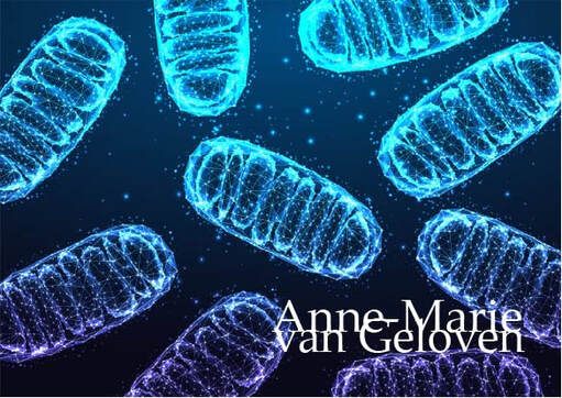
Mitochondria are the "powerhouses" or "lungs" of our cells and bioenergetic semi-autonomous organelles with their own genomes and genetic systems. [1] They are responsible for generating the energy that fuels a wide range of cellular processes in the skin, including cell signaling, pigmentation, wound healing, barrier integrity [2], metabolism and quality control. [3] Mitochondria exist in each cell of the body and are generally inherited exclusively from the mother. Their primary role is cellular respiration; a process converting the energy in nutrients (like glucose) into a usable form of energy called ATP or Adenosine Triphosphate. Mitochondria are particularly abundant in the skin, reflecting the skin's high metabolic demand. When the functionality of mitochondria is impaired or declines, it impacts skin's vitality, health and beauty. Mitochondrial dysfunction is 1 of the 12 hallmarks of skin ageing.
The skin is particularly susceptible to mitochondrial stress due to its constant exposure to environmental insults, such as UV radiation, pollution, and other oxidative stressors. These factors can damage mitochondrial DNA, leading to increased production of reactive oxygen species (ROS) and disrupting the delicate balance of cellular processes. [4] In aged post-mitotic cells, heavily lipofuscin-loaded lysosomes perform poorly, resulting in the enhanced accumulation of defective mitochondria, which in turn produce more reactive oxygen species causing additional damage (the mitochondrial-lysosomal axis theory). [5] Optimal mitochondrial function is indispensable for sustaining the specialized functions of each cell type, like keratinocyte differentiation, fibroblast ECM production, melanocytes melanin production and distribution, immune cell surveillance, sebocytes and adipocytes. [6] Mitochondrial dysfunction is both directly and indirectly linked to chronological ageing and photo-ageing. [7] As mitochondrial function declines, the skin's ability to regenerate and repair itself is decreased. [2] This results in visible signs of aging, such as wrinkles, loss of elasticity, dryness, uneven pigmentation, melasma, age spots, lipomas, impaired wound healing. [2-4-5-8-9] Mitochondrial dysfunction also has been implicated in skin conditions like acne, eczema, lupus, psoriasis, vitiligo, atopic dermatitis and even skin cancer. [10] Ageing is associated with changes in mitochondrial morphology, including [6] ▌Hyperfusion or increased fragmentation ▌Loss of mitochondrial connectivity [11-7] ▌Decline in the efficiency of oxidative phosphorylation, leading to reduced ATP production ▌Decline mitochondrial membrane potential (ΔΨM) ▌Compromised cellular energy metabolism ▌Reduced mitochondrial turnover (downregulated biogenesis) ▌Impaired mitochondrial quality control such as mitophagy (removal of damaged mitochondria through autophagy) [6] These alterations are related to the increased production of ROS exhibited by mitochondria during ageing, the accumulation of which causes oxidative damage to mitochondrial and cell components contributing to cellular senescence. [12] Good mitochondrial function or metabolism: [7] ▌Redox homeostasis: (the way of reducing oxidative stress) - mitochondrial respiration and ROS production are essential for keratinocyte differentiation ▌ATP production: Adenosine Triphosphate provides energy to drive and support many processes in living cells (and GTP) ▌Respiration: mitochondrial respiration is the most important generator of cellular energy ▌Biogenesis: allows cells to meet increased energy demands, to replace degraded mitochondria and is essential for the adaptation of cells to stress [6] ▌Calcium homeostasis ▌Cellular growth ▌Programmed cell death (apoptosis) reducing cell senescence [13] ▌Mitochondrial protein synthesis: mitochondria typically produce 13 proteins encoded by mitochondrial DNA (mtDNA) Dysfunctional Mitochondria: [7] ▌Oxidative stress ▌Decreased ATP levels ▌Dysfunctional OXPHOS: Oxidative phosphorylation, a metabolic pathway in which enzymes oxidize nutrients to release stored chemical energy in the form of ATP ▌Altered mitochondrial biogenesis ▌Calcium imbalance ▌Cell death Mitochondrial proteins Mitochondria contain >1,100 different proteins (MitoCoP) that often assemble into complexes and supercomplexes such as respiratory complexes and preprotein translocases. The chaperones Heat Shock Proteins HSP60-HSP10 are the most abundant mitochondrial proteins. [3] Small heat shock proteins form a chaperone system that operates in the mitochondrial intermembrane space. Depletion of small heat shock proteins leads to mitochondrial swelling and reduced respiration. [14] Mitochondrial hyperpigmentation Emerging research has shed light on the intricate relationship between mitochondrial dysfunction and the development of hyperpigmentation, a condition characterized by the overproduction and uneven distribution of melanin in the skin. One of the key mechanisms underlying this connection is the role of mitochondria in the regulation of melanogenesis, the process by which melanin is synthesized. Mitochondria are involved in the production of various cofactors and signaling molecules that are essential for the activity of tyrosinase, the rate-limiting enzyme in melanin synthesis. [15] When mitochondrial function is impaired, it can lead to an imbalance in the production and distribution of these cofactors and signaling molecules, ultimately resulting in the overproduction and uneven deposition of melanin in the skin. [15] This can manifest itself as age spots, melasma, and other forms of hyperpigmentation. The link between mitochondrial dysfunction and hyperpigmentation has been further supported by studies on genetic disorders that involve mitochondrial dysfunction, such as mitochondrial DNA depletion syndrome. In these conditions, patients often exhibit a range of pigmentary skin changes, including patchy hyper- and hypopigmentation, as well as reticular pigmentation. [16] Mitochondrial crosstalk and exosomes Mitochondria can crosstalk and move beyond cell boundaries. [17] Mitochondria-derived material might be transferred to neighboring cells in the form of cell-free mitochondria or included in extracellular vesicles [18-19]. This process supports cellular repair and contributes to vital mitochondrial functions. Besides restoring stressed cells and damaged tissues due to mitochondrial dysfunction, intercellular mitochondrial transfer also occurs under physiological and pathological conditions. [20] The transfer of active mitochondria from mesenchymal stem cells (MSCs) has been identified as a repair mechanism for rejuvenating damaged skin fibroblasts. [21] MITOCHONDRIAL SUPPORT Move According Martin Picard phD being physically active is a protective factor against almost everything health related. Exercise stimulates the production of mitochondria as more energy is required. Be hungry sometimes If there is too much supply of energy acquired via food leads to mass shrinking of mitochondria or fragmentation. Don´t over-eat, be calorie neutral and sometimes being calorie deficient is good for mitochondria. Maintain a healthy weight, preferably with a mediterranean diet containing phenolic and polyphenolic compounds (increase mitochondrial function and number) nitrate rich vegetables, soybeans and cacao beans. Mitohormesis In model organisms, lifespan can be improved by compromising mitochondrial function, which induces a hormetic response (“mitohormesis”), provided that this inhibition is partial and occurs early during development. Feel good Feeling good (positivity), especially at night, has a scientifically proven positive effect on mitochondrial health index, it is even a predictive factor. Q10 or Coenzyme Q10 (CoQ10) Q10 is part of the mitochondrial respiration chain and essential for cellular energy production. About 95% of our cellular energy is generated with support of Q10, which is produced by the human body itself. During skin ageing, both the cellular energy production and levels of Q10 are declined. Q10 is a powerful anti-oxidant [22], thus protecting cells from oxidative stress and damage and has proven to be able to "rescue" senescent cells by decreasing elevated senescent markers like p21 levels and β-Galactosidases positive cell numbers (in-vitro). Q10 is bio-active, increasing collagen type I and elastin production. [23] Q10 can be supplemented via nutrition, however also via topical application and is considered an evidence based active ingredient in skin care products. Ubiquinol (reduced form) shows higher bioavailability compared to ubiquinone (oxidized form). [23] Pyrroloquinoline quinone (PQQ) Q10 improves the energy in the mitochondria, however PQQ has shown to increase the number of mitochondria and a redox maestro. I´ve written a full post about this compound, which can be found as skincare ingredient and supplement. Read more about PQQ Glutathione Glutathione is formed in cell's cytoplasm from glutamic acid, cysteine and glycine. It is present in 2 forms: reduced (GSH) and oxidized (GSSG). Reduced GSH is an active anti-oxidant, while the presence of inactive GSSG is increased under oxidative stress. The ratio between GSH and GSSH is considered a measure of oxidative stress. Glutathione participates in redox reactions, acts as co-factor of many anti-oxidant enzymes and is the most important non-enzymatic anti-oxidant, essential for synthesis of proteins and DNA. Low Glutathione results in accelerated ageing and inflammatory skin diseases. Mitochondrial glutathione (mGSH) is the main line of defense for the maintenance of the appropriate mitochondrial redox environment to avoid or repair oxidative modifications leading to mitochondrial dysfunction and cell death. [24] Glutathione can be increased via supplementation via precursors cysteine or N-acetylcysteine (not recommended for pregnant women), a combination of Glycine and NAC (called GlyNAC) part of the popular "power of three" supplementation, or the reduced form of Glutathione itself, or increased via topical active ingredients like Licochalcone A. [25] I´ve written about GlyNAC in my post on autophagy. Nicotinamide NR nicotinamide ribosome which is the precursor of NMN nicotinamide mononucleotide which is the precursor of NAD+ nicotinamide adenine dinucleotide all could have a protective effect on mitochondria. Nicotinamide adenine dinucleotide is present in living organisms as ions NAD+ and NADP+ and in reduced forms NADH and NADPH. NADH is a cofactor of processes inside mitochondria: ▌ATP production ▌Activation of "youth proteins" sirtuins ▌Activation of PARP Poly (ADP-ribose) polymerase, a family of proteins involved in many cellular processes such as DNA repair, genomic stability and programmed cell death ▌Reduction of ROS (free radicals) NAD levels as lowered during ageing. [26] One of the fans of NMN supplementation is Harvard Professor David Sinclair, best known for his work on understanding why we age and how to slow its effects and also featured in my article about hormesis. There are about 14 studies done to date with NMN supplementation in humans, one of which was done by Professor Sinclair. NMN supplementation does raise NAD levels, however there aren't substantial proven health benefits, unless you are unhealthy. Resveratrol Although systemically Resveratrol promotes mitochondrial biogenesis. [27] Other data shows that UVA (14 J/cm(2)) along with resveratrol causes massive oxidative stress in mitochondria. As a consequence of oxidative stress, the mitochondrial membrane potential decreases which results in opening of the mitochondrial pores ultimately leading to apoptosis in human keratinocytes. [28] Magnesium Magnesium supplementation has been shown to improve mitochondrial function by increasing ATP production, decreasing mitochondrial ROS and calcium overload, and repolarizing mitochondrial membrane potential. There are many forms of Magnesium, however Citrate, Malate and Orotate are particularly good for energy. L-Carnitine Placebo-controlled trials have shown positive effects of L-Carnitine supplementation on both pre-frail subjects and elderly men. The effect is possibly mediated by counteracting age-related declining L-carnitine levels which may limit fatty acid oxidation by mitochondria. NEW Ergothioneine (EGT) Ergothioneine (EGT) is a sulfur-containing amino acid derivative known for its antioxidant properties, particularly in mitochondria. It is transported into cells and mitochondria via the OCTN1 transporter, where it helps reduce reactive oxygen species (ROS) and maintain cellular homeostasis [29]. EGT binds to and activates 3-mercaptopyruvate sulfurtransferase (MPST), enhancing mitochondrial respiration and exercise performance [30]. It also protects against oxidative stress and inflammation, potentially benefiting conditions like neurodegenerative diseases [31]. Melatonin Not much talked about when it comes to mitochondria, however should not be ignored as mitochondria can benefit significantly from melatonin supplementation. 1. Antioxidant protection: Melatonin acts as a powerful antioxidant within mitochondria, scavenging free radicals and reducing oxidative damage to mitochondrial DNA and proteins [32][34]. 2. Regulation of mitochondrial homeostasis: Melatonin helps maintain electron flow, efficiency of oxidative phosphorylation, ATP production, and overall bioenergetic function of mitochondria [32][34]. 3. Preservation of respiratory complex activities: Melatonin helps maintain the activities of mitochondrial respiratory complexes, which are crucial for energy production [32][34]. 4. Modulation of calcium influx: Melatonin regulates calcium influx into mitochondria, helping prevent calcium overload which can be damaging [32][34]. 5. Protection of mitochondrial permeability transition: Melatonin helps regulate the opening of the mitochondrial permeability transition pore, which is important for maintaining mitochondrial integrity [32][34]. 6. Enhancement of mitochondrial fusion: Melatonin promotes mitochondrial fusion, which is part of the quality control process for maintaining healthy mitochondria [33]. 7. Promotion of mitophagy: Melatonin enhances the removal of damaged mitochondria through mitophagy, helping maintain a healthy mitochondrial population [33]. 8. Reduction of nitric oxide generation: Melatonin decreases nitric oxide production within mitochondria, which can be damaging in excess [32][34]. 9. Selective uptake by mitochondria: Melatonin is selectively taken up by mitochondrial membranes, allowing it to exert its protective effects directly within these organelles [34]. 10. Support of mitochondrial biogenesis: Some studies suggest melatonin may promote the formation of new mitochondria [33]. The key antioxidants used by mitochondria are Glutathione (GSH), Glutathione peroxidase (GPx), Coenzyme Q10 (CoQ10), Superoxide dismutase (SOD), Melatonin, Vitamin C (ascorbate) and Vitamin E (α-tocopherol). Red light therapy By incorporating red light therapy into your skin care routine, you can help to counteract the damaging effects of mitochondrial dysfunction and support the skin's natural renewal processes. As we continue to explore the 12 hallmarks of ageing, I am confident that we will gain even more valuable insights and develop breakthrough innovations that will improve skin quality, health, beauty and vitality. Always consult a qualified healthcare professional or dermatologist to determine what the most suitable approach is for your particular skin condition and rejuvenation goals. Take care! Anne-Marie References
3/20/2024 Comments Telomeres: tiny caps with big impact![]()
Our DNA is as like precious book of life filled with information and instructions, with telomeres acting like the protective covers. Just as book covers get worn over time, our telomeres naturally shorten as we age. This shortening is like a biological clock, ticking away with each cell division.
Telomere shortening is considered one of the twelve key hallmarks of aging. Those hallmarks all play an important role in longevity, health-span, and skin quality, thus both health and beauty. Telomeres are the protective end-caps of chromosomes, similar to the plastic caps at the end of shoelaces. They maintain genomic stability and prevent chromosomal damage. Telomeres become slightly shorter each time a cell divides, and over time they become so short that the cell is no longer able to successfully divide. They shorten more rapidly in dermal fibroblasts compared to epidermal keratinocytes, hence there are significant differences amongst our cells. Telomeres in skin cells may be particularly susceptible to accelerated shortening because of both proliferation and DNA-damaging agents such as reactive oxygen species and sun exposure. [16]. When a cell is no longer able to divide due to telomere shortening, this can lead to
![]()
This consequently affects both health and beauty
FACTORS INFLUENCING TELOMERE SHORTENING Sleep quality Poor sleep quality significantly impacts telomere length:
INTERVENTIONS FOR TELOMERE PRESERVATION 1. Possible strategies to preserve telomere length
Telomerase is an enzyme that plays a crucial role in maintaining the length of telomeres and skin cell function. Telomerase is a ribonucleoprotein enzyme, meaning it contains both protein (TERT plus dyskerin) and RNA components (TER or TERC). Its primary function is to add repetitive DNA sequences (telomeres) to the ends of chromosomes, preventing them from shortening during cell division. Telomerase is active in embryonic stem cells, some adult stem cells, cancer cells, certain skin cells, specifically:
Poor sleep quality is associated with shorter telomere length. Studies have found significant associations between shortened telomere length and poor sleep quality and quantity, including obstructive sleep apnea [17]. Not feeling well rested in the morning was significantly associated with shorter telomere length in older adults [18]. Sleep loss and poor sleep quality may activate DNA damage responses and cellular senescence pathways [17]. Poor sleep can increase oxidative stress and inflammation, which may accelerate telomere shortening [17]. Disruption of circadian rhythms due to poor sleep may negatively impact telomere maintenance [17]. Improving sleep quality through lifestyle changes and sleep hygiene practices may help preserve telomere length. [19]
A study showed that diet, exercise, stress management, and social support could increase telomere length by approximately 10% over five years [20].
Adopt a plant-rich diet, such as the Mediterranean diet, which includes whole grains, nuts, seeds, green tea, legumes, fresh fruits (berries), vegetables (leafy greens), omega-3 fatty acids from sources like flaxseed and fish oil or fatty fish and foods rich in folate. This diet is rich in antioxidants and anti-inflammatory properties that help maintain telomere length [21]. 5. Fasting Fasting, especially intermittent fasting, has attracted interest for its potential impact on health, including telomere preservation. Multiple studies have shown that intermittent fasting (IF) and other fasting regimens can reduce markers of oxidative stress and inflammation. Research on animals has demonstrated that caloric restriction and intermittent fasting can boost telomerase activity and enhance telomere maintenance in specific tissues. A human study by Cheng et al. (2019) found a correlation between intermittent fasting and longer telomeres, by reducing PKA activity and IGF1 levels, which are crucial for regulating telomerase function. A study showed that 36 hours of fasting induced changes in DNA methylation and another one histone modifications, hence fasting has the potential to induce epigenetic changes. Important note: Be careful with a time-restricted eating schedule (often seen as a form of intermittent fasting, where you eat all meals within an 8 hour time-frame), especially women in menopause or people with a pre-existing heart condition. The American Heart Association presented data indicating that people with a pre-existing heart condition have a 91% higher risk of of death of a heart disease when following the time-restricted eating schedule with an 8 hour window, compared to those who eat within a 12-16 hours window. However, several experts have criticised the data, which aren´t published in a peer reviewed journal. When considering fasting, or a time-restricted eating schedule, especially for a longer period, talk to a qualified HCP first. 6. Exercise
EMERGING TECHNOLOGIES IN TELOMERE-TARGETING SKINCARE Small RNAs in skincare Small RNAs play a significant role in the effectiveness of telomere-targeting skincare by influencing skin regeneration and cellular processes. Recent research has highlighted their potential in enhancing wound healing and reducing scarring, which are critical aspects of maintaining healthy skin. Small RNAs, such as microRNAs, are involved in regulating gene expression related to skin aging and and show potential in telomere maintenance [29]. They can modulate the expression of genes that control cellular senescence, oxidative stress response, and inflammation, all of which are crucial for preserving telomere integrity and function [30].
RNAi technology in development RNAi-based skincare approaches could target genes involved in telomere maintenance or have effects on markers related to telomere biology:
RNA-based telomere extension is a method developed at Stanford University and uses modified RNA to extend telomeres in cultured human cells, allowing cells to divide more times than untreated cells [35]. IN OFFICE DERMATOLOGICAL TREATMENTS Aesthetic, regenerative treatments that support skin quality may indirectly support telomere preservation.
Telomere shortening questionable as stand-alone hallmark [36] Telomere length (TL) has long been considered one of the best biomarkers of aging. However, recent research indicates TL alone can only provide a rough estimate of aging rate and is not a strong predictor of age-related diseases and mortality. Other markers like immune parameters and epigenetic age may be better predictors of health status and disease risk. TL remains informative when used alongside other aging biomarkers like homeostatic dysregulation indices, frailty index, and epigenetic clocks. TL meets some criteria for an ideal aging biomarker (minimally invasive, repeatable, testable in animals and humans) but its predictive power for lifespan and disease is questionable. There is inconsistency in epidemiological studies on TL's association with aging processes and diseases. This has led to debate about TL's reliability as an aging biomarker. It's unclear if telomere shortening reflects a "mitotic clock" or is more a marker of cumulative stress exposure. TL is still widely used in aging research but there are ongoing questions about its usefulness as a standalone biomarker of biological age. As research in regenerative medicine advances, we're seeing promising developments in therapies targeting telomere biology for longevity, health and beauty. While telomere research is exciting, it's important to remember that it's just one part of a comprehensive approach to aging, and future treatments will likely combine multiple strategies to target preferably all 12 hallmarks for the best results. Always consult a qualified healthcare professional or dermatologist to determine what the most suitable approach is for you. . Take care! Anne-Marie
References
[1] Martin, H., Doumic, M., Teixeira, M.T. et al. Telomere shortening causes distinct cell division regimes during replicative senescence in Saccharomyces cerevisiae. Cell Biosci11, 180 (2021) [2] M. Borghesan, W.M.H. Hoogaars, M. Varela-Eirin, N. Talma, M. Demaria, A Senescence-Centric View of Aging: Implications for Longevity and Disease, Trends in Cell Biology, Volume 30, Issue 10, 2020, Pages 777-791, ISSN 0962-8924, [3] McHugh D, Gil J. Senescence and aging: Causes, consequences, and therapeutic avenues. J Cell Biol. 2018 Jan 2;217(1):65-77. [4] Oeseburg, H., de Boer, R.A., van Gilst, W.H. et al. Telomere biology in healthy aging and disease. Pflugers Arch - Eur J Physiol 459, 259–268 (2010) [5] Catarina M Henriques, Miguel Godinho Ferreira, Consequences of telomere shortening during lifespan, Current Opinion in Cell Biology, Volume 24, Issue 6, 2012 [6] Henriques CM, Ferreira MG. Consequences of telomere shortening during lifespan. Curr Opin Cell Biol. 2012 [7] Chaib, S., Tchkonia, T. & Kirkland, J.L. Cellular senescence and senolytics: the path to the clinic. Nat Med 28, 1556–1568 (2022) [8] Lei Zhang et al. Cellular senescence: a key therapeutic target in aging and diseases JCI The Journal of Clinical Investigation 2022 [9] Muraki K, Nyhan K, Han L, Murnane JP. Mechanisms of telomere loss and their consequences for chromosome instability. Front Oncol. 2012 Oct 4;2:135. [10] Marlies Schellnegger et al. Aging, 25 January 2024 Sec. Healthy Longevity Volume 5 - 2024 Unlocking longevity: the role of telomeres and it´s targeting interventions [11] Bär C, Blasco MA. Telomeres and telomerase as therapeutic targets to prevent and treat age-related diseases. F1000Res. 2016 Jan 20;5:F1000 Faculty Rev-89. [12] Kasiani C. Myers et al. Blood (2022) 140 (Supplement 1): 1895–1896. Gene therapies November 15 2022 Successful Ex Vivo Telomere Elongation with EXG-001 in a patients with Dyskeratosis Congenital Kasiani C. Myers et al. [13] Falckenhayn C, Winnefeld M, Lyko F, Grönniger E. et al. Identification of dihydromyricetin as a natural DNA methylation inhibitor with rejuvenating activity in human skin. Front Aging. 2024 Mar 4;4:1258184 [14] Minoretti P, Emanuele E. Clinically Actionable Topical Strategies for Addressing the Hallmarks of Skin Aging: A Primer for Aesthetic Medicine Practitioners. Cureus. 2024 Jan 19;16(1):e52548 [15] Guterres, A.N., Villanueva, J. Targeting telomerase for cancer therapy. Oncogene 39, 5811–5824 (2020). [16] Buckingham EM, Klingelhutz AJ. The role of telomeres in the ageing of human skin. Exp Dermatol. 2011 Apr;20(4):297-302. [17] Debbie Sabot, Rhianna Lovegrove, Peta Stapleton, The association between sleep quality and telomere length: A systematic literature review, Brain, Behavior, & Immunity - Health, Volume 28, 2023, 100577, ISSN 2666-3546 [18] Iloabuchi, Chibuzo et al. Association of sleep quality with telomere length, a marker of cellular aging: A retrospective cohort study of older adults in the United States Sleep Health: Journal of the National Sleep Foundation, Volume 6, Issue 4, 513 – 521 [19] Rossiello, F., Jurk, D., Passos, J.F. et al. Telomere dysfunction in ageing and age-related diseases. Nat Cell Biol 24, 135–147 (2022) [20] Elisabeth Fernandez Research September 16 2013 Lifestyle changes may lengthen telomeres, A measure of cell aging. Diet, Meditation, Exercise can improve key element of Immune cell aging, UCSF Scientist report [21] Martínez P, Blasco MA. Telomere-driven diseases and telomere-targeting therapies. J Cell Biol. 2017 Apr 3;216(4):875-887. [22] Guo, J., Huang, X., Dou, L. et al. Aging and aging-related diseases: from molecular mechanisms to interventions and treatments. Sig Transduct Target Ther 7, 391 (2022). [23] Hachmo Y, Hadanny A, Abu Hamed R, Daniel-Kotovsky M, Catalogna M, Fishlev G, Lang E, Polak N, Doenyas K, Friedman M, Zemel Y, Bechor Y, Efrati S. Hyperbaric oxygen therapy increases telomere length and decreases immunosenescence in isolated blood cells: a prospective trial. Aging (Albany NY). 2020 Nov 18;12(22):22445-22456 [24] Gutlapalli SD, Kondapaneni V, Toulassi IA, Poudel S, Zeb M, Choudhari J, Cancarevic I. The Effects of Resveratrol on Telomeres and Post Myocardial Infarction Remodeling. Cureus. 2020 Nov 14;12(11):e11482. [25] Widgerow AD, Ziegler ME, Garruto JA, Bell M. Effects of a Topical Anti-aging Formulation on Skin Aging Biomarkers. J Clin Aesthet Dermatol. 2022 Aug;15(8):E53-E60. PMID: 36061477; PMCID: PMC9436220. [26] Alt, C.; Tsapekos, M.; Perez, D.; Klode, J.; Stoffels, I. An Open-Label Clinical Trial Analyzing the Efficacy of a Novel Telomere-Protecting Antiaging Face Cream. Cosmetics 2022, 9, 95. [27] Cosmetics & Toiletries Telomere protection: Act on the origin of youth, June 3th 2015 Sederma [28] Yu Y, Zhou L, Yang Y, Liu Y. Cycloastragenol: An exciting novel candidate for age-associated diseases. Exp Ther Med. 2018 Sep;16(3):2175-2182. [29] Gerasymchuk M, Cherkasova V, Kovalchuk O, Kovalchuk I. The Role of microRNAs in Organismal and Skin Aging. Int J Mol Sci. 2020 Jul 25;21(15):5281. [30] Jacczak B, Rubiś B, Totoń E. Potential of Naturally Derived Compounds in Telomerase and Telomere Modulation in Skin Senescence and Aging. International Journal of Molecular Sciences. 2021; 22(12):6381. [31] Roig-Genoves, J.V., García-Giménez, J.L. & Mena-Molla, S. A miRNA-based epigenetic molecular clock for biological skin-age prediction. Arch Dermatol Res 316, 326 (2024). [32] Eline Desmet, Stefanie Bracke, Katrien Forier, Lien Taevernier, Marc C.A. Stuart, Bart De Spiegeleer, Koen Raemdonck, Mireille Van Gele, Jo Lambert, An elastic liposomal formulation for RNAi-based topical treatment of skin disorders: Proof-of-concept in the treatment of psoriasis, International Journal of Pharmaceutics, Volume 500, Issues 1–2, 2016, Pages 268-274, ISSN 0378-5173 [33] Oger E, Mur L, Lebleu A, Bergeron L, Gondran C, Cucumel K. Plant Small RNAs: A New Technology for Skin Care. J Cosmet Sci. 2019 May/Jun;70(3):115-126. PMID: 31398100. [34] Vimisha Dharamdasani, Abhirup Mandal, Qin M. Qi, Isabella Suzuki, Maria Vitória Lopes Badra Bentley, Samir Mitragotri, Topical delivery of siRNA into skin using ionic liquids, Journal of Controlled Release, Volume 323, 2020, Pages 475-482, ISSN 0168-3659 [35] Krista Conger January 2015 Stanford Medicine News Center Telomere extension turns back aging clock in cultured human cells, study finds [36] Alexander Vaiserman, Dmytro Krasnienkov Telemore length as marker of biological age: state-of-the-art, open issues and future perspectives Front. [37] Martínez P, Blasco MA. Telomere-driven diseases and telomere-targeting therapies. J Cell Biol. 2017 Apr 3;216(4):875-887 ![]()
In skin biology, senescence is a process by which a cell ages and permanently stops dividing but does not die. This is why they are also referred to as "zombie cells". Age-related accumulation of senescent cells is caused by of increased levels of senescence-inducing stressors and/or reduced elimination of senescent cells. Under normal physiological conditions, senescent cells play an important role maintaining cellular homeostasis and inhibiting proliferation of abnormal cells. However, over time, large numbers of zombie cells can build up in the skin and contribute to the overall reduction in skin's regenerative properties, impacting both its beauty and health.
There are 2 forms of cell senescence: Acute senescence: Senescent cells are produced in response to acute stressors to facilitate for example tissue repair, wound healing. They are cleared by our immune system. Chronic senescence: A not programmed process as response to prolonged stress or damage and these senescent cells are not cleared by our immune system, leading to the accumulation of zombie cells impacting our skin health and beauty. It has been suggested that inflammageing is mainly related to senescent cells and their associated SASP (Senescence Associated Secretory Phenotype) which increase in the body with age and contribute to inflammageing. Senescent cells cause inflammageing and inflammageing causes cell senescence. [1] Senescence can be triggered by a number of stress signals to the cell [1]:
Mechanisms of skin cell senescence:
The presence of senescent cells accelerates the ageing process due to their communication with nearby cells through various molecules: [18]
Fibroblast senescence could be the main driver of the skin ageing. [3] The increased number of senescent fibroblasts results in the production of SASPs rich in pro-inflammatory cytokines, including interleukin (IL)-1, IL-6, IL-8, IL-18, matrix metalloproteinases (MMPs), and a variety of other inflammatory chemokines [2] resulting in the breakdown of collagen, loss of elasticity and wrinkle formation. [3] Autophagy in dermal fibroblasts is essential for maintaining skin balance and managing the ageing process, particularly in response to external stressors like UV radiation and particulate matter (PM), by repairing cellular machineries. [4] Insufficient autophagy leads to an exaggerated skin inflammation triggered by inflammasome activation, resulting in accelerated ageing characteristics. When exposed to UVB (in vitro), skin cell types like fibroblasts and keratinocytes show DNA damage and increased senescence markers, such as increased SASPs. [3] Dermal fibroblasts also release insulin-like growth factor (IGF)-1, essential for epidermal cell proliferation and differentiation. [5] IGF-1 signalling in senescent fibroblasts is significantly decreased [6]. Inhibition of the IGF-1 pathway decreases collagen production in the dermis, causing epidermal thinning. Additionally, mitochondrial dysfunction and increased levels of superoxide anions prompt fibroblast ageing, thereby speeding up the skin ageing process. [5] Fibroblasts isolated from photo-aged skin produce a greater amount of pro-melanogenic growth factors. [14] Ageing-associated pigmentation has also been reported to be driven by (UVA-induced) fibroblast senescence. [15-16] Keratinocyte senescence The epidermis shows less impact of senescent keratinocytes due to their quicker turnover in comparison to fibroblasts. Senescent keratinocytes experience reduced ECM production and cell adhesions [8], along with elevated MMP expression in UV-induced senescence [9], and increased SASP levels, including pro-inflammatory cytokines. [10] Airborn particulate matter (PM2.5) can penetrate a disrupted skin barrier. PM2.5-induced ROS leads to epigenetic modification: reduced DNA methyltransferase, elevated DNA demethylase expression, p16INK4a promotor hypomethylation and therewith accelerated keratinocyte senescence. [11] Keratinocytes are the main type of cells that signal the need for melanogenesis. [12] UVR-induced DNA damage in keratinocytes activates melanogenesis. [13] Melanocyte senescence Senescent melanocytes express markers of inflammageing and dysfunctional telomeres. Senescent melanocyte SASPs induce telomere dysfunction and limit the proliferation of the surrounding cells, hence, senescent melanocytes affect and impair basal keratinocyte proliferation and contribute to epidermal atrophy. [17] STRATEGIES TO COMBAT CELL SENESCENCE PREVENTION Sunscreen: Protection against UV radiation combined with blue light defense (Licochalcone A: powerful anti-oxidant, Nrf2-Activator & increasing Glutathione + Colour pigments) and prevention + repair DNA damage (Glycyrrhetinic Acid) INTERVENTION Senotherapeutics can be classified into three development strategies: [25]
Skin care ingredients: [18]
Of course a healthy life-style and diet (consider also intermittent fasting) will support both your body & skin longevity and beauty Prevention and intervention of skin cell senescence offers a promising approach to improve skin health and beauty. Always consult a qualified healthcare professional or dermatologist to determine the most suitable approach for your particular skin condition and rejuvenation goals. Take care! Anne-Marie References
![]()
Many of the skin regenerating or rejuvenating treatments, like energy based devices in the doctors-office are based on the principle to cause controlled damage and therewith provocation of a skin rejuvenating repair response. One of the fascinating mechanisms behind laser "damage" is the heat shock response leading to increased production of regenerating heat shock proteins (HSPs). Heat shock proteins respond to heat stress, are crucial cellular defence mechanisms against stress (environmental and physiological), act as chaperones, aiding in protein folding, prevention of protein damage, cellular protection and repair, with other words HSPs play a crucial role in proteostasis. [1]
HEAT SHOCK PROTEINS AND OX-INFLAMMAGEING UV radiation and blue light cause oxidative stress and inflammation, and can overwhelm skin's own capacity to counteract the increased formation of reactive oxygen species (ROS) and inflammatory mediators. Chronic oxidative stress state and chronic low grade of inflammation are hallmarks of skin ageing and their combination can be called ox-inflammageing. Oxidative stress and inflammation alter cellular signal transduction pathways and thereby the expression of the ECM genes as well as the structure of the ECM proteins like collagen, fibronectin and elastin. Their reduced expression and increased degradation manifests eventually at the skin surface as wrinkles, loss of firmness, and elasticity. Heat shock proteins are chaperone proteins that facilitate the formation of the ECM and prevention of molecular oxidative damage or degradation and are classified based on their molecular weights.
HEAT SHOCK PROTEINS AND PROTEOME Proteostasis, or protein homeostasis, refers to the balance between protein synthesis (like collagen, fibronectin and elastin), folding, and degradation. As we age, this balance is disrupted, leading to the accumulation of misfolded and aggregated proteins [10]. Loss of proteostasis is another hallmark of aging and HSPs play a crucial role in maintaining proteostasis through several mechanisms: 1. Protein folding: HSPs assist in the proper folding of newly synthesised proteins and refolding of misfolded proteins [10][11]. 2. Protein degradation: HSPs collaborate with the ubiquitin-proteasome system and autophagy to target misfolded proteins for degradation [10][12]. 3. Stress response: Under stress conditions, HSPs are upregulated to protect cells from protein damage and maintain cellular functions [13][14]. HSP-70 and HSP-90 are particularly important in protein folding and refolding, while small HSPs are involved in preventing protein aggregation [11][14]. Several studies have provided evidence supporting the potential of HSPs as an intervention to improve proteostasis: lifespan extension: [15], neuroprotection (HSP70), stress resistance and cellular survival [13][14], protein aggregation prevention (small HSPs) [11][14], autophagy regulation and particularly HSP70 is crucial for cellular protein quality control [16]. STIMULATION OF REJUVENATING HEAT SHOCK PROTEINS Heat shock protein synthesis can be initiated not only by heat but also by many chemical and physical stimuli, such as heavy metals, amino acid analogues, oxidative stress, viral infection and UV and ionizing irradiation. [17] Exercise and hormesis: Mild stress induced by exercise or other hormetic interventions has been shown to upregulate HSPs and improve proteostasis. Laser Laser treatments have been shown to induce a heat shock response in the skin from epithelial cells to deeper connective tissues, leading to the production of heat shock proteins. This response is characterized by the temporary changes in cellular metabolism, release of growth factors, and increased cell proliferation and thus contribute to tissue regeneration and rejuvenation. [17] CBD It has been proven that a large number of genes belonging to the heat shock protein super-family were up-regulated following cannabidiol (CBD) treatment. [18] UV radiation Ultraviolet radiation (UV)‐induced cell death and sunburn cell formation can be inhibited by previous heat shock exposure and UV itself can induce HSP expression. However, levels of HSP-27 have been found to be elevated in sun‐protected aged skin indicating a link between HSP-27 expression and age‐dependent epidermal alterations. [19] I would recommend daily protection from UV radiation and blue light (or high energy visible light). Ultrasound Ultrasound exposure at different frequencies, intensities, and exposure times can induce HSP-72 expression. Higher ultrasound frequencies, such as 10 MHz, have been found to significantly increase HSP-72 levels. Additionally, increasing the temperature during ultrasound exposure has shown to enhance HSP-72 expression. Interestingly, ultrasound at 1 MHz was unable to induce HSP-72 significantly, while 10 MHz ultrasound induced HSP-72 after 5 minutes of exposure. [16] Radiofrequency Radiofrequency has been shown to increase HSP-70 and decrease melanin synthesis and tyrosinase activity. [20] RF-US treatment significantly increased levels of HSP47 proteins. [21] Red & near infra red light Although I've not seen much peer reviewed published evidence, red light and near infra red light therapy may release the HSPs in the skin if tissue reaches >42 - 45 degrees (even for 8 - 10 seconds). Nicotinamide Nicotinamide and its derivatives have been found to stimulate the expression of heat shock proteins, including HSP-27, HSP-47, HSP-70, and HSP-90 in the skin. These proteins play as mentioned before an essential role in collagen production, skin protection, skin health and rejuvenation. [6] NAD as nutrient interestingly has proven to tweak the epigenome by modulating DNMT1 enzymatic DNA methylation and cell differentiation. [22] In topical applications an ingredient called Dihydromyricetin also called Epicelline® has been successful in inhibiting DNMT1 enzyme activity biochemical assays. [23] Stimulation of heat shock proteins offers a promising and novel invasive, non invasive and topical approach for skin regeneration, rejuvenation, reduction of ox-inflammageing and prevention of loss of proteostasis. Always consult a qualified healthcare professional or dermatologist to determine the most suitable approach for your particular skin condition and rejuvenation goals. Take care! Anne-Marie References
![]()
Like epigenetics and exosomes, neurocosmetics represent a revolutionary approach for skin care incorporating neuroscience principles, leveraging the skin-brain connection to improve skin health and beauty. The term itself is a fusion of the words neuroscience and cosmetics. It differs from psychodermatology which like neurocosmetics connects the interaction between mind and skin, but in a different way. Some describe it as how simple sensory stimulation can improve our overall wellbeing and call it "mood beauty", however this doesn't do it justice as neurocosmetics go beyond mood boosting skincare.
DEFINITION NEUROCOSMETICS Dermatologist Professor Laurent Misery back in 2002 described that neurocosmetics are products which are supposed to modulate the neuro-immuno-cutaneous-system (NICS) function at an epidermal level. Skin cells can produce neuromediators, which are mediators for transmission of information between skin, immune and the nervous system. All skin cells express specific receptors for neuromediators and by binding of the neuromediator to its receptor, modulation of cell properties and skin functions are induced like cell differentiation and proliferation (renewal), pigmentation, etc. Hence, keratinocytes, Langerhans cells, melanocytes, endothelial cells, fibroblasts and the other cells of the skin are modulated and controlled by the nerves and in return skin is able to modulate neuronal activity and growth. [1] SKIN-BRAIN CONNECTION In an article from the International Journal of Novel Research and Developments, the skin-brain connection was described as a psychobiological concept that highlights how emotions, stress, and neurotransmitters impact skin health. Indicating that the skin acts as a neuroimmunoendocrine organ, emphasizing its sensitivity to neural signals and stress responses. [4] CUTANEOUS NERVOUS SYSTEM The skin a sophisticated sensory organ that allows you to interact with your environment through touch and feel. It contains a complex network of nerves that send information about sensations like pressure, pain, itch and temperature from the skin through the spinal cord to the brain [9]. The dynamic interactions between the skin and the nervous system is influenced by factors like stress and inflammation, which can impact skin health and ageing. [7] Nerves in the skin: These nerves are like tiny messengers that tell your brain about what your skin is feeling: pressure, heat or pain. Types of nerve fibers: Some are thick and wrapped in a protective coating, which helps them send messages quickly. Others are thin and slow but are very good at sending messages about pain or temperature changes. [3] Sensory receptors: These receptors can tell if something is touching the skin lightly or if there's a lot of pressure. They can also sense if something is hot, cold, or causing pain. [3] Autonomic nervous system: Part of the cutaneous nervous system helps control things that happen in the skin automatically, like sweating to regulate body temperature. [8] Nerve cells: There are about 20 different types of neurons in our skin. [10] The contribution of epidermal keratinocytes to NICS [3]
CUTANEOUS NEURO-AGEING Neuro-ageing is defined as the changes in the nervous system which cause continuous neurodegeneration due to oxidative stress, neuroinflammation or impaired neuromodulation. As skin ages, Aβ-toxin (increased by oxidative stress) accumulates at the nerve endings innervating the tissue, causing disrupted cellular communication, particularly affecting fibroblasts’ ability to produce collagen and extracellular matrix. On top there is a decrease of nerve growth factor (NGF) production, important for the development and maintenance of nerve cells. Different factors can lead to a drop in NGF production, resulting in malfunctioning keratinocytes and reduced lipolytic activity of adipocytes, visibly impacting skin hydration and firmness. [6] Skin nerve fibres are significantly reduced in number following UV irradiation and in ageing skin [5] and therefore neuro-protectors or targetting neurodegeneration can reduce stress manifestations and promote healthy cellular communication for optimal skin function. [3] Although not much is known regarding skin specific or topical neuroprotectors (most research was focussed on the brain), probably potent anti-oxidants, by significantly reducing oxidative stress from UV and blue light and anti-inflammatory ingredients may inhibit skin neuro-ageing and can be neuroprotective especially when combined with sunscreen and strengthening of the skin barrier. NEUROCOSMETIC VARIETY OF ACTIONS
THE FUTURE OF NEUROCOSMETICS The neurocosmetics market is booming, with a projected value of USD 2.69 billion by 2030. [11] The future of neurocosmetics holds promise for innovative ingredients and concepts that harness new neuroscientific insights to revolutionize skin care and sunscreen formulations, to cater to both physical and emotional aspects of skin health and beauty. Take care! Anne-Marie References
![]()
If you've scrolled through Instagram, you may have caught a glimpse of dermatologists raving about LED masks emitting red light, the secret, evidenced based weapon behind skin rejuvenation known as photo biomodulation. It uses low-powered light within the red to near-infrared range (wavelengths from 632 to 1064 nm) to induce a biological reaction aka stimulate cellular processes. The wonders of red light, also known as LLLT (low-level laser therapy), PBM (red light photo-biomodulation), or PBMT (photo-biomodulating therapy), extend far beyond non-invasive skin rejuvenation. I am not a fan of devices for home use, mostly because of lacking safety and/or efficacy, PBM definitely earned it's prominent spot in my skincare routine.
A summary of the benefts of red light with and without near infrared light for skin Numerous studies have demonstrated the effectiveness of red and infrared light therapy for skin rejuvenation. A combination of red light and near IR light has proven to stimulate the production of collagen (I & III) plus elastin production (Li WH et al Int J Cosmet Sci 2021), enhance mitochondrial ATP production, cell signaling, growth factor synthesis, rebalance ROS (reactive oxidative species) and reduce inflammation. Stem cells can be activated allowing tissue repair and healing. Wrinkle and scar reduction was observed and it can reduce UV damage both as treatment and prophylactic measure. In pigmentary disorders such as vitiligo, it can increase pigmentation by melanocyte proliferation and reduce depigmentation by inhibiting autoimmunity (Pinar Avci et al. Semin Cutan Med Surg. 2013 & Mitchell J Winkie et al. Review Photodermatol Photoimmunol Photomed A focused review of visible light therapies for vitiligo 2024). It has the potential to activate both keratinocytes (epidermis) and fibroblasts (epidermal junction and dermis). With consistent use, you can expect a reduction of lines and wrinkles, improvement of skin tone and texture. PBMT (when done effective and safe) will compliment both your skin rejuvenating and regenerating at home skincare regimen and in-office procedures or even post-surgical skin recovery. ATP ATP (adenosine triphosphate) is the primary source of energy for cellular processes and plays a crucial role in various biological functions. When red light with specific wavelengths (630 nm to 638 nm and 810 nm) is absorbed by the skin cells, it stimulates the mitochondria, which are the powerhouses of the cells responsible for ATP synthesis. This increase in ATP production is providing cells with more energy to carry out their functions effectively and has several beneficial effects on the skin like boosting cellular metabolism, promoting more efficient nutrient uptake and waste removal. The increased ATP levels facilitate collagen synthesis by fibroblasts, a vital component for skin structure, elasticity and firmness and reduction of lines and wrinkles.. ATP aids in the repair and regeneration of damaged skin cells. It accelerates the healing process, making it beneficial for wound healing, post-surgical recovery, and addressing skin issues such as acne scars. ROS (Reactive Oxidative Species) By modulating ROS levels, red light therapy helps reduce oxidative stress and its detrimental effects on the skin. ROS are highly reactive molecules that are naturally produced by cells as byproducts of metabolic processes. While low levels of ROS play important roles in cellular signaling and immune responses, excessive ROS can lead to oxidative stress and damage to cells and tissues. Restoring the balance of ROS result in improved skin health, reduced inflammation, and enhanced skin rejuvenation. Red light therapy has been shown to modulate reactive oxidative species (ROS) levels in the skin by promoting antioxidant defense mechanisms and reducing oxidative stress:
The difference between LLLT and PBM LLLT refers specifically to the use of lasers, which produce coherent, focussed and an intense beam of monochromatic light, while PBM has a broader range of light sources, may include laser as well as light-emitting diodes (LEDs) and other non-laser devices. LEDs are often used in PBM because they are cost effective, versatile and have the ability to cover large treatment areas. LLT uses higher power densities with more energy and has a shorter treatment duration in comparison to PBM to achieve desired therapeutic effects. While there are similarities in terms of mode of action", there is a difference of light source, treatment application and parameters. Based on consensus, PBM and PBMT are considered the correct way to describe this photonic specialty for therapeutic applications. In this post I will focus on PBM and specifically LEDs. A home device claiming to use cold near infrared laser light or Low-Level Laser Therapy is called LYMA laser. It is sold for several thousand euro´s. LED masks and LED panels LED masks specifically produced by the brand Omnilux (FDA cleared) are currently very popular for very good reasons; they are safe and effective when the LEDs emit the right wavelengths and used in the recommended frequency. Omnilux combines 2 therapeutically effective and complimentary wavelengths: 633nm and near-infrared 830 nm. Both wavelengths (more precise 630nm + 850nm) I would recommend to minimally look for in any red LED device, which will disqualify most LED masks and panels in the market! I've include some (not affiliated) links to devices below. Both masks and panels can be effective, however most panels are stronger in comparison to masks 60 mW/cm² vs mW/cm²), hence have the benefit of a shorter treatment time to get a similar result. Intensity and power of red light therapy devices are typically measured in terms of irradiance (measured in milliwatts per square centimeter, mW/cm²) and radiant flux (measured in watts, W), which quantify the amount of light energy emitted by the device. Wearing a mask during a hot summer or in a warmer climate will make you sweat and depending on the materials of the mask and straps, they may be very uncomfortable to wear. Panels have the benefit that they give a more even distribution of emitted light as masks are worn on the face and thus the LED bulbs are pushed on a small skin surface area, panels can cover a larger area (depending on their size) and are more versatile in use, as area's like neck, décolletage, or knees are easier to treat with a panel. With a mask you may be more mobile, although I would not recommend walking around while using the mask. My personal preference would be a panel for the reasons mentioned before and panels are more suitable (more hygienic) for family sharing. My son can use it after an intense workout to speed up his recovery and I like to use it for purposes beyond photo-biomodulation or skin rejuvenation, for example to improve my sleep. With a panel I get more "bang for my buck". ![]()
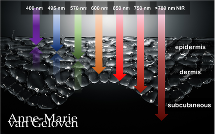
Red light and NIR (Near Infra Red light) have the ability to penetrate varying depths of the skin, resulting in distinct benefits, thus combinations of wavelengths will provide complementary effects.
630 nm Wavelength This wavelength is often used for its skin rejuvenation benefits. It has a relatively shallow penetration depth and is absorbed closer to the surface of the skin primarily affecting the epidermis. 630nm light is associated with increased circulation, reduce inflammation, improved skin tone & texture, aiding in the delivery of nutrients and oxygen to skin cells, and stimulating the production of collagen, leading to improved skin elasticity and a reduction of the appearance of fine lines & wrinkles. 660 nm Wavelength At 660nm, red light can penetrate a little deeper into the skin, reaching the dermis. It is known for its ability to stimulate collagen production, enhance cellular metabolism, and promote anti-inflammatory effects, helping to reduce redness and inflammageing. It also promotes wound healing, making it beneficial for post-surgical or post-trauma skin recovery. 810 nm Wavelength Improve healing & recovery & accelerate wound healing. 830 nm Wavelength Accelerate healing, reduce infection, improve aesthetic outcome following plastic surgery, increase endorfines (mood-enhancing), improve bone repair and growth. 850 nm Wavelength Improve general inflammation body, enhance muscle recovery, improve wound healing, reduced fine lines, wrinkles and hyperpigmentation. Always consult a qualified healthcare professional or dermatologist to determine if and what the most suitable red light therapy approach is for your particular skin condition and rejuvenation goals. Take care! References: Hamblin, Michael R. "Mechanisms and applications of the anti-inflammatory effects of photobiomodulation." AIMS biophysics 4.3 (2017): 337-361. Barolet, Daniel. Regulation of Skin Collagen Metabolism In Vitro Using a Pulsed 660 nm LED Light Source: Clinical Correlation with a Single-Blinded August 2009Journal of Investigative Dermatology 129(12):2751-9 Wunsch A, Matuschka K. (2014). A controlled trial to determine the efficacy of red and near-infrared light treatment in patient satisfaction, reduction of fine lines, wrinkles, skin roughness, and intradermal collagen density increase. Journal of Cosmetic and Laser Therapy, 16(5), 232-237. Avci P, et al. (2013). Low-level laser (light) therapy (LLLT) in skin: stimulating, healing, restoring. Seminars in Cutaneous Medicine and Surgery, 32(1), 41-52. Links to some devices which combine 630 nm and 850 nm: FDA-approved devices ensure safety and regulatory compliance, however the panels are more powerful: Omnilux(tm) Mask (FDA clearance) Very affordable panel (no FDA clearance) Affordable panel (no FDA clearance) ![]()
Glycation is one of the basic root causes of endogeneous (intrinsic) skin ageing and a very challenging one or almost impossible one to reverse. Glycation is an ageing reaction which begins in early life, developing clinical symptoms at around 30, and progressively accumulates in tissues and skin due to the glycated collagens that are difficult to be decomposed. Glycation occurs naturally in the body when sugars react with proteins and lipids to form advanced glycation end products (AGEs). AGEs can be exogenously ingested (through food consumption), inhaled via tobacco or endogenously produced and formed both intracellularly and extracellularly. AGE modifications lead to dermal stiffening, diminished contractile capacity of dermal fibroblasts, lack of elasticity in the connective tissues, contribute to hyperpigmentation and a yellowish skin appearance. The formation of AGEs is amplified through exogenous factors, e.g., ultraviolet radiation.
AGEs cause changes in the skin through 3 processes:
One study published in the Journal of Investigative Dermatology found that levels of AGEs were higher in the skin of older individuals compared to younger ones. The study also showed that there was a correlation between the level of AGEs and the severity of skin ageing. This suggests that inhibiting the production or accumulation of AGEs in the skin is a potential target for anti-ageing interventions or skin ageing management. AGEs are complex and heterogeneous, more than a dozen AGEs have been detected (however not all) in tissues and can be divided into three categories according to their biochemical properties. AGEs are formed through four pathways:
GLYCATION INHIBITION IS KEY AGEs can be crosslinked through side chains to form a substance of very high molecular weight, which is not easily degraded. The consequences from skin glycation are irreversible. This makes prevention or inhibition of the process the best potential strategy to maintain skin health and ageing skin management. One way to do this is by altering the diet to reduce the intake of sugars and carbohydrates, which are known to contribute to glycation. Several studies have found that reducing sugar intake can result in significant improvements in skin health, including reducing wrinkles and improving skin texture. ![]()
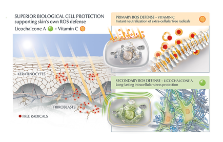
AGE inhibitors
Another potential strategy is the use of topical agents that inhibit the formation or accumulation of AGEs in the skin. One study published in the Journal of Cosmetic Science found that a cream containing carnosine, a peptide that inhibits glycation, improved skin elasticity and reduced the appearance of wrinkles in individuals with ageing skin. Skincare containing NAHP or Acetyl Hydroxyproline inhibits the formation of AGEs significantly (in vitro), most likely through a mechanism where NAHP competes with the proteins for the sugar. Finally, NAHP sacrifices itself in place of the proteins and gets (at least partially) glycated. NAHP also prevents loss of cellular contractile forces in a glycated in vitro dermis model and counteracts the diminished cell-matrix interaction that is caused by glyoxal-induced AGE formation. Take-aways from a study published in the International Journal of Cosmetic Science [2]: 1. NAHP significantly and dose-dependently inhibited the formation of advanced glycation end-products (AGEs) in a protein solution. 2. NAHP dose-dependently inhibited the formation of N-(carboxymethyl)lysine (CML), a specific AGE. 3. In fibroblast-populated collagen lattices, NAHP prevented the glycation-induced disturbance of fibroblast contractile capacity. 4. Ex vivo application of NAHP to skin explants decreased AGE fluorescence compared to glucose-treated samples. Anti-Oxidants I would suggest to combine those ingredients with an ingredient like Licochalcone A. Numerous high ranked publications support that Licochalcone A protects cells from oxidative stress mediated by e.g. UV and HEVIS (blue light) induced reactive oxidative species (ROS). Due to the activation and nuclear translocation of the transcription factor NrF2, the expression of anti-inflammatory, antioxidant and detoxifying enzymes are induced. These enzymes protect the skin cells (like keratinocytes and fibroblasts) from ROS-induced damage, like lipid peroxidation and DNA as well as protein damage. If Licochalcone A is combined with L-Ascorbic Acid, (the most active form of Vitamin C), it supporting skin's own collagen production, provides superior biological cell protection amongst other relevant benefits. Vitamin C (and E) has shown to inhibit protein glycation [4]. SPRAY TAN A study in Redox Biology indicates that sunless tanning with dihydroxyacetone (DHA) causes glycation and potential DNA damage in the epidermis [1].: 1. Glycation: DHA exposure led to the formation of advanced glycation end products (AGEs) in epidermal cells, confirmed by mass spectrometric detection of N-ε-(carboxyethyl)-l-lysine (CEL). 2. DNA damage: DHA induced a cellular stress response, including activation of stress-related genes and phosphoprotein signaling. While not directly measuring DNA damage, these responses are often associated with cellular stress that can lead to DNA damage. 3. Significance: The effects were observed at low millimolar concentrations of DHA, relevant to topical application. The stress response was rapid and pronounced, occurring within minutes of exposure. 4. Location: The effects were primarily observed in epidermal cells and reconstructs, with no mention of dermal effects. This study suggests that sunless tanning with DHA may not be as safe as previously thought, warranting further investigation into its long-term effects on skin health [1], however no need to panic as this negative effect is only seen in the top layer of the skin. DOUBLE TROUBLE: GLYCATION + UVB [3] The combination of GO-AGEs and UVB exposure has a more pronounced effect on skin inflammation and oxidative stress than either factor alone. This suggests a synergistic relationship between glycation and UV exposure in accelerating skin aging processes. 1. GO-AGEs combined with UVB irradiation significantly increased the secretion of pro-inflammatory cytokines (IL-1β, IL-6, IL-8) in skin cells compared to either GO-AGEs or UVB alone. 2. The GO-AGEs + UVB treatment group showed a more than three-fold increase in IL-6 and IL-8 mRNA levels compared to other groups. 3. GO-AGEs + UVB treatment induced significantly higher release of nitric oxide (NO) compared to other groups. 4. The combination of GO-AGEs and UVB enhanced reactive oxygen species (ROS) release, creating about 1.5 times more oxidative stress compared to control and other groups. 5. Cell viability was notably affected in the GO-AGEs, UVB, and GO-AGEs + UVB treatment groups compared to the control group. These findings are significant as they demonstrate that the combination of GO-AGEs and UVB exposure has a more pronounced effect on skin inflammation and oxidative stress than either factor alone. This suggests a synergistic relationship between glycation and UV exposure in accelerating skin aging processes. Use sunscreen daily. . GLYCATION AND SKIN HEALTH Acne In addition to its role in ageing, glycation in the skin has also been linked to a range of skin health problems. One study published in the Journal of Cosmetic Dermatology found that the level of AGEs in the skin was significantly higher in individuals with acne than in those without acne. The study also showed that treating acne with a topical antibiotic significantly reduced the levels of AGEs in the skin. Atopic Dermatitis Another study published in the Journal of Investigative Dermatology found that individuals with atopic dermatitis had higher levels of AGEs in their skin than healthy individuals. This suggests that glycation may play a role in the development of inflammatory skin conditions. Diabetes + Woundhealing The correlation between high sugar levels and skin ageing can be seen in diabetic patients, where one-third of this population has skin complications. A prominent feature of ageing human skin is the fragmentation of collagen fibers, which severely damages the structural integrity and mechanical properties of the skin. Elevated levels of MMP-1 and MMP-2 and higher crosslinked collagen in the dermis of diabetic skin lead to the accumulation of fragmented and crosslinked collagen, thereby impairing the structural integrity and mechanical properties of dermal collagen in diabetes. Collagen crosslinking makes it impossible for them to easily repair, resulting in reduced skin elasticity and wrinkles. Keratinocytes and fibroblasts are the main cells involved in wound healing, but due to the high glucose (HG) microenvironment in diabetics, the functional state of these cells is impaired, thereby accelerating cellular senescence (programmed cell death). Conclusion We can't completely stop the glycation process, therefore it's important that we inhibit it from a young age onwards, hence monitor the sugar intake of our children, use daily SPF and invest in good dermo-cosmetic products containing ingredients like NAHP and powerful anti-oxidants like L-Ascorbid Acid (Vitamin C is needed for the production of collagen) and Licochalcone A (also anti-inflammatory). Preventing signs of ageing, specifically caused by glycation is most effective. If your skin shows (advanced) signs of ageing, you can get visible improvement using skin component (hyaluron, collagen and elastin) bio-stimulating ingredients like Retinol, Bakuchiol, Arctiin, Creatine or Glycine Saponin. Consult your dermatologist if you wish to improve your skin's appearance or skin health issues. Take care Anne-Marie References [1] Perer J, Jandova J, Fimbres J, Jennings EQ, Galligan JJ, Hua A, Wondrak GT. The sunless tanning agent dihydroxyacetone induces stress response gene expression and signaling in cultured human keratinocytes and reconstructed epidermis. Redox Biol. 2020 Sep;36:101594. [2] Knoblich C, et al. N‐acetyl‐L‐hydroxyproline – A potent skin anti‐ageing active preventing advanced glycation end‐product formation in vitro and ex vivo. Int J Cosmet Sci. 2023;1-10. doi:10.1111/ics.12930 [3] Sultana, R., Parveen, A., Kang, MC. et al. Glyoxal-derived advanced glycation end products (GO-AGEs) with UVB critically induce skin inflammaging: in vitro and in silico approaches. Sci Rep 14, 1843 (2024). https://doi.org/10.1038/s41598-024-52037-z [3] Sadowska-Bartosz I, Bartosz G. Prevention of protein glycation by natural compounds. Molecules. 2015 Feb 16;20(2):3309-34. doi: 10.3390/molecules20023309. PMID: 25690291; PMCID: PMC6272653. Special thanks: Ph.D. dr Julia M. Weise Manager Biological Testing & Dorothea Schweiger Lab Manager Facial Skin Biology Beiersdorf HQ Hamburg 3/21/2023 Comments Exosomes in skin care and treatments![]()
Skin boosters using micro-injections with predominantly non-crosslinked hyaluron filler gels like Restylane® Vital, Juvéderm® VOLITE or Belotero® Revive are gaining popularity for very good reasons. Unlike traditional dermal fillers, they are not injected beneath the skin to volumise or shape the face. Instead, they are very fine dermal easily integrated "fillers" that are injected into the skin to hydrate, improve skin quality and give very natural results. They are also gently bio-stimulating, meaning they "stretch" the fibroblasts in the injected area and as a result this cell is producing more collagen. An effective bio-remodeling skin booster using 2 times 5 injection points (bio-aesthetic points - BAP) for a full-face treatment is Profhilo®. However, the recent K-beauty treatment via topical application or micro-injections with bio-remodeling exosomes is gaining popularity.
Exosomes are nano-sized cargos with a lipid bilayer structure carrying diverse biomolecules including lipids, proteins, and nucleic acids. These small extra cellular vesicles are secreted by most types of cells (skin relevant are the keratinocytes and fibroblasts) to communicate with each other. Exosomes circulate through bodily fluids and can transfer information. They can be either good or bad, however taken from a healthy young cell they will be sending the best messages. Studies have shown the efficacy of exosomes in skin ageing. They can facilitate skin remodeling (increasing collagen and decreasing senescent cells) leading to skin rejuvenation. Cells sleep because they don't get enough bio-stimulation: messages. Better messages is better skin architecture. This is why exosomes are so important. At the World Stem Cell Summit it used to be 90% about stem cells (they only life 28 days) and 10% about exosomes, now it is 50/50. The reason is called heterochronic parabiosis. 1. One of the most robust methods of improving the function of ageing tissues is that of heterochronic parabiosis,. The effect was shown in a study with a surgical procedure whereby a young and old mouse are joined together so the share one circulatory system. 2 This study according to dr Kate Goldie AMWC 2023 Monaco is proof that it is not the cells, but the messages they give that is transforming lots of different tissues, which has the ability to profoundly regenerate tissues. That is why people are so interested in exosomes. Exosomes taken from a very young cell give potentially the best messages as they "send the message" of youth. EV (Extra-cellular Vesicle) is the actual correct term as messages come as micro-vesicles and exosomes and form 2 different messages from the cell. 3 We start to understand active ingredients. In exosomes one of the most important ingredients is RNA and is part of the future of regenerative aesthetics. Messenger RNAs up-regulate and Micro-RNAs down-regulate. They physically go into the cell and change how the cells works. So we have to be cautious. In this study "The therapeutic and commercial landscape of stem cell vesicles in regenerative dermatology" dr Kate Goldie et al. assessed all available exosomes in the (UK) market. Most exosomes used in-office are extracted from human stem cells and frozen to keep them as stable. Unlike actual stem cells, exosomes don't have a nucleus and therefore they are safe to use. Exosome therapy is the application of topical exosomes after in-office treatments which disrupt the skin barrier, like laser resurfacing, chemical peelings or microneedling. Exosomes are also used in micro-injections as a stand-alone skin boosting treatment and in a few skin care products. Be aware that as usual, not all products are alike. The way exosomes are sourced (origin), size, their content (can be growth factors) and function determine largely their efficacy and the price of the product. One of the challenges is that we do not really know what is in the exosomes. They are like small packages with a lot of messengers. The use of exosomes looks promising for several indications: regenerative aesthetic medicine, healing, scar treatment, burns and atopic dermatitis, however their safety is not yet fully established and no official registration for their use granted. Take care 1. Cell Cycle. 2012 Jun 15; 11(12): 2260–2267. Heterochronic parabiosis for the study of the effects of aging on stem cells and their niches Irina M. Conboy 2. Heterochronic parabiosis reprograms the mouse brain transcriptome by shifting aging signatures in multiple cell types Methodios Ximerakis 3. J Cell Biol. 2013 Feb 18; 200(4): 373–383. Extracellular vesicles: Exosomes, microvesicles, and friends Graça Raposo et al ![]()
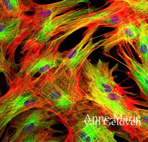
The fibroblast is one of the most important cells involved in ageing skin. You can find it in the lower layer of the epidermis and the dermis. It has many functions, one of which is the production of key components like hyaluron (filling + hydration), collagen (strength + structure) and elastin (flexibility + stretch). It particularly has to work hard to replenish hyaluronic acid or hyaluron as this filling component only has a half-life in the skin of several hours up to a day. Good quality collagen can last 15 years and elastin up to 70 years. It is also believed to be involved in the clean-up of dysfunctional components, like for example broken elastin, which is visible photodamage-damage and called solar elastosis. Fibroblast senescence (agedness) does also increase the risk of age spots. In proper ageing skin management, the fibroblast is a key target-cell.
Many aesthetic in-office treatments like ultrasound, radio-frequency, chemical peelings, laser etc. are based on causing controlled damage to the skin provoking wound-healing. This is the base of their rejuvenating or aesthetic impact. The number of new fibroblasts (myofibroblasts) is increased during the wound-healing process. Some injectables, like for example hyaluron-fillers cause the fibroblasts at the injection site to stretch and bio-stimulate collagen production. There are specific bio-stimulating injectable treatments. The most popular ones are Sculptra®, Radiesse®, Ellanse®, and a new one which combines hyaluron-filling and bio-stimulation is HArmonyCa®. As we age the fibroblast is undergoing some changes because of intrinsic and extrinsic factors. It loses it’s production power, it flattens, loses mechanical tension and therewith the ability to interact with other cells in the skin. It is becoming “tired and deaf”. My hypothesis was that injecting large droplets of hyaluron into the dermis might cause the fibroblast to become “lazy” via a negative feedback mechanism: when something is present in abundance, the fibroblast might not be stimulated enough to work hard to replenish it. This is not yet scientifically proven. It is important to keep the fibroblast in good shape and biologically active. We can stimulate it’s biological activity with skincare containing bio-stimulators, or ingredients which activate the production of important skin components by the fibroblast. On the other side we need to protect the cell from damage. Bio-stimulating active ingredients in skincare which have shown to particularly stimulate the fibroblast* are for example:
Protection from photo-damage we can achieve with a combination of sunscreen and anti-oxidants, more specifically Licochalcone A. Licochalcone A has a proven broad ability to protect the skin from damaging free-radicals or oxidative stress from UVA, UVB and HEVIS (High Energy Visible Light) affecting keratinocytes and fibroblasts. I am not yet aware of skincare ingredients which increase the number of (new) fibroblasts, like the semi or minimal invasive in-office treatments. It’s an interesting field to explore if this is possible without injury, inflammation or irritation. However, you probably get "more bang for your buck" by starting a a skincare routine with focus on bio-stimulation and protection of the fibroblast pre- and post minimal and semi invasive aesthetic treatments. This could be something we will proof with a clinical study. Take care *in vitro 12/15/2018 Comments Aquatic wrinkles![]()
After about spending some time in bathtub or in the pool, we can notice that our skin on particularly finger tops and toes start to wrinkle up. This wrinkling effect is believed to have a function.
When wet, things tend to be more slippery and our sophisticated skin is designed to counteract this by wrinkling up in a pattern optimised to provide a drainage network that improves grip, much like the tires on a car according to a study. Link to original publication. However, other studies would contradict that there would be a functional benefit for so called aquatic wrinkles. The osmosis theory Water molecules moving trough a semipermeable membrane from a low concentration area to a high concentration area is a process called osmosis. The shrinking and expanding effects of osmosis takes place simultaneously outer layer of the skin, causing wrinkles. The skin's outermost layer is also known as stratum corner could be responsible for this wrinkly reaction, The top layer of our skin consists of dead corneocytes. The longer these cells are attached to the skin, the bigger they are. The size of the corneocytes we actually use to objectively measure the skin's renewal and desquamation (shedding of cells) process. These dead (keratin containing) skin cells may absorb water and swell. The lower layer with living cells doesn't swell up. As top layer (which is increased in size) is still attached to the layer beneath, a wrinkly pattern is formed. The layer of dead skin cells is thicker at the palms of our hands and soles of our feet, the wrinkling effect is more evident. This response occurs more quickly in freshwater than seawater. Moreover, when we are exposed to water for a longer time, the water-repelling film on top of the outer layer of the skin may get impaired. The sympathetic nervous system / microcirculation theory It's known that there is a relationship between the wrinkling-effect and blood vessels constricting (narrowing) below the skin. When hands and feet are soaked in water, the nerve fibres in the skin shrink and the body temperature regulators loses volume. Therewith the top layer of the skin is pulled downward and the wrinkling pattern is formed. It is proven that wrinkling-effect response is impaired, if the nerves and/or blood vessels are damaged. Therewith the wrinkling effect can even be used to determine proper functioning of the sympathetic nervous system and/or skin's microcirculation. There is evidence that the wrinkling effect is impaired in patients suffering from diabetes: link to article. Regardless the cause, aquatic wrinkles disappear fast and the skin returns to normal once the water has evaporated. Take care. 7/15/2018 Comments Facial oils bad for skin?!![]()
Facial oils are a trending skin care product at the moment, loved and recommended by many "beauty guru's" and skin care experts. This is why I found it very interesting to read a comment written by a well respected dermatologist claiming that face oils would stifle skin renewal and exfoliation and would make skin dull over time. She must have a reason why she is saying this, and that's why I looked into this a little bit deeper.
To start with, I've done own research (not just me) with a facial oil, included many testers and found many benefits and no draw backs during the duration of the study. Moreover, I jumped out of my chair (literally) when I saw the visible results from the clinical photography, no joke! We've found that the oil (a combination of Argan oil and Lady's Thistle oil) improves moisture, elasticity and firmness, supports skin resilience, making the skin feel smooth and look more radiant. There was even a reduction of comedones detected. The results were published in a poster, accepted by the European Academy of Dermatology and Venereology in 2016. If you are an impatient person, and demand a fast answer, I can spill the tea right now: I've found no data to support that facial oils would stifle skin renewal and exfoliation, but the opposite. Moisturisers absolutely influence the skin barrier function and TEWL (transepidermal water loss - which is used to measure the skin barrier function). A good barrier function (confirmed by low TEWL), positively contributes to the skin cell renewal process, which includes skin exfoliation process. Very dry skin has an increased TEWL, and so does very well hydrated skin. There is simply more water on the skin surface to evaporate. A high TEWL with very well hydrated skin can therefore give the impression of an impaired barrier function and thus give a "false positive". This phenomenon is nicely explained in a publication by Marie Loden "Effect of moisturizers on epidermal barrier function". Looking at non fragrance plant oils also called fixed oils, there are many and they are all different, so it's impossible to generalise. Many plant oils, like almond, jojoba, soybean and avocado oils mostly remain on the skin surface. Even without penetrating deeper into the outer layer of the skin (called epidermis), the occlusive effect of plant oils will reduce water evaporation from the skin and help the growth of the cells of the top layer called keratinocytes. They actually support the skin barrier and therewith skin cell renewal. Part of the skin cell renewal is a process called desquamation, which is skin's natural exfoliation of dead skin cells. Helping this process will make skin appear more radiant and smooth, not duller. The benefits of plant oils are supported in many publications, one of which is found in the International Journal of Molecular Sciences anti inflammatory skin barrier repair effects by Tzu-Kai Lin 2017. Argan oil One of the most popular and well researched fixed oils is organ oil. It contains oleic and linoleic fatty acids. Both are part of our skin's natural intercellular lipid-enriched matrix or skin barrier. Linoleic acid (an omega 6 fatty acid) is in fact the most abundant polyunsaturated fatty acid. Our skin barrier is protecting our skin from water loss and penetration of external agressors. Thus the skin barrier keeps the good stuff in and bad stuff out. Linoleic acid plays a direct role in maintaining the integrity of this skin barrier. Some research shows that oleic acid may indeed disrupt the skin barrier and act as an permeability enhancer, helping other ingredients to penetrate deeper. When oleic acid is continuously applied, it could lead to barrier problems. Another ingredient in argan oil is tocopherol or vitamin E. Tocopherol is well known for it's antioxidative effect (neutralising damaging free radicals from pollution or sun which cause premature ageing) and lesser known for supporting the skin barrier. Daily topical application of argan oil (the finished product which contains multiple ingredients) has shown to improve skin elasticity (firmness), improve the skin hydration by restoring the barrier function and maintaining water-holding capacity. Furthermore it has a softening and relaxing effect on skin. Lady's Thistle oil The oil of the Milk Thistle plant (also known as Silybum Marianium) is a common ingredient in anti-aging skincare. It contains skin barrier supporting Linoleic acid and is known to nourish skin and improve radiance. Facial oils are certainly not for everybody, but in general skin will benefit from a cold pressed fixed plant oil or a mixture. Don't smother skin with oils, just apply a few drops by itself on the skin prior or after your moisturiser, or mix a few drops with your moisturiser of foundation. Hope you enjoy radiant skin & take care. ![]()
Understandably we want to get rid of pimples as soon as possible and sometimes apply harsh products to our skin in order to shrink them.
Depending on which ingredient is used, you might inflict injury to the skin and it's barrier which is very comparable to a mild burn. There is even a phenomenon called "toothpaste burn". During the healing process there is a risk of scarring. A study published by Tan J. et al in JJD 2017 shows that up to >87% of patients with mild to moderate acne reports atrophic scarring (sunken scar) to some degree. A healthy skin barrier and well hydrated skin will support a the healing process. However, as the barrier is impaired and the skin dried out, the skin's regeneration and healing process will take longer. Therefore be careful with "shrinking" pimples. The same applies for "popping" pimples, as this method by definition will cause injury to the skin. Picking and squeezing pimples will further irritate the skin tissue and delay proper healing. The risk of scarring is increased when the tissue is inflamed. A recent study of prevalence and risk factors of acne scarring confirmed that there is a relationship between the time between onset and effective treatment. Acne scars can be more difficult to treat than acne! It's better to seek expert advice if you have problematic skin. Take care. |
CategoriesAll Acne Age Clocks Ageing Aquatic Wrinkles Armpits Autophagy Biostimulators Blue Light & HEVIS Circadian Rhythms Cleansing Collagen CoQ10 Cosmetic Intolerance Syndrome Deodorant Dermaplaning Diabetes DNA Damage DNA Repair Dry Skin Epigenetics Evidence Based Skin Care Exfoliation Exosomes Eyes Face Or Feet? Facial Oils Ferroptosis Fibroblast Fingertip Units Gendered Ageism Glycation Growth Factors Gua Sha Hair Hair Removal Hallmark Of Aging Healthy Skin Heat Shock Proteins Hormesis Humidity Hyaluron Hyaluronidase Hypo-allergenic Indulging Jade Roller Keratinocytes Licochalcone A Lipid Peroxidation Luxury Skin Care Lymphatic Vessel Ageing Malar Oedema Menopause Mitochondrial Dysfunction Mood Boosting Skin Care Neurocosmetics Oily Skin Ox Inflammageing Peptides PH Balance Skin Photo Biomodulation Polynucleotides Proteasome Psoriasis Regeneration Regenerative Treatments Review Safety Scarring Sensitive Skin Skin Care Regimen Skin Flooding Skin Hydration Skin Senescence Skip-Care Sleep Slugging Sunscreen Tanning Under Eye Bags UV Index Vitamin C Vitamin D Well Ageing Skin Care Wound Healing Wrinkles
Archives
January 2025
|
Proudly powered by Weebly
Anne-Marie van Geloven © 2024 All rights reserved
|