Live your best life & take care
12/7/2024 Comments Collagen bankingCollagen banking is a proactive skincare strategy falling under the category prejuvenation aimed at preserving and stimulating collagen production to maintain youthful, firm and excellent skin quality over time. This approach can involve using various treatments, tweakments, products, supplements and lifestyle choices to boost collagen levels before significant signs of aging appear. The goal is to build a "reserve" or “bank” of collagen, ensuring skin remains resilient and less prone to wrinkles and sagging as natural collagen production declines and degradation increases with age. To start banking collagen as early as in your twenties makes sense, as the producing cell called the dermal fibroblast is still very healthy and active. Moreover as the loss is not yet so great (just a few percent loss), thus less invasive methods work well to maintain a youthful status quo. I´s never too late to start “banking” collagen, although when you are more mature, the word rejuvenation might be more suitable. There is no direct scientific evidence that collagen stimulation is more effective in your twenties than in your sixties. However, starting collagen stimulation earlier may be beneficial: ▌Collagen production begins to decline around age 25-30, decreasing by about 1% per year. ▌By the 50s and beyond, the collagen loss is greater >30%, becomes more noticeable and it´s always harder to get back what you lost than to maintain what you have. ▌Peak collagen levels occur at twenty years of age, thus maintaining what you have the highest achievable level. ▌Starting collagen stimulation treatments earlier may help prevent further collagen loss and require less invasive and number of treatments. WHAT IS COLLAGEN Collagen is the most abundant protein in the human body, making up about one-third of all proteins. 1. Location: Found in connective tissues, including skin, tendons, bones, and cartilage. 2. Function: Provides structural support, strength, and elasticity to tissues. 3. Production: Naturally produced by the body, but production decreases with age, starting around the mid-20s. Collagen plays a crucial role in maintaining skin elasticity, joint health, and overall tissue integrity. As collagen production declines with age, so does the skin quality, leading to visible signs of aging like wrinkles, loss of elasticity and firmness, and sagging skin. Collagen is one of the key target components for noticeable and effective skin rejuvenation or regeneration. There are at least 28 types of collagen in the human body, but the most abundant and relevant for skin are: [1] Type I Collagen: ▌Most abundant type in the skin, making up about 80-90% of skin's collagen. ▌Provides tensile strength and structure to the skin. ▌Maintains skin elasticity and firmness. Type III Collagen: ▌Found alongside Type I collagen in the skin, comprising about 8-12% of skin collagen. ▌Contributes to skin firmness and elasticity. ▌Important in early stages of wound healing and fetal development. Type IV Collagen: ▌Found in the basement membrane of the skin. ▌Provides support and filtration in the basement membranes. ▌Crucial for overall skin health and function. Type V and VI Collagen: ▌Present in smaller amounts in the skin. ▌Support skin health and collagen fibril formation. Collagen is primarily composed of three key amino acids: ▌Glycine: is the most abundant, contributing significantly to collagen's structure and stability ▌ Proline ▌ Hydroxyproline Proline and hydroxyproline are crucial for forming the triple-helix structure of collagen, which provides strength and flexibility. Additionally, essential amino acids like lysine play a critical role in collagen synthesis by forming hydroxylysine, important for stabilizing collagen fibers. A balanced intake of these amino acids is vital for maintaining healthy collagen levels in the body. COLLAGEN DECLINE Collagen production begins to diminish naturally in our mid-20s, decreasing by about 1% per year [2]. This decline becomes more pronounced in the 40s and 50s, contributing to visible signs of aging such as wrinkles and sagging skin [2]. Factors influencing collagen loss include genetic predisposition (DNA), changes in epigenetic pattern (influenced by environment), hormonal changes (especially post-menopause), and fibroblast aging [2][3]. Environmental factors like UV exposure and pollution, and lifestyle decisions like smoking, and poor diet, poor sleep and stress further accelerate collagen degradation [4]: 1. UV exposure stimulates the production of matrix metalloproteinases (MMPs), enzymes that break down collagen in the skin. 2. Smoking constricts blood vessels in the skin, depriving it of oxygen and nutrients crucial for collagen production. It also increases MMP production and generates free radicals that damage collagen fibers. 3. Poor diet, particularly high sugar consumption, can lead to glycation, a process that makes collagen dry, brittle, and weak. COLLAGEN DEGRADATION Collagen degradation is a complex process involving several key enzymes, primarily from the matrix metalloproteinase (MMP) family, along with other proteases. The degradation of collagen as one of the components of the ECM (extracellular matrix) is a very important process in the development, morphogenesis, tissue remodeling, and repair. 1. Matrix Metalloproteinases (MMPs): Typical structure of MMPs consists of several distinct domains. MMP family can be divided into six groups: collagenases, gelatinases, stromelysins, matrilysins, membrane-type MMPs, and other non-classified MMPs [6]. ▌Collagenases: MMP-1, MMP-8, and MMP-13 are responsible for cleaving fibrillar collagen into smaller fragments [6][7]. ▌Gelatinases: MMP-2 and MMP-9 further degrade denatured collagen (gelatin) into smaller peptides [8]. ▌Stromelysins: MMP-3 and MMP-10 degrade non-collagen ECM components but can also activate other MMPs [7]. ▌Matrilysins: MMP-7 and MMP-26 contribute to ECM remodeling by degrading various substrates, including collagen [7]. 2. Proteases Serine proteases: ▌Elastase: Degrades elastin and can enhance the activity of MMPs, contributing to collagen breakdown [7]. Cysteine proteases: ▌Cathepsins: Particularly cathepsin K, which degrades collagen in bone and cartilage tissues [9]. Aspartic proteases: ▌These enzymes participate in the breakdown of ECM proteins under specific conditions, although their role in direct collagen degradation is less prominent compared to MMPs [7]. Papain-like cysteine proteases: ▌Known for its ability to degrade collagen under acidic conditions, often used in studies related to scar tissue remodeling [9]. These enzymes work together to regulate collagen turnover, ensuring proper tissue remodeling and repair while preventing excessive degradation that can lead to tissue damage and aging. DISORGANISED COLLAGEN In young skin, collagen fibrils are abundant, tightly packed, and well-organized, displaying characteristic d-bands. This organization provides structural integrity and elasticity to the skin [10]. In contrast, aged skin shows fragmented and disorganized collagen fibrils. These fibrils are rougher, stiffer, and harder, contributing to the skin's reduced elasticity and increased fragility [10]. The disorganization in more mature skin is primarily due to the breakdown of collagen by matrix metalloproteinases (MMPs) and non-enzymatic processes like glycation, which lead to structural changes and impair skin function [10][3]. IMPACT OF GLYCATION ON COLLAGEN Glycation is a non-enzymatic process where sugars bind to proteins like collagen, leading to the formation of advanced glycation end-products (AGEs). This process causes collagen fibers to become stiff, disorganized, and less functional, contributing to skin aging and reduced elasticity [11][12]. I wrote a full blogpost on skin glycation, however not specific about collagen with a surprising effect of spray tan. Read more. Prevention and treatment of glycation [13][14][15] 1. Dietary modifications: ▌Reduce intake of refined sugars and high-AGE foods (e.g., fried and processed foods). ▌Consume antioxidant-rich foods such as fruits, vegetables, and green tea to combat oxidative stress. 2. Lifestyle changes: ▌Regular exercise helps maintain stable blood sugar levels ▌Adequate hydration supports skin health. 3. Cooking methods: ▌Use moist heat methods like steaming or poaching to minimize AGE formation. 4. Skincare: ▌Use products with anti-glycation agents like carnosine or NAHP or Acetyl Hydroxyproline. ▌Inhibitors of protein glycation include antioxidants, such as vitamin C and vitamin E commonly found in skincare. COLLAGEN PRODUCTION Collagen production is a multi-step process involving both intracellular and extracellular activities.
SKINCARE INGREDIENTS THAT STIMULATE COLLAGEN PRODUCTION 1. Vitamin A and derivatives Retinoids (Retinol = cosmetic ingredient, Tretinoin = prescription strenght) Retinoids increase collagen production by promoting fibroblast activity and reducing collagenase activity, which breaks down collagen. This is a dose-dependant effect. The regeneration or renewal from the epidermis is boosted so efficently that the lipid production can´t keep up, hence this is one of the reasons why many experience dry skin symptoms at the start and irritation. Lipids are like the morter between the bricks of the skin barrier. When the barrier is not intact, water from the skin can evaporate and irritants can penetrate. To reduce this unwanted effect, you can slowly introduce this ingredient into your skincare regimen and start with a low dose or formulations with lower irritation potential. Vitamin A, specifically prescription strenght is considered the most evidence based topical ingredient. 2. Vitamin C (Ascorbic Acid) Vitamin C, also known as ascorbic acid, plays a crucial role in collagen synthesis and maintenance, significantly influencing skin health and structural integrity. Because it is such an important ingredient and this post would add up to a 30 minutes read, I´ve dedicated a new full article on the role of vitamin C in collagen production, degradation and various forms of vitamin C. Click here. 3. Peptides There are many different peptides fround in skincare formulation. We can identify the following main types by function: 1. Carrier peptides: These help deliver trace elements like copper and manganese necessary for wound healing and enzymatic processes. 2. Signal peptides: These stimulate collagen, elastin, and other protein production by sending "messages" to specific cells. 3. Neurotransmitter-inhibiting peptides: These claim to work similarly to Botulinumtoxin, reducing muscle contractions that lead to expression lines. 4. Enzyme-inhibitor peptides: These block enzymes that break down collagen and other important skin proteins. 5. Antimicrobial peptides: These provide a defense against harmful microorganisms on the skin. 6. Antioxidant peptides: These help protect the skin from oxidative stress and free radical damage. Collagen stimulating peptides Mode of Action: Collagen peptides potentially stimulate fibroblast proliferation and increase the expression of collagen and elastin genes, enhancing the structural integrity of the skin [17][18]. While many peptides are too large to penetrate the skin effectively, some collagen-stimulating peptides have shown evidence of skin penetration and efficacy in skincare formulations. 1. Penetration-enhancing techniques: Various methods have been developed to improve peptide penetration into the skin, including chemical modification, use of penetration enhancers, and encapsulation in nanocarriers [19]. 2. Specific evidence based peptides: ▌GHK (Glycyl-L-histidyl-L-lysine): This copper peptide has shown ability to penetrate the skin and stimulate collagen production [20]. ▌KTTKS (Lysine-threonine-threonine-lysine-serine): When modified with palmitic acid (palmitoyl pentapeptide-4), this peptide demonstrated improved skin penetration and collagen-stimulating effects [20]. ▌GEKG (Glycine-glutamic acid-lysine-glycine): Studies have shown this tetrapeptide can penetrate the skin when used with appropriate delivery systems [21]. GEKG significantly induces collagen production at both the protein and mRNA levels in human dermal fibroblasts [22]. GEKG is derived from extracellular matrix (ECM) proteins and has been shown to enhance gene expression responsible for collagen production up to 2.5-fold, boosts collagen, hyaluronic acid, and fibronectin production by dermal fibroblasts [22]. ▌Palmitoyl Pentapeptide Mode of Action: Act as signaling molecules to stimulate collagen production by mimicking broken down collagen fragments signaling fibroblasts to produce more collagen [17][18]. Their efficacy can vary depending on the specific formulation, percentage and delivery method used. More about peptides click here 4. Glycine Saponins ▌Mode of action: Glycine saponins are known to stimulate hyaluronic acid, collagen and elastin synthesis in the skin (in vitro). 5. Creatine ▌Mode of action: Creatine is a popular supplement used by bio-hackers. However there are benefits for this ingredient in topical applications too. In vitro (cells) it has shown to increase collagen type I by +24%, collagen type IV + 11% and elastin +36% vs untreated control. 7. Growth factors ▌Mode of action: Growth factors like TGF-β stimulate collagen production by activating fibroblasts and promoting cellular regeneration.TGF-β has been shown to enhance the production of types I and III collagens by cultured normal human dermal fibroblasts, with a 2-3-fold increase in collagen production compared to control cells [23]. 8. Bakuchiol [24] This ingredient is underestimated and misnamed as “phyto-retinol” as it stimulates (like retinol) pro-collagen production with less irritation potential. However this is where the comparison stops. It is a potent anti-oxidant, stimulates fibronectin (component in the ECM which keeps it nice and organized) ex-vivo and so much more. Researchers have found that bakuchiol outperforms retinol in inhibiting the activity of two crucial matrix metalloproteinase enzymes, MMP-1 and MMP-12, which are responsible for the breakdown of collagen and elastin in the skin. The study emphasizes that bakuchiol not only mimics some of the beneficial effects of retinol but also offers a gentler option for those with sensitive skin or those who may be pregnant or breastfeeding, where Retinol (and absolutely Tretinoin) use is often discouraged. Bakuchiol doesn’t seem to act via the retinoic acid receptors, which isn’t that surprising if you compare its structure to retinol and tretinoin, while bakuchiol superficially resembles them, its six-membered ring is aromatic and flat, and oxygen is on the other end of the molecule. 9. Alpha Hydroxy Acids (AHAs) and Beta Hydroxy Acids (BHAs)
Play significant roles in skincare, particularly in promoting skin health and rejuvenation. Their mechanisms of action include stimulating collagen production, through different pathways. Alpha Hydroxy Acids (AHAs) AHAs, such as glycolic acid and lactic acid, are primarily known for their exfoliating properties. They work by breaking down the bonds that hold dead skin cells together, promoting cell turnover and revealing fresher skin beneath. However, AHAs also have a direct impact on collagen production: 1. Direct stimulation: Studies have shown that treatments with AHAs lead to increased skin thickness and density of collagen in the dermis, suggesting a direct enhancement of collagen production [25][26][27]. 2. Mechanisms of action: AHAs promote the production of glycosaminoglycans (GAGs) and improve the quality of elastic fibers, which are vital for maintaining skin structure and elasticity [26][27]. Beta Hydroxy Acids (BHAs) BHAs, with salicylic acid being the most common example, are oil-soluble acids that penetrate deeper into pores. While their primary function is to exfoliate and clear out clogged pores, they also contribute to collagen production: 1. Indirect: The exfoliation process initiated by BHAs can lead to increased cell turnover, which indirectly supports collagen production by creating an environment conducive to skin regeneration [28]. By removing dead skin cells and promoting new cell growth, BHAs help maintain a healthier skin matrix. 2. Anti-inflammatory effects: BHAs possess anti-inflammatory properties that can reduce redness and irritation in the skin. This reduction in inflammation can create a more favorable environment for collagen synthesis over time [28]. 10. Niacinamide (Vitamin B3) Scientific studies have demonstrated that niacinamide can significantly enhance collagen production and inhibit matrix metalloproteinases (MMPs), which are enzymes responsible for collagen degradation. 1. Increased collagen production: Research indicates that topical application of niacinamide leads to a notable increase in collagen synthesis. A study found that fibroblasts treated with niacinamide exhibited more than a 50% increase in collagen production, highlighting its effectiveness in rejuvenating skin structure [29]. 2. Inhibition of MMPs: Niacinamide has also been shown to inhibit the activity of MMPs, particularly MMP-1 and MMP-12. These enzymes break down collagen and elastin, contributing to skin aging. By reducing MMP activity, niacinamide helps maintain skin elasticity and firmness [30]. 3. Mechanistic insights: The mechanisms behind niacinamide's effects include its role in enhancing cellular energy metabolism and reducing oxidative stress. Niacinamide influences the activity of enzymes critical for cellular function, such as sirtuins and poly(ADP-ribose) polymerases (PARP), which are essential for DNA repair and cellular health [31]. Additionally, niacinamide has been shown to increase levels of antioxidant enzymes like superoxide dismutase, further protecting against oxidative damage that can lead to collagen breakdown [32]. IN-OFFICE TREATMENTS STIMULATING COLLAGEN PRODUCTION This innovative field of regenerative medicine showcases a variety of treatment options, each with unique characteristics and potential benefits. It's essential to recognize that the effectiveness of collagen-stimulating treatments can differ from person to person. For the best outcomes, a combination of methods may be suggested. A qualified healthcare professional can evaluate your individual needs and goals to determine the most suitable treatment approach for you. 1. INJECTABLE TREATMENTS ▌Sculptra (Poly-L-lactic acid): Stimulates collagen type I production through neocollagenesis, creating a controlled inflammatory response that activates fibroblasts [40]. This treatment is often referred to as biostimulation or chemical biostimulation. Key points about Sculptra and collagen stimulation: 1. Injection depth: Sculptra is typically injected into the deep dermis or subcutaneous layers, not the superficial dermis [41]. 2. Collagen production: The microparticles in Sculptra stimulate fibroblasts to produce new collagen, leading to gradual volume restoration [41]. 3. Mechanism: Sculptra works through a process called neocollagenesis, where the poly-L-lactic acid microparticles induce a controlled inflammatory response, stimulating collagen production [42]. 4. Treatment classification: This approach is classified as biostimulation or chemical biostimulation, as it uses a biocompatible material to stimulate the body's natural collagen production [42]. 5. Onset of results: Unlike hyaluronic acid fillers, Sculptra's effects are not immediate. Results typically begin to show around 12 weeks after treatment and continue to improve over 6 to 12 months [43]. 6. Treatment sessions: Most patients require 2 to 3 treatment sessions spaced 4 to 6 weeks apart to achieve optimal results [43]. Sculptra primarily stimulates Type I collagen production in the skin. According to peer-reviewed research: 1. Type I Collagen: Sculptra has been shown to increase Type I collagen production by 66.5% after 3 months of treatment [44]. 2. Efficacy: Sculptra's collagen-stimulating effects can last up to 25 months or about 2 years [44]. ▌Sculptra works differently than traditional fillers or treatments like lasers and microneedling. It acts as a bio-activator, triggering the body's natural collagen production over time [44]. ▌The gradual collagen production stimulated by Sculptra can lead to more natural-looking and longer-lasting results compared to some other treatments [44]. ▌Radiesse (Calcium Hydroxylapatite): Provides immediate volume and stimulates collagen type I and mostly type III production over time through a scaffold effect. ▌Exosomes: Derived from stem cells (or other sources), they promote healing and collagen synthesis through growth factor release. ▌Mode of action: Deliver growth factors and cytokines to fibroblasts, enhancing collagen production and repair processes [38]. ▌Efficacy: Emerging evidence suggests improved skin rejuvenation outcomes. ▌Polynucleotides: These biopolymers enhance skin hydration and stimulate collagen production via cellular signaling. ▌Mode of action: Injected polynucleotides enhance fibroblast activity and collagen synthesis by providing nucleic acids that support cell repair and regeneration [37]. ▌Efficacy: Improves skin hydration and elasticity over time. ▌Hyaluronic Acid fillers: While primarily volumizing, they can also promote collagen synthesis indirectly by hydrating the skin. 2. ENERGY-BASED TREATMENTS ▌Ultherapy: Uses micro-focused ultrasound to create thermal coagulation points, stimulating collagen remodeling. ▌Mode of action: Uses focused ultrasound energy to heat deeper layers of the skin, stimulating collagen production through mechanical stretching of fibroblasts [36]. ▌Efficacy: Clinically shown to lift and tighten skin over several months post-treatment. ▌HIFU (High-Intensity Focused Ultrasound): Similar to Ultherapy, it targets deeper layers of skin to induce collagen synthesis through thermal effects. ▌SoftWave therapy is a non-invasive shockwave treatment that uses a patented technology to promote natural healing at the cellular level. It operates by generating therapeutic energy waves through a high-energy electrical discharge in water, which creates pressure waves that stimulate blood flow and activate the body’s healing processes. This method is particularly effective for addressing chronic pain, sports injuries, and conditions like arthritis by enhancing tissue regeneration and reducing inflammation. ▌Tissue regeneration: The therapy enhances blood supply to tissues, facilitating faster recovery from injuries. It stimulates the production of collagen and activates resident stem cells, which are crucial for tissue repair. ▌No downtime: Treatments are quick, typically lasting between 10 to 15 minutes, and patients can resume their normal activities immediately afterward with minimal side effects. This makes it a convenient option for those seeking effective pain management without the need for surgery or medication. ▌Radiofrequency (RF) treatments: Includes devices like Thermage and Morpheus8, which deliver RF energy to stimulate collagen production through thermal effects. ▌Mode of action: Delivers heat to the dermis, causing collagen fibers to contract (tightening) and stimulating new collagen production [35]. ▌Efficacy: Enhances skin firmness and elasticity with visible results after a few sessions. ▌Tixel: Tixel sessions involve a non-invasive skin rejuvenation treatment that utilizes Thermo-Mechanical Ablation (TMA) technology. The Tixel device features a heated titanium tip that creates controlled micro-channels in the skin, stimulating collagen production and promoting healing. ▌Duration: Each session lasts between 20 to 45 minutes, depending on the treatment area and specific skin concerns. ▌Areas treated: Effective for fine lines, wrinkles, acne scars, sun damage, and skin laxity, particularly around delicate areas like the eyes and neck. ▌Downtime: Minimal downtime is required, with some redness and sensitivity similar to a mild sunburn lasting up to three days. ▌Results: Improvements can be seen after one session, but optimal results typically require 3 to 6 sessions spaced several weeks apart. 3. LASER TREATMENTS ▌Ablative lasers (e.g., CO2 Laser): Vaporize tissue and stimulate significant collagen remodeling. ▌Non-ablative lasers: Deliver heat to stimulate collagen without damaging the surface of the skin. ▌Mode of action: Uses laser energy to create controlled thermal damage, promoting collagen remodeling and synthesis [34]. ▌Efficacy: Proven to improve skin tone, texture, and reduce wrinkles with a series of treatments. ▌HALO treatments refer to a type of hybrid fractional laser therapy designed to improve skin texture, tone, and overall appearance. The HALO laser combines two types of wavelengths: 1. Ablative (2940 nm): Targets the epidermis (outer skin layer) to address surface issues like fine lines, sun spots, and uneven texture. 2. Non-ablative (1470 nm): Penetrates deeper into the dermis to stimulate collagen production and treat deeper skin concerns. ▌Customizable treatments: Each session can be tailored to individual skin needs, allowing for varying levels of intensity and downtime. ▌Minimal downtime: Patients typically experience mild redness and peeling for a few days, with many returning to normal activities quickly. ▌Results: Improvements in skin clarity, reduction of fine lines, and enhanced radiance can often be seen within a week, with optimal results developing over time. HALO treatments are suitable for all skin types and are often recommended for those seeking significant anti-aging benefits without extensive recovery time. Intense Pulsed Light (IPL) ▌Mode of action: Uses broad-spectrum light to induce controlled thermal injury, stimulating collagen synthesis as part of the skin's repair mechanism [39]. ▌Efficacy: Effective for reducing pigmentation and improving overall skin texture. 4. MICRONEEDLING ▌Traditional microneedling: Creates micro-injuries to stimulate the body’s natural healing response and collagen production by activating fibroblasts [33]. ▌Efficacy: Studies show significant improvements in skin texture and elasticity after multiple sessions. ▌Microneedling with RF: Combines traditional microneedling with RF energy for enhanced results. 5. THREAD LIFTING ▌PDO Threads: Absorbable threads that lift the skin while simultaneously stimulating collagen production as they dissolve. 6. SKIN BOOSTERS: BIO-STIMULATORS ▌Profhilo: A hyaluronic acid-based treatment that hydrates the skin and stimulates collagen and elastin production. ▌Ellanse: A biostimulator that provides immediate volume and stimulates long-term collagen type I and type III production. 7. LIGHT THERAPY ▌LED Light Therapy (LLT): Uses specific wavelengths of light to promote cellular activity and stimulate collagen production. OTHER TREATMENTS ▌Micro-Coring™ technology Ellacor is a non-surgical skin tightening treatment called Micro-Coring™ technology to improve the appearance of moderate to severe wrinkles and skin laxity, particularly in the mid and lower face. This innovative procedure uses hollow needles to remove microscopic plugs of skin, stimulating the body’s natural healing response, which promotes collagen and elastin production. ▌Procedure: Up to 12,000 micro-cores can be removed in a session, with each core being less than 0.5 mm in diameter, minimizing the risk of scarring. ▌Treatment duration: Sessions typically last around 30 minutes, and multiple treatments may be needed for optimal results. ▌Recovery: Most patients experience mild redness and swelling but can usually resume normal activities within a few days. Ellacor offers a unique alternative to traditional surgical methods, providing significant skin rejuvenation without thermal injury or extensive downtime. ▌Pulsed Radiofrequency (PRF) and Platelet-Rich Plasma (PRP) are emerging treatments in regenerative aesthetics, particularly for their roles in enhancing collagen production and promoting tissue healing. Pulsed Radiofrequency (PRF) is a technique that utilizes electromagnetic fields to induce thermal and electrical changes in tissues, which can promote healing and regeneration. PRP is an autologous preparation derived from a patient's blood, enriched with platelets and growth factors that facilitate tissue repair. 1. Mechanism of Action: ▌ PRF generates a pulsed electromagnetic field that enhances cellular activity and promotes healing through the release of growth factors from platelets [45][46]. ▌PRP contains a high concentration of platelets that release various growth factors, such as platelet-derived growth factor (PDGF) and vascular endothelial growth factor (VEGF), which are essential for tissue regeneration [46][47]. 2. Collagen production: ▌Both PRF and PRP stimulate fibroblast activity, leading to increased collagen synthesis. Studies have shown that the application of PRP can significantly enhance collagen production in various tissues [48][49]. 3. Clinical applications: ▌PRF has been effectively used in pain management and regenerative medicine, particularly for conditions like chronic pain due to peripheral tissue damage [45]. ▌PRP has gained popularity in dermatology and plastic surgery for its ability to accelerate wound healing and improve skin texture [47][48]. 4. Combination therapy: ▌The combination of PRF and PRP has shown synergistic effects, enhancing the activation of platelets and improving clinical outcomes in regenerative applications [45]. This approach may lead to better tissue repair compared to either treatment alone. 5. Safety profile: ▌ Both treatments are considered safe due to their autologous nature, minimizing risks associated with immune reactions or disease transmission [46][47]. 6. Efficacy duration: ▌The effects of both therapies can be long-lasting; studies indicate that the benefits of PRP can persist for several months post-treatment, depending on the condition being treated [48][49]. OVERSTIMULATION Many of the collagen stimulating methods used are by “controlled damage proking repair”. While collagen is generally beneficial, excessive damage, repair and stimulation or abnormal production can lead to fibrosis or scarring. Read more. Prevent potential adverse effects: 1. Use FDA-approved devices and treatments 2. Seek treatment from qualified professionals 3. Follow recommended treatment intervals 4. Avoid overtreatment or combining too many modalities simultaneously or with very short periods in between Collagen loss is a continuous process which is significantly impacted by sunlight, environment and lifestyle (sleep, stress, exercise, low alcohol, no smoking, diet). There are simple steps you can take to slow down or even reverse this process, for example with daily use of a broadspectrum sunscreen and a tailored skincare routine with vitamin C, peptides, growth factors or supplementation with collagen powder in case your diet (especially vegetarians) doesn´t provide enough building blocks to produce collagen. Always consult a qualified healthcare professional to determine what the most suitable approach is for your skin health and beauty. Take care Anne-Marie References [1] Ricard-Blum, S. (2011). The collagen family. Cold Spring Harbor Perspectives in Biology, 3(1), a004978. https://doi.org/10.1101/cshperspect.a004978 [2] Shuster S, Black MM, McVitie E. "The influence of age and sex on skin thickness, skin collagen and density." British Journal of Dermatology. 1975;93(6):639-643. doi:10.1111/j.1365-2133.1975.tb05113.x. [3] Varani J, Dame MK, Rittie L, Fligiel SE, Kang S, Fisher GJ, Voorhees JJ. Decreased collagen production in chronologically aged skin: roles of age-dependent alteration in fibroblast function and defective mechanical stimulation. Am J Pathol. 2006 Jun;168(6):1861-8. doi: 10.2353/ajpath.2006.051302. PMID: 16723701; PMCID: PMC1606623. [4] Farage MA, Miller KW, Elsner P, Maibach HI. Aging Clin Exp Res. 2008;20(3):195-204. doi:10.1007/BF03020230. [6] Jabłońska-Trypuć, A., Matejczyk, M., & Rosochacki, S. (2016). Matrix metalloproteinases (MMPs), the main extracellular matrix (ECM) enzymes in collagen degradation, as a target for anticancer drugs. Journal of Enzyme Inhibition and Medicinal Chemistry, 31(sup1), 177–183. https://doi.org/10.3109/14756366.2016.1161620 [7] Ledwoń P, Papini AM, Rovero P, Latajka R. Peptides and Peptidomimetics as Inhibitors of Enzymes Involved in Fibrillar Collagen Degradation. Materials (Basel). 2021 Jun 10;14(12):3217. doi: 10.3390/ma14123217. PMID: 34200889; PMCID: PMC8230458. [8] Reilly DM, Lozano J. Skin collagen through the lifestages: importance for skin health and beauty. Plast Aesthet Res. 2021;8:2. http://dx.doi.org/10.20517/2347-9264.2020.153 [9] Sys Rev Pharm 2021;12(03):676-684 A multifaceted review journal in the field of pharmacy Does Papain Enzyme Improve Collagen Degradation? Herman Y. L. Wihastyoko et al. [10] He T, Fisher GJ, Kim AJ, Quan T. Age-related changes in dermal collagen physical properties in human skin. PLoS One. 2023 Dec 8;18(12):e0292791. doi: 10.1371/journal.pone.0292791. PMID: 38064445; PMCID: PMC10707495. Age-related changes in dermal collagen physical properties in ... https://pmc.ncbi.nlm.nih.gov/articles/PMC10707495/ [11]Trujillo, J., & Galligan, J. J. (2024). An overview on glycation: molecular mechanisms, impact on biomolecules, and related diseases. Glycoconjugate Journal. https://doi.org/10.1007/s10719-024-10254-y [12]Sadowska-Bartosz, I., & Bartosz, G. (2022). Accumulation of Advanced Glycation End-Products in the Body and Its Prevention. Nutrients, 14(19), 4072. https://doi.org/10.3390/nu14194072 [13] Sadowska-Bartosz, I., & Bartosz, G. (2015). Prevention of protein glycation by natural compounds. Molecules, 20(2), 3309-3334. [14] Uribarri, J., et al. (2015). Dietary advanced glycation end products and their role in health and disease. Advances in Nutrition, 6(4), 461-473. [15] Guilbaud, A., et al. (2016). How can diet affect the accumulation of advanced glycation end-products in the human body? Foods, 5(4), 84. [16] Wu, M., Cronin, K., & Crane, J. (2023). Biochemistry, Collagen Synthesis. In StatPearls [Internet]. StatPearls Publishing. Available from: https://www.ncbi.nlm.nih.gov/books/NBK507709/ [17] Edgar, S., Hopley, B., Genovese, L. et al. Effects of collagen-derived bioactive peptides and natural antioxidant compounds on proliferation and matrix protein synthesis by cultured normal human dermal fibroblasts. Sci Rep 8, 10474 (2018). https://doi.org/10.1038/s41598-018-28492-w [18] Frontiers | Collagen peptides affect collagen synthesis and the expression of collagen, elastin, and versican genes in cultured human dermal fibroblasts https://www.frontiersin.org/journals/medicine/articles/10.3389/fmed.2024.1397517/full [19] International Journal of Cosmetic Science Skin permeability, a dismissed necessity for anti-wrinkle peptide performance Seyedeh Maryam Mortazavi, Hamid Reza Moghimi First published: 18 March 2022 https://doi.org/10.1111/ics.12770 [20] Pickart L, et al. GHK Peptide as a Natural Modulator of Multiple Cellular Pathways in Skin Regeneration. Biomed Res Int. 2015;2015:648108. doi:10.1155/2015/648108. [21] Binder L, et al. Dermal peptide delivery using enhancer molecules and colloidal carrier systems--A comparative study of a cosmetic peptide. Int J Pharm. 2018;557:36-46. doi:10.1016/j.ijpharm.2018.08.019. [22] https://pubmed.ncbi.nlm.nih.gov/21692860/ Farwick M, Grether-Beck S, Marini A, Maczkiewitz U, Lange J, Köhler T, Lersch P, Falla T, Felsner I, Brenden H, Jaenicke T, Franke S, Krutmann J. Bioactive tetrapeptide GEKG boosts extracellular matrix formation: in vitro and in vivo molecular and clinical proof. Exp Dermatol. 2011 Jul;20(7):602-4. doi: 10.1111/j.1600-0625.2011.01307.x. PMID: 21692860. [23] Ignotz, R. A., & Massagué, J. (1986). Transforming growth factor-beta stimulates the expression of fibronectin and collagen and their incorporation into the extracellular matrix. Journal of Biological Chemistry, 261(9), 4337-4345. [24] Bluemke, A., Ring, A. P., Immeyer, J., Hoff, A., Eisenberg, T., Gerwat, W., Meyer, F., Breitkreutz, F., Klinger, S., Brandner, L. M., Sandig, J. M., Seifert, G., Segger, M., Rippke, D., Schweiger, F., & Dorothea, R. (2022). Multidirectional activity of bakuchiol against cellular mechanisms of facial ageing – Experimental evidence for a holistic treatment approach. International Journal of Cosmetic Science, 44(5), 558-570. doi:10.1111/ics.12784. [25] Ditre CM, Griffin TD, Murphy GF, Sueki H, Telegan B, Johnson WC, Yu RJ, Van Scott EJ. Effects of alpha-hydroxy acids on photoaged skin: a pilot clinical, histologic, and ultrastructural study. J Am Acad Dermatol. 1996 Feb;34(2 Pt 1):187-95. doi: 10.1016/s0190-9622(96)80110-1. PMID: 8642081. [26] Almeman, A. A. (2024). Evaluating the Efficacy and Safety of Alpha-Hydroxy Acids in Dermatological Practice: A Comprehensive Clinical and Legal Review. Clinical, Cosmetic and Investigational Dermatology, 17, 1661–1685. doi:10.2147/CCID.S453243. [27] Karwal, K.; Mukovozov, I. Topical AHA in Dermatology: Formulations, Mechanisms of Action, Efficacy, and Future Perspectives. Cosmetics 2023, 10, 131. https://doi.org/10.3390/cosmetics10050131 [28] He, X.; Wan, F.; Su, W.; Xie, W. Research Progress on Skin Aging and Active Ingredients. Molecules 2023, 28, 5556. https://doi.org/10.3390/molecules28145556 [29] Bissett, D. L., Oblong, J. E., & Matts, P. J. (2004). Niacinamide: A B vitamin that improves the appearance of aged skin. *Journal of Cosmetic Dermatology*, 3(1), 1-7. doi:10.1111/jocd.12004. [30] Hakozaki, T., Minwalla, Z., & Zhuang, J. (2002). The effect of niacinamide on reducing cutaneous pigmentation and suppression of melanosome transfer. *British Journal of Dermatology*, 147(20), 20-31. [31] Huang, Y., Zhang, Y., & Chen, N. (2024). Mechanistic insights into the multiple functions of niacinamide: A narrative review. *PMC*. doi:10.1007/s12325-024-02045-0. [32] Kumar, S., & Gupta, R. (2024). Niacinamide: A versatile ingredient in dermatology and cosmetology. *PMC*. doi:10.1007/s12325-024-02046-z. [33] Alam, M., Han, S., Pongprutthipan, M., Disphanurat, W., Kakar, R., Nodzenski, M., Pace, N., Kim, N., Yoo, S., Veledar, E., Poon, E., & West, D. P. (2014). Efficacy of a needling device for the treatment of acne scars: A randomized clinical trial. JAMA Dermatology, 150(8), 844-849. https://doi.org/10.1001/jamadermatol.2013.8687 [34] Zhang, Y., Li, H., Wang, J., & Wang, Y. (2023). Dynamic panoramic presentation of skin function after fractional CO2 laser. Journal of Cosmetic Dermatology, 22(8), 3098-3105. https://doi.org/10.1111/jocd.16445 [35] Fabi, S. G., & Sundaram, H. (2013). The role of radiofrequency in skin tightening. Journal of Clinical and Aesthetic Dermatology, 6(9), 35-42. Retrieved from https://www.ncbi.nlm.nih.gov/pmc/articles/PMC3799110/ [36] Sullivan, P. K., & Heller, M. M. (2017). The role of ultrasound in skin rejuvenation: A review of the literature. Journal of Cosmetic Dermatology, 16(1), 18-25. https://doi.org/10.1111/jocd.12279 [37] Pérez, M. R., & Gutiérrez, J. M. (2021). Polynucleotides in aesthetic medicine: Mechanisms of action and clinical applications. Journal of Cosmetic Dermatology, 20(10), 3090-3096. https://doi.org/10.1111/jocd.14189 [38] Liu, Y., Wang, Y., & Zhang, H. (2023). Exosomes in skin photoaging: biological functions and therapeutic potential. Stem Cells Translational Medicine, 12(1), 34-45. https://doi.org/10.1002/sctm.22-0145 [39] Sadick, N. S., & Matarasso, A. (2019). Skin Rejuvenation Using Intense Pulsed Light. JAMA Dermatology, 155(1), 43-50. https://doi.org/10.1001/jamadermatol.2018.3795 [40] DeLorenzi, C., & Cohen, J. L. (2015). Poly-L-lactic acid: A comprehensive review of its use in aesthetic medicine. Journal of Cosmetic Dermatology, 14(4), 293-301. https://doi.org/10.1111/jocd.12176 [41] Vleggaar, D., & Bauer, U. (2004). Facial enhancement and the European experience with Sculptra™ (poly-l-lactic acid). Journal of Drugs in Dermatology, 3(5), 542-547. [42] Goldberg, D., Guana, A., Volk, A., & Daro-Kaftan, E. (2013). Single-arm study for the characterization of human tissue response to injectable poly-L-lactic acid. Dermatologic Surgery, 39(6), 915-922. [43] Lowe, N. J., Maxwell, C. A., & Patnaik, R. (2005). Adverse reactions to dermal fillers: review. Dermatologic Surgery, 31(s4), 1616-1625. [44] Werschler, W. P., et al. (2020). "Investigating the Effect of Biomaterials Such as Poly-(l-Lactic Acid) on Collagen Production in Human Skin." Journal of Cosmetic Dermatology, 19(3), 675-683. [45] Michno et al. (2023). "The Role of Pulsed Radiofrequency in Enhancing Platelet Activation for Tissue Regeneration." *Journal of Pain Research*. [PMC10302511](https://pmc.ncbi.nlm.nih.gov/articles/PMC10302511/). [46] Mishra et al. (2016). "Platelet Rich Plasma: A Short Overview of Certain Bioactive Components." *Bioactive Components in Regenerative Medicine*. [PMC5329835](https://pmc.ncbi.nlm.nih.gov/articles/PMC5329835/). [47] Karpie et al. (2022). "Platelet-Rich Plasma in Plastic Surgery: A Systematic Review." *Therapeutic Advances in Psychopharmacology*. [Karger](https://karger.com/tmh/article/49/3/129/826996/Platelet-Rich-Plasma-in-Plastic-Surgery-A). [48] Lopez-Vidriero et al. (2010). "The Utility of Platelet-Rich Plasma in Modern Orthopedic Practices: A Review of the Literature." *Orthopedic Reviews*. [Scholastica HQ](https://journaloei.scholasticahq.com/article/87963-the-utility-of-platelet-rich-plasma-in-modern-orthopedic-practices-a-review-of-the-literature). [49] Hall et al. (2009). "Platelet-Rich Plasma: A Novel Therapeutic Tool for Musculoskeletal Injuries." *Sports Medicine*. [Reumatologia Clinica](https://www.reumatologiaclinica.org/en-platelet-rich-plasma-a-new-articulo-S2173574312001554).
Comments
Autophagy was initially classified under "altered proteostasis" as part of the hallmarks of aging. However as autophagy is involved in various other aspects of aging, such as DNA repair and metabolism, it's now seen as an "integrative hallmark". Autophagy is the cell´s way of cleaning up and recycling it´s own parts to maintain health and efficiency [1] by breaking down various parts of the cell, such as proteins, fats, and small structures called organelles. This breakdown happens in special compartments within the cell called lysosomes, which contain enzymes that can digest these cellular components. Impaired autophagy is a cause of aging and not just a consequence. When the efficiency of autophagy declines, it contributes to the accumulation of damaged cellular components, affecting other hallmarks of aging and the progression of health and beauty (skin health) problems [1][2].
SIMPLIFIED HOW AUTOPHAGY WORKS
▌Initiation: The process begins when a cell is under stress, such as during nutrient deprivation or oxidative stress. ▌Formation of the autophagosome: A double-membrane structure called a phagophore forms and expands to engulf damaged or unnecessary cellular components. ▌Encapsulation: The phagophore completely surrounds the targeted cellular material, forming a sealed vesicle called an autophagosome. ▌Fusion with lysosome: The autophagosome travels through the cell and fuses with a lysosome, forming an autophagolysosome - see picture below ▌Breakdown and recycling: Inside the autophagolysosome, lysosomal enzymes break down the captured cellular material into basic building blocks like amino acids, fatty acids, and nucleotides. ▌Reuse of materials: The broken-down components are released back into the cell's cytoplasm, where they can be reused to build new cellular structures or generate energy. THREE MAIN TYPES OF AUTOPHAGY (illustration) A. Macroautophagy: The most common form, involving the formation of autophagosomes that engulf cellular components and fuse with lysosomes for degradation. B. Chaperone-mediated autophagy: Selective degradation of specific proteins with the help of chaperone proteins. D. Microautophagy: Direct engulfment of cytoplasmic material by lysosomes. Various impairments in these autophagy mechanisms can occur: ▌Autophagosome formation ▌Decreased lysosomal function ▌Impaired fusion of autophagosomes with lysosomes ▌Accumulation of non-degradable material in lysosomes These impairments lead to the accumulation of damaged cellular components, contributing to the aging process. CONSEQUENCES DECLINE AUTOPHAGIC ACTIVITY
GENERAL CAUSES IMPAIRED AUTOPHAGY - HEALTH
Disruption of key regulatory pathways Autophagy is tightly regulated by several molecular pathways, and disruption of these can impair the process: ▌Nutrient sensing pathways: Inhibition of AMPK and SIRT1 or activation of mTOR can suppress autophagy initiation [1][5]. ▌Mutations affecting proteins like ULK1, Atg13, or other autophagy-related genes can disrupt autophagosome formation [5]. ▌Dysregulation of transcription factor TFEB, which controls expression of autophagy and lysosomal genes, can impair the process [1][5]. Defects in autophagosome formation or maturation Problems with the machinery involved in forming or maturing autophagosomes can impair autophagy: ▌Disruption of membrane sources like the ER or mitochondria can affect autophagosome formation [6]. ▌Mutations affecting proteins involved in autophagosome-lysosome fusion, like Dynein, can block completion of autophagy [6]. Lysosomal dysfunction Since lysosomes are crucial for the degradation step of autophagy, lysosomal defects can severely impair the process: ▌Lysosomal storage disorders, like Pompe disease, directly impair the degradative capacity of lysosomes [1]. ▌Accumulation of undegraded material in lysosomes can overwhelm their function over time [1]. Cellular stress and damage Various cellular stressors can both induce and potentially overwhelm autophagy: ▌Oxidative stress and mitochondrial dysfunction can both trigger and potentially impair autophagy if severe [7][8]. ▌Accumulation of protein aggregates, as seen in neurodegenerative diseases, can overwhelm autophagic capacity [6][7]. Metabolic imbalances Disruptions in cellular metabolism can impair autophagy: ▌Chronic exposure to excess nutrients, like in obesity or alcoholic liver disease, can suppress autophagy through mTOR activation [1][5]. ▌Energy deficits can potentially impair autophagy if severe enough to disrupt basic cellular functions [5]. In many cases, impaired autophagy results from a combination of these factors, often creating a vicious cycle where initial dysfunction leads to further cellular stress and damage, progressively worsening autophagic impairment over time [1][7][8]. This is particularly evident in age-related and neurodegenerative diseases, where multiple factors converge to disrupt cellular homeostasis and autophagic function. SKIN AGING Autophagy impairment contributes significantly to skin aging through multiple mechanisms: [9] ▌Reduced collagen and elastin production by fibroblasts ▌Accumulation of damaged ECM components ▌Altered keratinocyte differentiation and reduced barrier function (thinning) ▌Reduced stem cell function and altered cellular metabolism ▌Accumulation of cellular damage and reduced stress resistance Impaired autophagy in fibroblasts and keratinocytes leads to wrinkles and reduced skin elasticity [10][11] and more visible signs of aging skin. SKIN SPECIFIC CAUSES IMPAIRED AUTOPHAGY - BEAUTY Autophagy decline was observed in both intrinsic and extrinsic skin aging [12]. Oxidative stress and environmental factors Skin cells are constantly exposed to environmental stressors that can impair autophagy: ▌Ultraviolet (UV) radiation is a major factor that can disrupt autophagy in skin cells, particularly keratinocytes and melanocytes [13][14]. ▌Reactive oxygen species (ROS) generated from various environmental factors can deactivate key autophagy regulators like Akt and mTORC1, leading to impaired autophagy initiation [15]. Aging and senescence As skin cells age, their autophagic capacity tends to decline: ▌Premature skin aging is associated with decreased autophagy in various skin cell types [13]. ▌Senescence of mesenchymal cells in the dermis is linked to impaired autophagy and contributes to skin aging [14]. Dysregulation of autophagy pathways Several molecular pathways can become dysregulated, leading to impaired autophagy: ▌Mutations or alterations in autophagy-related genes (ATGs) can disrupt the formation of autophagosomes and impair the process [15][16]. ▌Dysfunction of the mTORC1 signaling pathway, a key regulator of autophagy, can lead to autophagy impairment [17]. Cellular energy imbalances Disruptions in cellular metabolism can impair autophagy in skin cells: ▌Low cellular energy levels (high AMP/ATP ratio) can abnormally trigger AMPK activation, disrupting normal autophagy regulation [17]. ▌Nutrient imbalances can affect mTORC1 activity, which is crucial for proper autophagy function [17]. Inflammatory processes Chronic inflammation in the skin can interfere with normal autophagy: ▌Inflammatory skin conditions like psoriasis and atopic dermatitis are associated with impaired autophagy in various skin cell types [16][17]. Lysosomal dysfunction Since lysosomes are crucial for the final stages of autophagy, their dysfunction can severely impair the process: ▌Accumulation of undegraded material in lysosomes, which can occur with aging or in certain skin conditions, can overwhelm lysosomal function and impair autophagy completion [15][14]. ROLE OF UV AND BLUE LIGHT IN AUTOPHAGY IMPAIRMENT IMPLICATIONS FOR SKIN HEALTH AND PHOTOAGING UV Radiation and autophagy: UV exposure has a complex effect on autophagy in skin cells. Acute UV exposure activates autophagy as a protective mechanism. This process helps degrade oxidized lipids and metabolic wastes, potentially slowing photoaging. However, chronic UV exposure leads to autophagy impairment and accelerated skin aging [13]. UV radiation modulates several signaling pathways involved in regulating autophagy: [14] [18] 1. mTOR (mechanistic target of rapamycin): A negative regulator of autophagy 2. AMPK (AMP-activated protein kinase): Promotes autophagy 3. PI3K/Akt pathway: Influences autophagy regulation 4. p53: Plays a role in UV-induced autophagy response UV exposure also affects the expression and activity of autophagy-related genes like Atg5, Atg7, and LC3 [14]. The UV-induced DNA damage and oxidative stress contribute significantly to autophagy dysfunction over time. Blue light and autophagy: ▌Blue light induces approximately 50% of the oxidative stress in skin cells compared to UV. ▌It penetrates deeper into the skin, affecting both epidermal keratinocytes and dermal fibroblasts. ▌Prolonged exposure may lead to autophagy impairment, contributing to premature skin aging and pigmentation issues. Molecular mechanisms and key players: [14] Several molecular mechanisms and key players are involved in the UV-autophagy relationship:
AUTOPHAGY AND DNA REPAIR Autophagy plays a crucial role in maintaining cellular homeostasis and genomic stability, particularly in skin health and DNA repair [19]. When UVB radiation hits our skin, it activates AMPK, which in turn boosts the autophagy process in our cells [18]. This mechanism is essential for repairing various types of DNA damage, including broken DNA strands, small structural changes, and errors that occur during DNA replication [20]. Autophagy positively regulates the recognition of DNA damage by nucleotide excision repair (NER) and enhances the repair of UV-induced lesions, particularly through the removal of oxidized proteins and lipids [21]. By responding to various DNA lesions and regulating multiple aspects of the DNA damage response (DDR), autophagy helps maintain the integrity of our genetic material and promotes overall skin health. IMPACT ON SKIN CELLS The skin, being the largest organ, is significantly affected by impaired autophagy, which impacts various skin cells differently, leading to visible signs of aging such as wrinkles, reduced skin thickness, and pigmentation changes. Ethnic differences in autophagy capacity may influence susceptibility to skin damage [12]. Autophagy has different effects in three categories of skin cells: [13] ▌stem cells: autophagy supports self-renewal and quiescence. Declining autophagy can lead to stem cell loss over time. ▌short-lived differentiating cells: like keratinocytes, autophagy contributes to differentiation processes like cornification but is less impacted by aging. ▌long-lived differentiated cells (hair follicles and sweat glands): autophagy maintains cell survival and function. Decreased autophagy leads to accumulation of damaged components. The roles of autophagy in skin aging are complex and cell type-specific [13]. Keratinocytes Keratinocytes, the primary cell type in the epidermis, rely heavily on autophagy for differentiation and barrier function [16]. Different autophagy proteins showed distinct localization patterns in the epidermis [12]. LC3 and ATG9L1 were enriched in granular layers, while ATG5-ATG12 and ATG16L1 were in basal/spinous layers [12]. Autophagy plays a critical role in keratinocyte cornification, the process by which these cells form the outermost layer of the skin. Autophagy protects keratinocytes against UV-induced DNA damage and inflammation, potentially slowing photoaging [13]. Impaired autophagy in keratinocytes can lead to: ▌Reduced barrier function ▌Increased susceptibility to environmental stressors [14] ▌Altered epidermal differentiation ▌Accumulation of damaged proteins and organelles ▌Increased DNA damage, senescence, and aberrant lipid composition after oxidative stress [14][22]. mTOR inhibition directly promoted keratinocyte differentiation [12]. Fibroblasts Dermal fibroblasts are responsible for producing extracellular matrix (ECM) components, including collagen and elastin. Fibroblast autophagy helps clear lipofuscin (age pigment) and damaged proteins that accumulate with age. Autophagy impairment in fibroblasts can result in: ▌Reduced proteostasis and ECM production (collagen and elastin production) [13] ▌Accumulation of senescent cells and DNA damage [13] ▌Increased matrix metalloproteinase (MMP) activity, leading to ECM degradation ▌Altered cellular metabolism and energy production ▌Accumulation of autophagosomes, resulting in the deterioration of dermal integrity and skin fragility [10][11] These changes contribute to the formation of wrinkles and loss of skin elasticity [14]. Melanocytes Melanocytes, responsible for skin pigmentation, are particularly sensitive to autophagy impairment [13]. Autophagy defects disturb melanosome biogenesis and transport, leading to pigmentation disorders. Autophagy-deficient melanocytes display a senescence-associated secretory phenotype (SASP), contributing to inflammation and pigmentation changes [23]. Declining melanocyte autophagy may contribute to age-related pigmentation changes and hair graying. The consequences of impaired autophagy: ▌Accumulation of damaged melanosomes ▌Altered melanin production and distribution ▌Increased susceptibility to oxidative stress, inflammation and senescence ▌Pigmentation disorders like vitiligo and hyperpigmentation Stem cells Skin stem cells, including those in hair follicles and the interfollicular epidermis, rely on autophagy for maintenance and function. Impaired autophagy in stem cells can lead to: ▌Reduced self-renewal capacity ▌Altered differentiation potential ▌Accumulation of damaged cellular components ▌Premature stem cell exhaustion These effects contribute to reduced skin regeneration and repair capacity with age [14]. Sweat glands and sebaceous glands Autophagy is essential for normal sebum production in sebaceous glands (long-lived cells) and in sweat glands suppresses accumulation of lipofuscin ("age pigment") during aging and maintains gland function [13]. Autophagy plays a crucial role in the function of sweat glands and sebaceous glands. Impairment can result in: ▌Reduced sweat production, affecting thermoregulation ▌Altered sebum composition and production - can affect skin barrier function and contribute to conditions like acne [24] ▌Increased susceptibility to infections and skin disorders Merkel cells Autophagy regulates serotonin signaling in Merkel cells and may impact age-related changes in touch sensation [13]. Hair follicles In hair follicles, (long lived cells) autophagy promotes hair growth [14] and may counteract age-related hair loss when pharmacologically activated [13]. PIGMENTATION Dysregulation of autophagy in melanocytes affects melanin synthesis and transfer, leading to pigmentation disorders [23]. Autophagy activity correlates with skin lightness measurements and plays a role in melanosome degradation in keratinocytes . autophagy proteins like LC3, p62, ATG9L1, ATG5-ATG12 and ATG16L1 were decreased in hyperpigmented skin, while mTORC1 activity was increased in hyperpigmented elbow skin [12]. Autophagy impairment can lead to various pigmentation disorders: [12] ▌Hyperpigmentation: Accumulation of damaged melanosomes and altered melanin distribution ▌Hypopigmentation: Potential link to vitiligo through increased melanocyte sensitivity to oxidative stress ▌Uneven skin tone: Dysregulation of melanin production and transfer to keratinocytes Restoring autophagy (inhibiting mTORC1 with Torin 1) improved both pigmentation (maintaining skin color uniformity) and epidermal differentiation (barrier function) [12] and could be a therapeutic approach for photoaging and hyperpigmentation. PIGMENTATION ISSUES 1. Senile Lentigo (Age Spots): Studies have shown that autophagy declines in hyperpigmented skin areas such as senile lentigocompared to even-toned skin [12]. This decline in autophagy is associated with increased melanin deposition and melanocyte proliferation in the epidermis [13]. The impaired autophagy in these areas also correlates with reduced levels of late epidermal differentiation markers like filaggrin and loricrin [13]. 2. Photoaging: Ultraviolet (UV) radiation, a major cause of photoaging, affects autophagy in skin cells. While UV exposure initially increases autophagy as a protective mechanism, chronic exposure leads to impaired autophagic function. This impairment contributes to the accumulation of damaged cellular components and oxidized proteins, accelerating the photoaging process [14][12]. 3. Xerotic hyperpigmentation: In areas of skin with severe xerosis (dry skin) and hyperpigmentation, an exacerbated decline in autophagy has been observed. This decline is accompanied by severe dehydration and barrier defects, showing correlations with deteriorating skin physiological conditions [13]. The impaired autophagy in these areas contributes to both pigmentation abnormalities and compromised epidermal differentiation. These examples demonstrate that impaired autophagy is associated with various aspects of skin aging, including pigmentation changes, barrier function decline, and altered epidermal differentiation. The decline in autophagic activity appears to be both a result of aging processes and a contributing factor to the progression of skin aging symptoms [12][13][14]. SOLAR ELASTOSIS Solar elastosis is characterized by the accumulation of abnormal elastotic material (broken elastin fibres due to sun damage) in the dermis. While not directly linked to impaired autophagy, the loss of autophagy and/or it's housekeeping partner proteasome could be a contributing factor. 1. Autophagy is crucial for cellular homeostasis: Autophagy is described as "an essential cellular process that maintains balanced cell life" and is responsible for "clearing surplus or damaged cell components notably lipids and proteins" [12]. 2. Impaired autophagy in photoaging: Loss of autophagy leads to both photodamage and the initiation of photoaging in UV exposed skin [12][18]. 3. UV radiation affects autophagy: UV exposure can both stimulate and impair autophagy, depending on the circumstances. For example, repeated UVA radiation negatively affects the autophagy process in fibroblasts due to modifications in lysosomal functioning [25]. 4. Accumulation of damaged components: When autophagy is impaired, there's a reduced ability to clear damaged cellular components. This could include broken down elastin fibres. The proteasome and autophagy work closely together in cleaning up and recycling proteins like elastin. 5. Chronic inflammation: Photoaging is characterized by a chronic inflammatory response, which can be exacerbated by defects in autophagy. In turn, defects in autophagy have also been shown to cause severe inflammatory reaction in the skin [12]. AUTOPHAGY FAT CELLS Autophagy in fat cells, or adipocytes, plays a significant role in regulating adipose tissue biology and metabolism. 1. Role in adipose tissue biology: Autophagy is crucial for maintaining cellular homeostasis in adipose tissue by degrading and recycling cellular components. It influences adipogenic differentiation and affects the size and function of adipose tissue depots [26]. 2. Influence of obesity: In obesity, autophagy is often altered. Adipocytes in obese individuals show increased autophagic activity, which is associated with enhanced lipid mobilization and metabolic activity [27]. This process can be influenced by proinflammatory cytokines, leading to selective degradation of lipid droplet proteins like Perilipin 1 [27]. 3. Adipocyte browning: Autophagy is involved in the browning of white adipose tissue, which is associated with increased energy expenditure and protection against obesity [28]. Suppression of autophagy can block adipogenesis and lipid accumulation, indicating its role in fat storage and metabolism [28]. 4. Response to fasting: During fasting, autophagy is upregulated in adipose tissue to promote fat breakdown and support metabolic processes like ketogenesis [29]. This response involves the regulation of genes that influence autophagic activity. 5. Regulation by mTOR: The mTOR signaling pathway is a major regulator of autophagy in adipocytes. Under conditions of nutrient deprivation or stress, mTOR activity is inhibited, leading to the activation of autophagy [17]. AUTOPHAGY AND INSULIN RESISTANCE Activation of autophagy is beneficial for improving insulin sensitivity without compromising insulin production [30][31]. Impaired autophagy as a cause of insulin resistance 1. Accumulation of cellular debris: When autophagy is impaired, damaged organelles and proteins accumulate in cells, leading to cellular stress and inflammation that can contribute to insulin resistance [32]. 2. ER stress: Autophagy inhibition can cause severe endoplasmic reticulum stress in adipocytes, which can suppress insulin receptor signaling and contribute to peripheral insulin resistance [33]. 3. Mitochondrial dysfunction: Impaired autophagy can lead to accumulation of damaged mitochondria, which can disrupt cellular metabolism and contribute to insulin resistance [32]. 4. Reduced insulin signaling: Knockdown of autophagy genes like Atg7 in adipocytes can reduce insulin-stimulated phosphorylation of insulin receptor subunits and IRS-1, directly impairing insulin signaling [33]. Insulin resistance as a cause of impaired autophagy 1. Hyperinsulinemia: Chronic exposure to high insulin levels, as seen in insulin-resistant states, can suppress autophagy through activation of mTORC1 and inhibition of FoxO1 [30]. 2. Nutrient excess: The excess nutrients associated with obesity and insulin resistance can inhibit autophagy through mTORC1 activation [32][33]. 3. Altered gene expression: Insulin resistance can downregulate the expression of genes encoding major autophagy components, further impairing autophagic function [34]. Bidirectional relationship The relationship between insulin resistance and impaired autophagy often creates a vicious cycle: 1. Initial insulin resistance can lead to suppression of autophagy. 2. Impaired autophagy then exacerbates cellular stress and dysfunction. 3. This cellular dysfunction further worsens insulin resistance. 4. The cycle continues, progressively worsening both conditions [32][33]. Tissue-specific effects The relationship between autophagy and insulin sensitivity can vary depending on the tissue: 1. In insulin-responsive tissues like muscle, liver, and adipose tissue, moderate activation of autophagy can improve insulin sensitivity by reducing cellular stress and inflammation [30][32]. 2. In pancreatic β-cells, however, excessive autophagy can reduce insulin storage and secretion, potentially worsening glucose intolerance despite improved peripheral insulin sensitivity [30]. PREVENTION AND TREATMENT OPTIONS Targeting nutrient-sensing pathways (mTORC1, AMPK, SIRT1) can enhance autophagic activity and mitigate age-related cellular damage [4][35][36] The most efficient and evidence-based methods to improve autophagy are: 1. Intermittent fasting (IF): ▌The 16/8 method (16 hours fasting, 8 hours eating window) is commonly recommended [37][38]. ▌Alternate-day fasting and the 5:2 diet (5 days normal eating, 2 days restricted calories) are also effective [38][39]. ▌Fasting periods of 18-72 hours show increasing benefits for autophagy [37]. Fasting a lot is not recommended for women in their reproductive age, the use of geroprotectors (a few mentioned under point 6) are more suitable. 2. Calorie restriction (CR): [4] ▌Reducing daily calorie intake by 10-40% can trigger autophagy [38]. ▌Long-term calorie restriction increases the expression of autophagy-related genes [40]. 3. Exercise: [4] ▌Both aerobic exercise and resistance training stimulate autophagy [37][41]. ▌Aerobic exercise (lower intensity, longer duration) may be more effective for autophagy than high-intensity exercise [37]. 4. Ketogenic diet: ▌A high-fat, low-carb diet can mimic fasting effects and trigger autophagy [41]. 5. Sleep: ▌Good quality sleep supports autophagy, as it follows the sleep-wake cycle [41]. 6. Specific nutrients and supplements: ▌Spermidine (naturally occurring in our body and food) has been shown to enhance autophagy [40][42] and is on top of the list. ▌Resveratrol, found in red wine and grapes, may induce autophagy [40] (in very high doses), however there are contradicting study outcomes. ▌Curcumin (from turmeric) has shown potential in animal studies [41]. ▌Green tea contains compounds that may support autophagy [40]. ▌GlyNAC - more information below 7. Stress management: ▌Chronic stress can interfere with autophagy, so stress reduction techniques like meditation or yoga may be beneficial [38]. 8. Pharmacological Interventions: ▌Several antidiabetic medicines and other pharmacological agents are being explored to modulate autophagy and slow aging [3][4]. ▌Genetic approaches to upregulate autophagy-related genes (e.g., ATG7, BECN1) are being investigated as potential therapeutic strategies for neurodegenerative diseases [35][43]. 9. Hormetic stress activates autophagy: Hormesis influences and activates autophagy through various mechanisms, contributing to cellular stress resistance and potential health benefits. ▌Hormesis appears to be executed by a variety of physiological cellular processes, including autophagy that cooperatively interact and converge [44]. ▌Hormetic heat shock activates autophagy in human RPE cells [45]. Heat shock factor 1 (HSF1) plays a role in hormetic autophagy activation [46=73]. HHS enhances the expression of fundamental autophagy-associated genes in ARPE-19 cells through the activation of HSF1 [45]. ▌Inhibition of mTOR (mechanistic target of rapamycin) is a key pathway for hormetic autophagy activation. Inhibition of mTOR (specifically dephosphorylation of mTOR complex 1) triggers augmented autophagy [44]. ▌Hormetic autophagy contributes to stress resistance, longevity, and improved proteostasis [46]. 10. Sunscreen: I promote the use of sunscreens, particularly ones with the natural compounds Licochalcone A (powerful anti-oxidant, Nrf2 activator, Glutathione stimulator and MMP1 inhibitor) [47][48][49][50] and Glycerrhetinic Acid (supports DNA repair) [51]. The regular use of sunscreen can decrease the risk of impaired autophagy in skin: ▌Reduction of oxidative stress: By blocking UV rays, sunscreen helps prevent the generation of excessive ROS, which can impair autophagy [18]. ▌Prevention of DNA damage: Sunscreen protects skin cells from UV-induced DNA damage, which can interfere with autophagy-related gene expression [18][21]. ▌Maintenance of cellular homeostasis: By reducing overall UV-induced stress on skin cells, sunscreen helps maintain the balance necessary for proper autophagy function [21]. Several studies have demonstrated the link between UV protection and autophagy preservation. A study published in the Journal of Investigative Dermatology showed that UV radiation can dysregulate autophagy in skin cells, and that protecting against UV exposure can help maintain normal autophagy function [21]. Research published in the International Journal of Molecular Sciences highlighted that sunscreen use can prevent UV-induced damage to autophagy-related proteins and pathways in skin cells [18]. A review in Frontiers in Pharmacology discussed how sunscreen, as part of a comprehensive photoprotection strategy, can help preserve autophagy function in skin by reducing overall UV-induced cellular stress [21]. By using sunscreen regularly, individuals can significantly reduce their risk of impaired autophagy in skin cells, contributing to overall skin health and slowing the photoaging process. 11. Red light therapy: Red light therapy, particularly at a wavelength of 660 nm, has been shown to promote autophagy, the cellular process of cleaning out damaged cells and regenerating healthier ones. Studies indicate that this therapy can enhance autophagy in various contexts, such skin health [57]. Additionally, red light therapy is often used in combination with fasting to further boost cellular repair processes associated with autophagy. Red light activates autophagy in retinal cells: Studies have shown that red light exposure can activate multiple steps of the autophagy process in retinal pigment epithelium (RPE) cells. It increases autophagy-related proteins and promotes the formation of autophagosomes [58]. 12. Polynucleotides: 1. DNA damage response: DNA damage can trigger autophagy as a protective mechanism. Polynucleotides, particularly damaged DNA, can activate autophagy pathways [59]. 2. RNA-mediated regulation: Certain RNA molecules, such as microRNAs and long non-coding RNAs, can modulate autophagy-related gene expression and signaling pathways [59]. 13. Exosomes: Exosomes have a complex relationship with autophagy: 1. Autophagy regulation: Exosomes can carry proteins and RNAs that influence autophagy in recipient cells. For example, some exosomal microRNAs can target autophagy-related genes [59]. 2. Protein content alteration: Autophagy modulators can significantly alter the protein content of phosphatidylserine-positive extracellular vesicles (PS-EVs), including exosomes, produced by cancer cells [59]. 3. Signaling molecules: Exosomes can contain important signaling molecules like SQSTM1 and TGFβ1 pro-protein, which are involved in autophagy regulation [59]. 4. Intercellular communication: Exosomes derived from cells treated with autophagy modulators can influence the metabolism and phenotype of recipient cells [59]. 5. Autophagy-related protein transport: Exosomes can carry autophagy-related proteins like LC3-II, potentially transferring autophagic capabilities between cells [59]. The relationship between exosomes and autophagy is bidirectional. Autophagy can also influence exosome production and content. The specific effects may vary depending on the cell type, physiological context, and the particular polynucleotides or exosomes involved. GLYNAC AND AUTOPHAGY GlyNAC, a combination of glycine and N-acetylcysteine, has shown promising effects on various aspects of cellular health, including autophagy. Glutathione synthesis and oxidative stress GlyNAC supplementation has been shown to improve glutathione (GSH = body's master antioxidant) synthesis and reduce oxidative stress [52][53][54]. GSH is a crucial antioxidant that plays a role in regulating autophagy and DNA repair. By improving GSH levels, GlyNAC may indirectly support autophagic processes [52][53]. Aging hallmarks GlyNAC supplementation has been shown to improve multiple hallmarks of aging, including mitochondrial dysfunction, oxidative stress, and inflammation [52][53][54].[55].These improvements may indirectly support autophagic processes, as these hallmarks are interconnected with autophagy regulation [1][2]. Direct evidence on autophagy While direct evidence of GlyNAC's effect on autophagy is limited, some studies provide insights: 1. In a study on HIV patients, GlyNAC supplementation improved mitophagy markers, suggesting a potential role in enhancing selective autophagy of mitochondria [53]. 2. N-acetylcysteine, a component of GlyNAC, has been shown to induce autophagy in various cellular models, potentially through its antioxidant properties and effects on mTOR signaling [56]. Potential mechanisms The potential mechanisms by which GlyNAC might influence autophagy include: 1. Reduction of oxidative stress, which can promote autophagy induction [52][53][54]. 2. Improvement of mitochondrial function, which is closely linked to mitophagy regulation [7][8][52][53]. 3. Modulation of nutrient-sensing pathways, such as mTOR, which are key regulators of autophagy [53][56]. Future directions While the evidence suggests that GlyNAC supplementation may have beneficial effects on cellular processes related to autophagy, more direct research is needed to fully elucidate its impact on autophagic flux and regulation. By improving autophagy, we're not just investing in our appearance, but in the fundamental processes that keep our body healthy. Always consult a qualified healthcare professional to determine what the most suitable approach is for your needs and rejuvenation or regeneration goals. Take care! Anne-Marie
References:
[1] Aman, Y., et al. (2021). Autophagy in healthy aging and disease. Nature Aging, 1(8), 634-650. [2] Rubinsztein, D. C., et al. (2011). Autophagy and aging. Cell, 146(5), 682-695. [3] Kaushik, S. et al. (2021). Autophagy and the hallmarks of aging. Ageing Research Reviews, 72. [4] Kitada, M., & Koya, D. (2021). Autophagy in metabolic disease and ageing. Nature Reviews Endocrinology, 17, 647 - 661. [5] Ichimiya T et al. Autophagy and Autophagy-Related Diseases: A Review. Int J Mol Sci. 2020 [6] Budini M. et al. Front. Mol. Neurosci. Autophagy and Its Impact on Neurodegenerative Diseases: New Roles for TDP-43 and C9orf72 (2017). [7] Niture S. et al. Int. J. Hepatol. Emerging Roles of Impaired Autophagy in Fatty Liver Disease and Hepatocellular Carcinoma (2021) [8] Edens B.M. et al. Front. Cell. Neurosci. Impaired Autophagy and Defective Mitochondrial Function in Motor Neuron Degeneration (2016) [9] Jeong D et al. The Role of Autophagy in Skin Fibroblasts, Keratinocytes, Melanocytes, and Epidermal Stem Cells. J Invest Dermatol. 2020 [10] Kim H et al. (2018). Autophagy in Human Skin Fibroblasts: Impact of Age. International Journal of Molecular Sciences, 19 [11] Tashiro K. et al. Biochem. Biophys. Res. Commun. Age-related disruption of autophagy in dermal fibroblasts modulates ECM (2014) [12] Wang M et al. Autophagy: Multiple Mechanisms to Protect Skin from Ultraviolet Radiation-Driven Photoaging. Oxid Med Cell Longev. 2019 [13] Murase D. et al. Int. J. Mol. Sci. Autophagy Declines with Premature Skin Aging Altering Skin Pigmentation and Epidermal Diff. (2020). [14] Eckhart Leopold, Tschachler Erwin , Gruber Florian Autophagic Control of Skin Aging Frontiers in Cell and Developmental Biology 2019 [15] Lin Y. et al. Front. Immunol. The multifaceted role of autophagy in skin autoimmune disorders (2024). [16] Kim HJ, Park J, Kim SK, Park H, Kim JE, Lee S. Autophagy: Guardian of Skin Barrier. Biomedicines. 2022 [17] Klapan K, Simon D, Karaulov A, Gomzikova M, Rizvanov A, Yousefi S, Simon HU. Autophagy and Skin Diseases. Front Pharmacol. 2022 [18] Ma J et al. Autophagy plays an essential role in ultraviolet radiation-driven skin photoaging. Front Pharmacol. 2022 [19] Gomes LR, Menck CFM, Leandro GS. Autophagy Roles in the Modulation of DNA Repair Pathways. Int J Mol Sci. 2017 [20] Umar S.A. et al. RSC Adv. Integrating DNA damage response and autophagy in UV-B induced skin photo-damage (2020). [21] Zhong X. et al. Medicine. Role of autophagy in skin photoaging: A narrative review (2024). [22] Song X. et al. Redox Biol. Autophagy deficient keratinocytes show increased DNA damage and senescence after oxidative stress (2016). [23] Ni C. et al. Int. J. Biochem. Cell Biol. Autophagy deficient melanocytes display SASP with oxidized lipid mediators (2016). [24] Rossiter H. et al. Exp. Dermatol. Inactivation of autophagy changes sebaceous gland morphology and function (2018). [25] Gromkowska-Kępka K.J. et al. J. Cosmet. Dermatol. The impact of ultraviolet radiation on skin photoaging: in vitro studies review (2021). [26] Romero M, Zorzano A. Role of autophagy in the regulation of adipose tissue biology. Cell Cycle. 2019 [27] Ju L. et al. Cell Death Dis. Obesity-associated inflammation triggers autophagy-lysosomal response in adipocytes (2019) [28] Seung-Hyun Ro et al. Front. Physiol., 28 January 2019 Autophagy in Adipocyte Browning: Emerging Drug Target for Intervention in Obesity [29] Furthering fat loss in the fasting response - EurekAlert! Peer reviewed publication Osaka University 2022 [30] Yamamoto S et al. Autophagy Differentially Regulates Insulin Production and Insulin Sensitivity. Cell Rep. 2018 [31] Ning Wang et al. Autophagy: Playing an important role in diabetes and its complications, Medicine in Drug Discovery 2024 [32] Frendo-Cumbo S. et al. Front. Cell Dev. Biol. Communication Between Autophagy and Insulin Action (2021). [33] Budi Y.P. et al. PeerJ. Autophagy's role in high-fat diet-induced insulin resistance in mouse adipose tissues (2022). [34] Kezhong Zhang; “NO” to Autophagy: Fat Does the Trick for Diabetes. Diabetes 1 February 2018 [35] Uddin M. et al. Front. Aging Neurosci. Autophagy and Alzheimer's Disease: Mechanisms to Therapeutic Implications (2018). [36] Cheon S. et al. Exp. Neurobiol. Autophagy, Cellular Aging and Age-related Human Diseases (2019) [37] Al-Bari M. et al. Int. J. Mol. Sci. Targeting Autophagy with Natural Products for Cancer Therapy (2021) - dr Erik Berg Youtube 2023 [38] InsideTracker. Autophagy Fasting: What You Should Know Before Starting Your Fast (2024) [39] Life MD Autophagy Fasting: What You Need to Know Before Starting Jeffrey Vacek, DNP, FNP-C 2023 [40] DECODE AGE Autophagy: Definition, Process, causes & Supplements Dec 20, 2023 By Madhulatha Kesam Reddy Naga [41]. MedicineNet How Do You Trigger Autophagy? Medical Author: Dr. Jasmine Shaikh, MD Medical Reviewer: Pallavi Suyog Uttekar, MD [42] Hofer, S.J.et al. Spermidine is essential for fasting-mediated autophagy and longevity. Nat Cell Biol 26, 1571–1584 (2024) [43] Tan C. et al. Neurobiol. Aging. Autophagy in aging and neurodegenerative diseases (2014) [44] Moore MN. Lysosomes, Autophagy, and Hormesis in Cell Physiology, Pathology, and Age-Related Disease. Dose Response. 2020 [45] Amirkavei M et al. Hormetic Heat Shock Enhances Autophagy through HSF1 in Retinal Pigment Epithelium Cells. Cells. 2022 [46] Kumsta C. et al. Nat. Commun. Hormetic heat stress and HSF-1 induce autophagy in C. elegans (2017) [47] Mann T. et al. Photodermatol. Photoimmunol. Photomed. HEVIS induces skin oxidative stress: Protective effects of Licochalcone A (2019) [48] Lim H.W. et al. J. Am. Acad. Dermatol. Impact of visible light on skin health: Antioxidants in skin protection (2022) [49] Ladewig S. et al. EADV Poster. Licochalcone A protects against HEV light-induced ROS and MMP-1 expression in vitro (2018) [50] Kühn J. et al. Exp. Dermatol. Licochalcone A activates Nrf2 and reduces cutaneous oxidative stress in vivo (2014) [51] Hong M. et al. J. Invest. Dermatol. Glycyrrhetinic Acid: Modulator of Skin Pigmentation and DNA-Repair (2009) [52] Kumar P. et al. Clin. Transl. Med. GlyNAC supplementation improves multiple aging-related deficits in older adults (2021) [53] Kumar P. et al. Clin. Transl. Sci. GlyNAC supplementation improves multiple aging-related deficits in older adults (2020) [54] Kumar P. et al. Antioxidants. GlyNAC improves mitochondrial function and insulin resistance in type 2 diabetes (2022) [55].Kumar P. et al. Nutrients. GlyNAC supplementation increases lifespan and corrects aging-related defects in mice (2021) [56] Sun Y. et al. CNS Neurosci. Ther. N-acetylcysteine induces mitochondria-dependent apoptosis in glioma cells (2016) [57] Yang KL et al. In vitro anti-breast cancer studies of LED red light therapy through autophagy. Breast Cancer. 2021 [58] Pinelli R. et al. Antioxidants. Light pulses and phytochemicals promote autophagy to counter oxidative stress in AMD (2023) [59] Hanelova K. et al. Cell Commun. Signal. Autophagy modulators affect signaling molecules in PS+ extracellular vesicles (2023)
Blue light, is also known as high-energy visible (HEV) light and is the most energetic part of the visible light spectrum (380 - 700 nm) with wavelengths ranging from indigo or ultramarine light 420-440 nanometers, blue light 450-495 nanometers to cyan light 480 - 520 nanometers. Blue light has lower energy than ultraviolet (UV) radiation (280–400 nm) and can reach further into the dermis, up to the depth of 1 mm. [1] Sunlight is the primary natural source of blue light. Up to 50% of the damaging oxidative stress in human skin is generated in the VIS spectrum and the other 50% by UV light [2], contributing to premature ageing, ox-inflammageing and hyperpigmentation like age spots.
Blue light from electronic devices The use of electronic devices has led to increased exposure to artificial blue light sources, however the amount of blue light emitted during the conventional use of electronic devices is by far not enough to trigger harmful skin effects. If you sit in front of a monitor uninterrupted for a week at a distance from the screen of approximately 30 cm, this would be the same as the blue light intensity of spending one minute outside on a sunny day in Hamburg Germany at around midday at midsummer. If you hold a smartphone right next to the skin, the intensity does increase, but it would still take approximately 10 hours of uninterrupted use to match the effect on the skin of just one minute of sunlight. The emissions from electronic devices are barely noticeable in comparison to natural blue light directly from the sun and are, thus negligible. However, blue light or HEV light from sunlight can be harmful for skin. Dr Ludger Kolbe Chief Scientist for Photobiology and his team at Beiersdorf AG did pioneering research regarding the harmful effects of HEVIS. [3-4] I would also like to take the opportunity to debunk an important myth at the start of this article as infrared or near infrared light does not induce damaging free radicals (even in high amounts), there is no such thing "infra-ageing" as a result or IR and in fact red light photobiomodulation supports skin rejuvenation. Read more Direct effects of blue light and HEV Light on skin Blue light and HEV light can have both beneficial and detrimental effects on the skin. The most significant direct effects are mediated through their interaction with chromophores, such as flavins, porphyrins, and opsins, which can trigger the overproduction of reactive oxygen species (ROS), reactive nitrogen species (RNS). and hyperpigmentation. Reactive oxygen and nitrogen species cause DNA damage and modulate the immune response. [1] This oxidative stress can lead to: Photo-ageing: Exposure to blue light and HEV light can induce premature skin aging, causing wrinkles, fine lines, and loss of elasticity. Hyperpigmentation: Blue light and HEV light can stimulate melanin production, leading to uneven skin tone and the development of age spots or other forms of hyperpigmentation. DNA damage: The ROS and RNS generated by blue light and HEV light can cause DNA damage, plus potentially increase the risk of skin cancer. Inflammation: The oxidative stress triggered by blue light and HEV light can cause an inflammatory response in the skin, exacerbating conditions like acne, eczema, and psoriasis. Molecular and physiological mechanisms of direct blue light effects on the skin [1]
Indirect effects of blue light and HEV Light on skin Blue light and HEV light can also have indirect effects on the skin by disrupting the body's circadian rhythms. This occurs via both the central mechanism, which involves stimulation of light-sensing receptors located in the retina, and via the peripheral mechanism, which involves direct interaction with skin cells. By disrupting the normal circadian rhythm, blue light can negatively affect the skin's natural overnight repair and regeneration processes. [1] The circadian rhythm has been shown to affect multiple cellular and physiological processes occurring in the skin:
Molecular mechanisms of indirect effects of blue light on the skin [1]
Ideal daytime & nighttime skin care regimen When considering cosmetic interventions, a strategy of daytime protection plus defense and night-time repair may be optimal. The skin's own repair mechanisms, such as base excision repair and nucleotide excision repair, attempt to mitigate blue light induced DNA damage. [12] Daytime protection plus defense Of course prevention and/or reduction of blue light exposure from sunlight is key. Reduce the time spent on electronic devices, especially before bedtime, can help minimize the disruption of circadian rhythms and the indirect effects of blue light and HEV light on the skin. Against premature ageing and hyperpigmentation an evidence based effective approach could be the daily use of tinted broad-spectrum sunscreen preferably containing Licochalcone A (the most effective anti-oxidant reducing damaging free radical activity from both UV and blue light and moreover protects against collagenase MMP-1 expression) strengthening skin's biological defense [4-5-6-7], while iron oxides in colour pigments provide physical protection against blue light. Against hyperpigmentation there are (tinted) sunscreens which on top contain the most potent human tyrosinase inhibitor found in dermatological skin care called Thiamidol® [8-9] and one of the 3 ingredients in the "new Kligman Trio" (NT) [18] and Glycyrrhetinic Acid which supports skin's DNA repair and skin pigmentation [10] and inhibits hyaluronidase activity (HYAL1). Most regular sun filters used in sunscreen don't offer any protection against blue light, however according to the website of BASF the chemical UV filters Tinosorb® A2B and Tinosorb® M can reduce the exposure to blue light. [11] Ectoin or ectoine has shown positive effects against high-energy visible light by decreasing the levels of OPN3 or Opsin-3, a photoreceptor involved in light perception, after HEVL exposure, suggesting role in mitigating light-induced stress on skin cells. Although ectoin does not act as an anti-oxidant or provide a physical barrier, it effectively preserves cellular integrity and function under HEVL stress conditions. [19] However, ectoine exhibits a complex effect on DNA damage, protecting against some forms of radiation-induced damage while potentially enhancing structural changes in DNA under certain conditions. [20] More data would be needed. Scattering and absorption of blue light [5] The penetration depth of visible light is influenced by the reflection, scattering, and absorption mediated not only by the skin’s physical barrier but also by the VL chromophores in the skin and Fitzpatrick skin or photo-type (FST). The primary VL-scatter and absorption molecules in the skin include hemoglobin, melanin, bilirubin, carotene, lipids, and other structures, including cell nuclei and filamentous proteins like keratin and collagen. Melanin and keratins are the primary VL absorbers and scatterers in the epidermis, while hemoglobin is the dominant absorber, and collagen is the major VL scatter in the dermis. Melanin's absorption spectrum ranges from 200 to 900 nm, with the peak absorption varying based on melanin moiety. This means that individuals with darker skin types, which have higher melanin content, are more prone to hyperpigmentation from blue light or VIS due to the greater absorption and scattering of VIS in their skin on top of the previously mentioned higher levels of tyrosinase–DCT complexes leading to increased melanogenesis, leading to both transient and long-lasting pigmentation [13], dependent upon the total dose and exacerbation of melasma especially in individuals with FSTs III to VI. Blue light tanning Recent data demonstrate synergistic effects between VL and UV-A on erythema and pigmentation. VL-induced pigmentation is more potent and more sustained than UVA1-induced pigmentation in darker skin tones.Typically, three mechanisms are involved in the responsive reaction of melanocytes to VL, with increased melanin content: immediate pigment darkening (IPD), persistent pigment darkening (PPD), and delayed tanning (DT). [15] Read more. VL can also exacerbate post inflammatory hyperpigmentation (study with FST IV and V). [16] Blue light therapy While the detrimental effects of blue light and HEV light on the skin have been well-documented, these wavelengths have also shown promise in the treatment of certain skin conditions. In controlled clinical settings, blue light has been used to: Treat Acne: Blue light can reduce the growth of Propionibacterium acnes, the bacteria responsible for acne, and has an anti-inflammatory effect. Manage Psoriasis and Atopic Dermatitis: Blue light has been found to have an anti-inflammatory and antiproliferative effect, making it potentially beneficial for the treatment of these chronic inflammatory skin diseases. Reduce Itch: Some studies have suggested that blue light may help alleviate the severity of itching in certain skin conditions. Vitiligo: Blue light therapy via LEDs can stimulate repigmentation in patients with vitiligo with minimal adverse events, however larger studies are needed. [17] The optimal protocols for blue light therapy are still being developed, and the long-term safety of this treatment modality requires further investigation and should not be initiated without HCP recommendation and monitoring. Overall, the research suggests that prolonged or excessive exposure to high-energy blue light, can have negative long-term effects on skin structure, function, and appearance in all phototypes. As our understanding of the individual variations in skin's response to blue light exposure deepens, the development of personalised or tailored effective solutions become increasingly more tangible. Always consult a qualified healthcare professional or dermatologist to determine what the most suitable approach is for your particular skin condition and rejuvenation goals. Take care! Anne-Marie
References
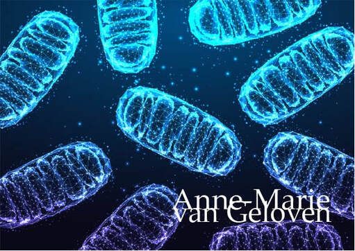
Mitochondria are the "powerhouses" or "lungs" of our cells and bioenergetic semi-autonomous organelles with their own genomes and genetic systems. [1] They are responsible for generating the energy that fuels a wide range of cellular processes in the skin, including cell signaling, pigmentation, wound healing, barrier integrity [2], metabolism and quality control. [3] Mitochondria exist in each cell of the body and are generally inherited exclusively from the mother. Their primary role is cellular respiration; a process converting the energy in nutrients (like glucose) into a usable form of energy called ATP or Adenosine Triphosphate. Mitochondria are particularly abundant in the skin, reflecting the skin's high metabolic demand. When the functionality of mitochondria is impaired or declines, it impacts skin's vitality, health and beauty. Mitochondrial dysfunction is 1 of the 12 hallmarks of skin ageing.
The skin is particularly susceptible to mitochondrial stress due to its constant exposure to environmental insults, such as UV radiation, pollution, and other oxidative stressors. These factors can damage mitochondrial DNA, leading to increased production of reactive oxygen species (ROS) and disrupting the delicate balance of cellular processes. [4] In aged post-mitotic cells, heavily lipofuscin-loaded lysosomes perform poorly, resulting in the enhanced accumulation of defective mitochondria, which in turn produce more reactive oxygen species causing additional damage (the mitochondrial-lysosomal axis theory). [5] Optimal mitochondrial function is indispensable for sustaining the specialized functions of each cell type, like keratinocyte differentiation, fibroblast ECM production, melanocytes melanin production and distribution, immune cell surveillance, sebocytes and adipocytes. [6] Mitochondrial dysfunction is both directly and indirectly linked to chronological ageing and photo-ageing. [7] As mitochondrial function declines, the skin's ability to regenerate and repair itself is decreased. [2] This results in visible signs of aging, such as wrinkles, loss of elasticity, dryness, uneven pigmentation, melasma, age spots, lipomas, impaired wound healing. [2-4-5-8-9] Mitochondrial dysfunction also has been implicated in skin conditions like acne, eczema, lupus, psoriasis, vitiligo, atopic dermatitis and even skin cancer. [10] Ageing is associated with changes in mitochondrial morphology, including [6] ▌Hyperfusion or increased fragmentation ▌Loss of mitochondrial connectivity [11-7] ▌Decline in the efficiency of oxidative phosphorylation, leading to reduced ATP production ▌Decline mitochondrial membrane potential (ΔΨM) ▌Compromised cellular energy metabolism ▌Reduced mitochondrial turnover (downregulated biogenesis) ▌Impaired mitochondrial quality control such as mitophagy (removal of damaged mitochondria through autophagy) [6] These alterations are related to the increased production of ROS exhibited by mitochondria during ageing, the accumulation of which causes oxidative damage to mitochondrial and cell components contributing to cellular senescence. [12] Good mitochondrial function or metabolism: [7] ▌Redox homeostasis: (the way of reducing oxidative stress) - mitochondrial respiration and ROS production are essential for keratinocyte differentiation ▌ATP production: Adenosine Triphosphate provides energy to drive and support many processes in living cells (and GTP) ▌Respiration: mitochondrial respiration is the most important generator of cellular energy ▌Biogenesis: allows cells to meet increased energy demands, to replace degraded mitochondria and is essential for the adaptation of cells to stress [6] ▌Calcium homeostasis ▌Cellular growth ▌Programmed cell death (apoptosis) reducing cell senescence [13] ▌Mitochondrial protein synthesis: mitochondria typically produce 13 proteins encoded by mitochondrial DNA (mtDNA) Dysfunctional Mitochondria: [7] ▌Oxidative stress ▌Decreased ATP levels ▌Dysfunctional OXPHOS: Oxidative phosphorylation, a metabolic pathway in which enzymes oxidize nutrients to release stored chemical energy in the form of ATP ▌Altered mitochondrial biogenesis ▌Calcium imbalance ▌Cell death Mitochondrial proteins Mitochondria contain >1,100 different proteins (MitoCoP) that often assemble into complexes and supercomplexes such as respiratory complexes and preprotein translocases. The chaperones Heat Shock Proteins HSP60-HSP10 are the most abundant mitochondrial proteins. [3] Small heat shock proteins form a chaperone system that operates in the mitochondrial intermembrane space. Depletion of small heat shock proteins leads to mitochondrial swelling and reduced respiration. [14] Mitochondrial hyperpigmentation Emerging research has shed light on the intricate relationship between mitochondrial dysfunction and the development of hyperpigmentation, a condition characterized by the overproduction and uneven distribution of melanin in the skin. One of the key mechanisms underlying this connection is the role of mitochondria in the regulation of melanogenesis, the process by which melanin is synthesized. Mitochondria are involved in the production of various cofactors and signaling molecules that are essential for the activity of tyrosinase, the rate-limiting enzyme in melanin synthesis. [15] When mitochondrial function is impaired, it can lead to an imbalance in the production and distribution of these cofactors and signaling molecules, ultimately resulting in the overproduction and uneven deposition of melanin in the skin. [15] This can manifest itself as age spots, melasma, and other forms of hyperpigmentation. The link between mitochondrial dysfunction and hyperpigmentation has been further supported by studies on genetic disorders that involve mitochondrial dysfunction, such as mitochondrial DNA depletion syndrome. In these conditions, patients often exhibit a range of pigmentary skin changes, including patchy hyper- and hypopigmentation, as well as reticular pigmentation. [16] Mitochondrial crosstalk and exosomes Mitochondria can crosstalk and move beyond cell boundaries. [17] Mitochondria-derived material might be transferred to neighboring cells in the form of cell-free mitochondria or included in extracellular vesicles [18-19]. This process supports cellular repair and contributes to vital mitochondrial functions. Besides restoring stressed cells and damaged tissues due to mitochondrial dysfunction, intercellular mitochondrial transfer also occurs under physiological and pathological conditions. [20] The transfer of active mitochondria from mesenchymal stem cells (MSCs) has been identified as a repair mechanism for rejuvenating damaged skin fibroblasts. [21] MITOCHONDRIAL SUPPORT Move According Martin Picard phD being physically active is a protective factor against almost everything health related. Exercise stimulates the production of mitochondria as more energy is required. Be hungry sometimes If there is too much supply of energy acquired via food leads to mass shrinking of mitochondria or fragmentation. Don´t over-eat, be calorie neutral and sometimes being calorie deficient is good for mitochondria. Maintain a healthy weight, preferably with a mediterranean diet containing phenolic and polyphenolic compounds (increase mitochondrial function and number) nitrate rich vegetables, soybeans and cacao beans. Mitohormesis In model organisms, lifespan can be improved by compromising mitochondrial function, which induces a hormetic response (“mitohormesis”), provided that this inhibition is partial and occurs early during development. Feel good Feeling good (positivity), especially at night, has a scientifically proven positive effect on mitochondrial health index, it is even a predictive factor. Q10 or Coenzyme Q10 (CoQ10) Q10 is part of the mitochondrial respiration chain and essential for cellular energy production. About 95% of our cellular energy is generated with support of Q10, which is produced by the human body itself. During skin ageing, both the cellular energy production and levels of Q10 are declined. Q10 is a powerful anti-oxidant [22], thus protecting cells from oxidative stress and damage and has proven to be able to "rescue" senescent cells by decreasing elevated senescent markers like p21 levels and β-Galactosidases positive cell numbers (in-vitro). Q10 is bio-active, increasing collagen type I and elastin production. [23] Q10 can be supplemented via nutrition, however also via topical application and is considered an evidence based active ingredient in skin care products. Ubiquinol (reduced form) shows higher bioavailability compared to ubiquinone (oxidized form). [23] Pyrroloquinoline quinone (PQQ) Q10 improves the energy in the mitochondria, however PQQ has shown to increase the number of mitochondria and a redox maestro. I´ve written a full post about this compound, which can be found as skincare ingredient and supplement. Read more about PQQ Glutathione Glutathione is formed in cell's cytoplasm from glutamic acid, cysteine and glycine. It is present in 2 forms: reduced (GSH) and oxidized (GSSG). Reduced GSH is an active anti-oxidant, while the presence of inactive GSSG is increased under oxidative stress. The ratio between GSH and GSSH is considered a measure of oxidative stress. Glutathione participates in redox reactions, acts as co-factor of many anti-oxidant enzymes and is the most important non-enzymatic anti-oxidant, essential for synthesis of proteins and DNA. Low Glutathione results in accelerated ageing and inflammatory skin diseases. Mitochondrial glutathione (mGSH) is the main line of defense for the maintenance of the appropriate mitochondrial redox environment to avoid or repair oxidative modifications leading to mitochondrial dysfunction and cell death. [24] Glutathione can be increased via supplementation via precursors cysteine or N-acetylcysteine (not recommended for pregnant women), a combination of Glycine and NAC (called GlyNAC) part of the popular "power of three" supplementation, or the reduced form of Glutathione itself, or increased via topical active ingredients like Licochalcone A. [25] I´ve written about GlyNAC in my post on autophagy. Nicotinamide NR nicotinamide ribosome which is the precursor of NMN nicotinamide mononucleotide which is the precursor of NAD+ nicotinamide adenine dinucleotide all could have a protective effect on mitochondria. Nicotinamide adenine dinucleotide is present in living organisms as ions NAD+ and NADP+ and in reduced forms NADH and NADPH. NADH is a cofactor of processes inside mitochondria: ▌ATP production ▌Activation of "youth proteins" sirtuins ▌Activation of PARP Poly (ADP-ribose) polymerase, a family of proteins involved in many cellular processes such as DNA repair, genomic stability and programmed cell death ▌Reduction of ROS (free radicals) NAD levels as lowered during ageing. [26] One of the fans of NMN supplementation is Harvard Professor David Sinclair, best known for his work on understanding why we age and how to slow its effects and also featured in my article about hormesis. There are about 14 studies done to date with NMN supplementation in humans, one of which was done by Professor Sinclair. NMN supplementation does raise NAD levels, however there aren't substantial proven health benefits, unless you are unhealthy. Resveratrol Although systemically Resveratrol promotes mitochondrial biogenesis. [27] Other data shows that UVA (14 J/cm(2)) along with resveratrol causes massive oxidative stress in mitochondria. As a consequence of oxidative stress, the mitochondrial membrane potential decreases which results in opening of the mitochondrial pores ultimately leading to apoptosis in human keratinocytes. [28] Magnesium Magnesium supplementation has been shown to improve mitochondrial function by increasing ATP production, decreasing mitochondrial ROS and calcium overload, and repolarizing mitochondrial membrane potential. There are many forms of Magnesium, however Citrate, Malate and Orotate are particularly good for energy. L-Carnitine Placebo-controlled trials have shown positive effects of L-Carnitine supplementation on both pre-frail subjects and elderly men. The effect is possibly mediated by counteracting age-related declining L-carnitine levels which may limit fatty acid oxidation by mitochondria. NEW Ergothioneine (EGT) Ergothioneine (EGT) is a sulfur-containing amino acid derivative known for its antioxidant properties, particularly in mitochondria. It is transported into cells and mitochondria via the OCTN1 transporter, where it helps reduce reactive oxygen species (ROS) and maintain cellular homeostasis [29]. EGT binds to and activates 3-mercaptopyruvate sulfurtransferase (MPST), enhancing mitochondrial respiration and exercise performance [30]. It also protects against oxidative stress and inflammation, potentially benefiting conditions like neurodegenerative diseases [31]. Melatonin Not much talked about when it comes to mitochondria, however should not be ignored as mitochondria can benefit significantly from melatonin supplementation. 1. Antioxidant protection: Melatonin acts as a powerful antioxidant within mitochondria, scavenging free radicals and reducing oxidative damage to mitochondrial DNA and proteins [32][34]. 2. Regulation of mitochondrial homeostasis: Melatonin helps maintain electron flow, efficiency of oxidative phosphorylation, ATP production, and overall bioenergetic function of mitochondria [32][34]. 3. Preservation of respiratory complex activities: Melatonin helps maintain the activities of mitochondrial respiratory complexes, which are crucial for energy production [32][34]. 4. Modulation of calcium influx: Melatonin regulates calcium influx into mitochondria, helping prevent calcium overload which can be damaging [32][34]. 5. Protection of mitochondrial permeability transition: Melatonin helps regulate the opening of the mitochondrial permeability transition pore, which is important for maintaining mitochondrial integrity [32][34]. 6. Enhancement of mitochondrial fusion: Melatonin promotes mitochondrial fusion, which is part of the quality control process for maintaining healthy mitochondria [33]. 7. Promotion of mitophagy: Melatonin enhances the removal of damaged mitochondria through mitophagy, helping maintain a healthy mitochondrial population [33]. 8. Reduction of nitric oxide generation: Melatonin decreases nitric oxide production within mitochondria, which can be damaging in excess [32][34]. 9. Selective uptake by mitochondria: Melatonin is selectively taken up by mitochondrial membranes, allowing it to exert its protective effects directly within these organelles [34]. 10. Support of mitochondrial biogenesis: Some studies suggest melatonin may promote the formation of new mitochondria [33]. The key antioxidants used by mitochondria are Glutathione (GSH), Glutathione peroxidase (GPx), Coenzyme Q10 (CoQ10), Superoxide dismutase (SOD), Melatonin, Vitamin C (ascorbate) and Vitamin E (α-tocopherol). Red light therapy By incorporating red light therapy into your skin care routine, you can help to counteract the damaging effects of mitochondrial dysfunction and support the skin's natural renewal processes. As we continue to explore the 12 hallmarks of ageing, I am confident that we will gain even more valuable insights and develop breakthrough innovations that will improve skin quality, health, beauty and vitality. Always consult a qualified healthcare professional or dermatologist to determine what the most suitable approach is for your particular skin condition and rejuvenation goals. Take care! Anne-Marie References
Many people associate a tan with health, beauty and an active lifestyle. Although a moderate dose of solar radiation is indispensable for our health, unfortunately, there is no such thing as a real "healthy tan" or "healthy sun-kissed glow" as it is always a visible sign of skin damage. Tanning is a response by the skin to exposure to ultraviolet (UV) radiation (and HEV or Blue Light), either from natural sunlight or artificial sources like tanning beds which leads to photo-ageing, pigmentary disorders (like age spots or hyperpigmentation) and immunosuppression, hence skin cancer. When skin is exposed to sunlight: UV rays and high energy visible light (HEV) or also called Blue Light (the most energetic region of HEV), it produces more melanin, a pigment that darkens the skin as a (partial) protective mechanism to prevent further damage. The amount of artificial blue light emitted during the conventional use of electronic devices is not enough to trigger harmful skin effects. (Click here to read more)
MELANIN Melanin is only produced by cells called melanocytes, mostly distributed in the epidermal-dermal junction. Melanocytes contain specialized organelles called melanosomes to store and produce melanin. Melanosomes are transferred from the melanocytes to the neighboring keratinocytes, which are the most abundant cells in the epidermis. One melanin-forming melanocyte surrounded by 36 keratinocytes and a Langerhans cell is called the melano-epidermal unit. [1.2] Melanocytes use the amino acid tyrosine to produce melanin and protect epidermal keratinocytes and dermal fibroblasts from the damaging effects of solar radiation.. [13] The are two melanin pigment classes:
Differences in skin pigmentation do not result from differences in the number of melanocytes in the skin, as one might assume, but from differences in the melanogenic activity (melano-competence), the type of melanin produced in melanosomes (the ratio between eumelanin and pheomelanin differs per Fitzpatrick phototype) and the size, number and packaging of melanosomes, with melanin content of melanosomes ranging from 17.9% to 72.3%. [7] The amount of melanin is never enough for adequate photoprotection, and a "base tan" does not prevent sunburn. Particularly darker phototypes are more sensitive for the damaging effects of Blue Light. Both eumelanin and pheomelanin production are promoted by UV radiation and Blue Light and therefore sunscreens offering a combination of both UV (A + B) protection and Blue Light defense are recommended for all phototypes. TANNING PROCESS The skin's tanning process occurs in four distinct phases: [3]
ROLE OF UVA, UVB AND BLUE LIGHT One of the most important acute effects of UVR is DNA damage. UVA and UVB show different properties regarding their biological effects on the skin. [7] Shorter wavelengths (nm) correspond to higher energy. Infrared does not induce oxidative stress. Read more UVA radiation (320-400 nm) penetrates deeper into the skin and can induce indirect DNA damage by the generation of reactive oxygen species (ROS), leading to premature skin aging. UVA, in contrast to UVB is not filtered by window glass, is able to penetrate deeper into the skin and reach the dermis. They are present constantly, with relatively equal intensity, during all daylight hours throughout the year. It has been estimated that 50% of exposure to UVA occurs in the shade. UVA rays are less intense than UVB, but there are 30 to 50 times more of them. To produce the same erythemal response, approximately 1000 times more UVA dose is needed compared with UVB. [7] The bulbs used in tanning beds emit mostly UVA. UVB radiation (280-320 nm) is less prevalent than UVA, primarily affects the outermost layers of the skin, causing direct DNA damage (more potent than UVA) and triggers inflammatory responses that lead to increased melanin production. UVB radiation fluctuates throughout the day, are at their strongest at noon. and are more cytotoxic and mutagenic than UVA. The action spectrum for UV-induced tanning and erythema are almost identical, but UVA is more efficient in inducing tanning whereas UVB is more efficient in inducing erythema (redness). Dark skin is twice as effective compared to light skin in inhibiting UVB radiation penetration. [7] UVB helps the skin to produce Vitamin D. Blue light (400-500 nm) visible light accounts for 50% of sunlight [11] and can contribute to immediate, delayed, continuous and long-lasting pigmentation by activating melanocyte-specific photoreceptors and increasing melanin synthesis, particularly in individuals with darker (melano-competent) skin types [9], cause DNA damage [10] and generate damaging reactive oxygen species in both the epidermis and the dermis. [12] The effects may last longer than those induced by UVA and UVB radiation. Blue Light can penetrate even deeper than UVA and reach the hypodermis. Blue light therapy is used to target acne causing bacteria and inflammation, however the risks might outweigh the benefits especially in darker phototypes and it might worsen acne marks. EPIDERMIS AND DERMIS Both dermal fibroblasts and epidermal keratinocytes play a crucial role in regulating skin pigmentation and tanning response. [13 15] In comparison to epidermal tanning, dermal tanning is less visible, however more immediate. Dermal fibroblasts secrete various paracrine factors that regulate melanocyte function, survival, and melanin production. Factors like hepatocyte growth factor (HGF), nerve growth factor (NGF), stem cell factor (SCF), and basic fibroblast growth factor (bFGF) stimulate melanogenesis and pigmentation [14 15] Fibroblast senescence and altered secretory profiles in conditions like melasma contribute to abnormal pigmentation by stimulating melanogenesis. [15] Epidermal keratinocytes produce factors like α-melanocyte stimulating hormone (α-MSH) and Wnt1 that activate melanogenic pathways in melanocytes, leading to increased melanin synthesis and transfer to keratinocytes. [15 16]. Keratinocyte-derived exosomes can enhance melanin production by melanocytes. [16] Differences in autophagic activity between various keratinocytes also influences pigmentation. [15] MicroRNAs MicroRNAs are small, non-coding RNA molecules that regulate gene expression by binding to messenger RNA (mRNA) and typically suppressing protein production, for example collagen. They are classified as epigenetic modulators. Several miRNAs have been identified as differentially expressed in aged skin compared to young skin, including: - miR-383, miR-145, miR-34a (upregulated in sun-exposed aged skin) - miR-6879, miR-3648, miR-663b (downregulated in sun-exposed aged skin) [17] Enjoy the sun, however protect your (and your children's) skin from a photo-damaging tan to remain skin health and beauty. Sunless self-tanning products containing dihydroxyacetone (DHA) or Erythrulose provide a safe alternative to achieve a "sun-kissed" glow. You can use after-sun skin care which helps to rehydrate, reduce damage of "sun-stressed" skin and support it's repair. Always consult a qualified healthcare professional or dermatologist to determine what the most suitable approach is for your particular skin condition and rejuvenation goals. Take care! Anne-Marie
References
3/20/2024 Comments Telomeres: tiny caps with big impact
Our DNA is as like precious book of life filled with information and instructions, with telomeres acting like the protective covers. Just as book covers get worn over time, our telomeres naturally shorten as we age. This shortening is like a biological clock, ticking away with each cell division.
Telomere shortening is considered one of the twelve key hallmarks of aging. Those hallmarks all play an important role in longevity, health-span, and skin quality, thus both health and beauty. Telomeres are the protective end-caps of chromosomes, similar to the plastic caps at the end of shoelaces. They maintain genomic stability and prevent chromosomal damage. Telomeres become slightly shorter each time a cell divides, and over time they become so short that the cell is no longer able to successfully divide. They shorten more rapidly in dermal fibroblasts compared to epidermal keratinocytes, hence there are significant differences amongst our cells. Telomeres in skin cells may be particularly susceptible to accelerated shortening because of both proliferation and DNA-damaging agents such as reactive oxygen species and sun exposure. [16]. When a cell is no longer able to divide due to telomere shortening, this can lead to
This consequently affects both health and beauty
FACTORS INFLUENCING TELOMERE SHORTENING Sleep quality Poor sleep quality significantly impacts telomere length:
INTERVENTIONS FOR TELOMERE PRESERVATION 1. Possible strategies to preserve telomere length
Telomerase is an enzyme that plays a crucial role in maintaining the length of telomeres and skin cell function. Telomerase is a ribonucleoprotein enzyme, meaning it contains both protein (TERT plus dyskerin) and RNA components (TER or TERC). Its primary function is to add repetitive DNA sequences (telomeres) to the ends of chromosomes, preventing them from shortening during cell division. Telomerase is active in embryonic stem cells, some adult stem cells, cancer cells, certain skin cells, specifically:
Poor sleep quality is associated with shorter telomere length. Studies have found significant associations between shortened telomere length and poor sleep quality and quantity, including obstructive sleep apnea [17]. Not feeling well rested in the morning was significantly associated with shorter telomere length in older adults [18]. Sleep loss and poor sleep quality may activate DNA damage responses and cellular senescence pathways [17]. Poor sleep can increase oxidative stress and inflammation, which may accelerate telomere shortening [17]. Disruption of circadian rhythms due to poor sleep may negatively impact telomere maintenance [17]. Improving sleep quality through lifestyle changes and sleep hygiene practices may help preserve telomere length. [19]
A study showed that diet, exercise, stress management, and social support could increase telomere length by approximately 10% over five years [20].
Adopt a plant-rich diet, such as the Mediterranean diet, which includes whole grains, nuts, seeds, green tea, legumes, fresh fruits (berries), vegetables (leafy greens), omega-3 fatty acids from sources like flaxseed and fish oil or fatty fish and foods rich in folate. This diet is rich in antioxidants and anti-inflammatory properties that help maintain telomere length [21]. 5. Fasting Fasting, especially intermittent fasting, has attracted interest for its potential impact on health, including telomere preservation. Multiple studies have shown that intermittent fasting (IF) and other fasting regimens can reduce markers of oxidative stress and inflammation. Research on animals has demonstrated that caloric restriction and intermittent fasting can boost telomerase activity and enhance telomere maintenance in specific tissues. A human study by Cheng et al. (2019) found a correlation between intermittent fasting and longer telomeres, by reducing PKA activity and IGF1 levels, which are crucial for regulating telomerase function. A study showed that 36 hours of fasting induced changes in DNA methylation and another one histone modifications, hence fasting has the potential to induce epigenetic changes. Important note: Be careful with a time-restricted eating schedule (often seen as a form of intermittent fasting, where you eat all meals within an 8 hour time-frame), especially women in menopause or people with a pre-existing heart condition. The American Heart Association presented data indicating that people with a pre-existing heart condition have a 91% higher risk of of death of a heart disease when following the time-restricted eating schedule with an 8 hour window, compared to those who eat within a 12-16 hours window. However, several experts have criticised the data, which aren´t published in a peer reviewed journal. When considering fasting, or a time-restricted eating schedule, especially for a longer period, talk to a qualified HCP first. 6. Exercise
EMERGING TECHNOLOGIES IN TELOMERE-TARGETING SKINCARE Small RNAs in skincare Small RNAs play a significant role in the effectiveness of telomere-targeting skincare by influencing skin regeneration and cellular processes. Recent research has highlighted their potential in enhancing wound healing and reducing scarring, which are critical aspects of maintaining healthy skin. Small RNAs, such as microRNAs, are involved in regulating gene expression related to skin aging and and show potential in telomere maintenance [29]. They can modulate the expression of genes that control cellular senescence, oxidative stress response, and inflammation, all of which are crucial for preserving telomere integrity and function [30].
RNAi technology in development RNAi-based skincare approaches could target genes involved in telomere maintenance or have effects on markers related to telomere biology:
RNA-based telomere extension is a method developed at Stanford University and uses modified RNA to extend telomeres in cultured human cells, allowing cells to divide more times than untreated cells [35]. IN OFFICE DERMATOLOGICAL TREATMENTS Aesthetic, regenerative treatments that support skin quality may indirectly support telomere preservation.
Telomere shortening questionable as stand-alone hallmark [36] Telomere length (TL) has long been considered one of the best biomarkers of aging. However, recent research indicates TL alone can only provide a rough estimate of aging rate and is not a strong predictor of age-related diseases and mortality. Other markers like immune parameters and epigenetic age may be better predictors of health status and disease risk. TL remains informative when used alongside other aging biomarkers like homeostatic dysregulation indices, frailty index, and epigenetic clocks. TL meets some criteria for an ideal aging biomarker (minimally invasive, repeatable, testable in animals and humans) but its predictive power for lifespan and disease is questionable. There is inconsistency in epidemiological studies on TL's association with aging processes and diseases. This has led to debate about TL's reliability as an aging biomarker. It's unclear if telomere shortening reflects a "mitotic clock" or is more a marker of cumulative stress exposure. TL is still widely used in aging research but there are ongoing questions about its usefulness as a standalone biomarker of biological age. As research in regenerative medicine advances, we're seeing promising developments in therapies targeting telomere biology for longevity, health and beauty. While telomere research is exciting, it's important to remember that it's just one part of a comprehensive approach to aging, and future treatments will likely combine multiple strategies to target preferably all 12 hallmarks for the best results. Always consult a qualified healthcare professional or dermatologist to determine what the most suitable approach is for you. . Take care! Anne-Marie
References
[1] Martin, H., Doumic, M., Teixeira, M.T. et al. Telomere shortening causes distinct cell division regimes during replicative senescence in Saccharomyces cerevisiae. Cell Biosci11, 180 (2021) [2] M. Borghesan, W.M.H. Hoogaars, M. Varela-Eirin, N. Talma, M. Demaria, A Senescence-Centric View of Aging: Implications for Longevity and Disease, Trends in Cell Biology, Volume 30, Issue 10, 2020, Pages 777-791, ISSN 0962-8924, [3] McHugh D, Gil J. Senescence and aging: Causes, consequences, and therapeutic avenues. J Cell Biol. 2018 Jan 2;217(1):65-77. [4] Oeseburg, H., de Boer, R.A., van Gilst, W.H. et al. Telomere biology in healthy aging and disease. Pflugers Arch - Eur J Physiol 459, 259–268 (2010) [5] Catarina M Henriques, Miguel Godinho Ferreira, Consequences of telomere shortening during lifespan, Current Opinion in Cell Biology, Volume 24, Issue 6, 2012 [6] Henriques CM, Ferreira MG. Consequences of telomere shortening during lifespan. Curr Opin Cell Biol. 2012 [7] Chaib, S., Tchkonia, T. & Kirkland, J.L. Cellular senescence and senolytics: the path to the clinic. Nat Med 28, 1556–1568 (2022) [8] Lei Zhang et al. Cellular senescence: a key therapeutic target in aging and diseases JCI The Journal of Clinical Investigation 2022 [9] Muraki K, Nyhan K, Han L, Murnane JP. Mechanisms of telomere loss and their consequences for chromosome instability. Front Oncol. 2012 Oct 4;2:135. [10] Marlies Schellnegger et al. Aging, 25 January 2024 Sec. Healthy Longevity Volume 5 - 2024 Unlocking longevity: the role of telomeres and it´s targeting interventions [11] Bär C, Blasco MA. Telomeres and telomerase as therapeutic targets to prevent and treat age-related diseases. F1000Res. 2016 Jan 20;5:F1000 Faculty Rev-89. [12] Kasiani C. Myers et al. Blood (2022) 140 (Supplement 1): 1895–1896. Gene therapies November 15 2022 Successful Ex Vivo Telomere Elongation with EXG-001 in a patients with Dyskeratosis Congenital Kasiani C. Myers et al. [13] Falckenhayn C, Winnefeld M, Lyko F, Grönniger E. et al. Identification of dihydromyricetin as a natural DNA methylation inhibitor with rejuvenating activity in human skin. Front Aging. 2024 Mar 4;4:1258184 [14] Minoretti P, Emanuele E. Clinically Actionable Topical Strategies for Addressing the Hallmarks of Skin Aging: A Primer for Aesthetic Medicine Practitioners. Cureus. 2024 Jan 19;16(1):e52548 [15] Guterres, A.N., Villanueva, J. Targeting telomerase for cancer therapy. Oncogene 39, 5811–5824 (2020). [16] Buckingham EM, Klingelhutz AJ. The role of telomeres in the ageing of human skin. Exp Dermatol. 2011 Apr;20(4):297-302. [17] Debbie Sabot, Rhianna Lovegrove, Peta Stapleton, The association between sleep quality and telomere length: A systematic literature review, Brain, Behavior, & Immunity - Health, Volume 28, 2023, 100577, ISSN 2666-3546 [18] Iloabuchi, Chibuzo et al. Association of sleep quality with telomere length, a marker of cellular aging: A retrospective cohort study of older adults in the United States Sleep Health: Journal of the National Sleep Foundation, Volume 6, Issue 4, 513 – 521 [19] Rossiello, F., Jurk, D., Passos, J.F. et al. Telomere dysfunction in ageing and age-related diseases. Nat Cell Biol 24, 135–147 (2022) [20] Elisabeth Fernandez Research September 16 2013 Lifestyle changes may lengthen telomeres, A measure of cell aging. Diet, Meditation, Exercise can improve key element of Immune cell aging, UCSF Scientist report [21] Martínez P, Blasco MA. Telomere-driven diseases and telomere-targeting therapies. J Cell Biol. 2017 Apr 3;216(4):875-887. [22] Guo, J., Huang, X., Dou, L. et al. Aging and aging-related diseases: from molecular mechanisms to interventions and treatments. Sig Transduct Target Ther 7, 391 (2022). [23] Hachmo Y, Hadanny A, Abu Hamed R, Daniel-Kotovsky M, Catalogna M, Fishlev G, Lang E, Polak N, Doenyas K, Friedman M, Zemel Y, Bechor Y, Efrati S. Hyperbaric oxygen therapy increases telomere length and decreases immunosenescence in isolated blood cells: a prospective trial. Aging (Albany NY). 2020 Nov 18;12(22):22445-22456 [24] Gutlapalli SD, Kondapaneni V, Toulassi IA, Poudel S, Zeb M, Choudhari J, Cancarevic I. The Effects of Resveratrol on Telomeres and Post Myocardial Infarction Remodeling. Cureus. 2020 Nov 14;12(11):e11482. [25] Widgerow AD, Ziegler ME, Garruto JA, Bell M. Effects of a Topical Anti-aging Formulation on Skin Aging Biomarkers. J Clin Aesthet Dermatol. 2022 Aug;15(8):E53-E60. PMID: 36061477; PMCID: PMC9436220. [26] Alt, C.; Tsapekos, M.; Perez, D.; Klode, J.; Stoffels, I. An Open-Label Clinical Trial Analyzing the Efficacy of a Novel Telomere-Protecting Antiaging Face Cream. Cosmetics 2022, 9, 95. [27] Cosmetics & Toiletries Telomere protection: Act on the origin of youth, June 3th 2015 Sederma [28] Yu Y, Zhou L, Yang Y, Liu Y. Cycloastragenol: An exciting novel candidate for age-associated diseases. Exp Ther Med. 2018 Sep;16(3):2175-2182. [29] Gerasymchuk M, Cherkasova V, Kovalchuk O, Kovalchuk I. The Role of microRNAs in Organismal and Skin Aging. Int J Mol Sci. 2020 Jul 25;21(15):5281. [30] Jacczak B, Rubiś B, Totoń E. Potential of Naturally Derived Compounds in Telomerase and Telomere Modulation in Skin Senescence and Aging. International Journal of Molecular Sciences. 2021; 22(12):6381. [31] Roig-Genoves, J.V., García-Giménez, J.L. & Mena-Molla, S. A miRNA-based epigenetic molecular clock for biological skin-age prediction. Arch Dermatol Res 316, 326 (2024). [32] Eline Desmet, Stefanie Bracke, Katrien Forier, Lien Taevernier, Marc C.A. Stuart, Bart De Spiegeleer, Koen Raemdonck, Mireille Van Gele, Jo Lambert, An elastic liposomal formulation for RNAi-based topical treatment of skin disorders: Proof-of-concept in the treatment of psoriasis, International Journal of Pharmaceutics, Volume 500, Issues 1–2, 2016, Pages 268-274, ISSN 0378-5173 [33] Oger E, Mur L, Lebleu A, Bergeron L, Gondran C, Cucumel K. Plant Small RNAs: A New Technology for Skin Care. J Cosmet Sci. 2019 May/Jun;70(3):115-126. PMID: 31398100. [34] Vimisha Dharamdasani, Abhirup Mandal, Qin M. Qi, Isabella Suzuki, Maria Vitória Lopes Badra Bentley, Samir Mitragotri, Topical delivery of siRNA into skin using ionic liquids, Journal of Controlled Release, Volume 323, 2020, Pages 475-482, ISSN 0168-3659 [35] Krista Conger January 2015 Stanford Medicine News Center Telomere extension turns back aging clock in cultured human cells, study finds [36] Alexander Vaiserman, Dmytro Krasnienkov Telemore length as marker of biological age: state-of-the-art, open issues and future perspectives Front. [37] Martínez P, Blasco MA. Telomere-driven diseases and telomere-targeting therapies. J Cell Biol. 2017 Apr 3;216(4):875-887
Hair is a powerful factor in how we're perceived by others and even how we see ourselves. It plays a significant role in the perception of youth and attractiveness. Studies have shown that hair style, color, and quality can significantly affect how old we look and how attractive we're considered [1]. Research suggests that hair is one of the most defining characteristics of our appearance, with the potential to make us look years younger or older [1]. From an evolutionary perspective, lustrous hair has long been associated with youth, health, and fertility [1]. Culturally, hair has been a symbol of beauty and status across societies for centuries [2].
HAIR GENETICS BEYOND MATERNAL INHERITANCE We have approximately 5 million hair follicles distributed across our bodies, with only about 100,000 located on the scalp [3][4]. Contrary to popular belief, hair characteristics are not solely inherited from one's mother. Human genetic makeup consists of 23 pairs of chromosomes, including the sex-determining X and Y chromosomes [5]. Females typically have two X chromosomes (with one usually inactivated through a process called X-chromosome inactivation), while males have one X and one Y chromosome [6]. Our hair's characteristics, including texture, color, and growth patterns, are determined by about 600 genes [7]. Interestingly, only 11% of these genes are located on the X chromosome [8]. The majority of genes influencing hair traits are found on autosomes (non-sex chromosomes), contributing to the inheritance patterns observed in families [9]. For instance, genes like EDAR and FGFR2 have been associated with hair thickness in Asian populations, while TCHH has been linked to hair texture in individuals of Northern European ancestry [10]. Research has identified several genes on the X chromosome that play a role in male pattern baldness, including the androgen receptor (AR) gene. Telomere length in hair follicle stem cells correlates with hair growth capacity and may be a biomarker for hair follicle aging. The complexity of hair genetics extends beyond sex chromosomes, involving multiple autosomal genes, environmental factors, hence epigenetics, and this is great news as changes in epigenetic patterns are partially reversible! Epigenetics Epigenetics refers to heritable changes in gene expression that occur without alterations in the DNA sequence itself [11]. Environmental factors, diet, lifestyle, chronic stress, sleep, circadian rhythms, physical activity, aging and even social interactions can influence gene expression through four main epigenetic mechanisms:
These epigenetic mechanisms can significantly impact hair biology
Example of change in epigenetic pattern Ever wondered why hair starts growing in odd places as we age? It is a good example of epigenetic changes. As we get older, changes in our epigenome can cause regions of our DNA that are normally silent (due to histone modifications) to become readable. In essence, we're becoming more like our ancient ancestors! This is why some people start growing more hair in places like ears and noses as they age. Epigenetic changes can thus silence or activate hair growth-related genes, potentially contributing to hair loss or promoting regeneration. Thus, the future of our hair health is literally (at least partially) in our hands today!. Lifestyle changes and hair regrowth Lifestyle modifications have demonstrated impacts on hair regrowth, particularly in early stages of hair loss and for prevention. 1. Nutrition: A balanced diet rich in proteins, vitamins (especially biotin, vitamins A, C, and D), and minerals (iron, zinc) has been associated with improved hair growth [20]. Supplementation with these nutrients has shown benefits in treating telogen effluvium and other hair loss conditions [21]. 2. Stress Management: Chronic stress can lead to telomere shortening and premature hair follicle aging. Stress reduction techniques like meditation and yoga have been linked to increased telomerase activity, potentially benefiting hair growth. 3. Exercise: Regular physical activity improves blood circulation to the scalp, potentially enhancing nutrient delivery to hair follicles. A study found that moderate exercise was associated with increased expression of hair growth-related genes. 4. Sleep: Adequate sleep is crucial for maintaining healthy hair growth cycles. Sleep deprivation has been linked to increased oxidative stress and inflammation, which can negatively impact hair follicles. Studies have shown promising results in targeting epigenetic mechanisms for hair loss treatment
In office therapies 1. Low-Level Laser Therapy (LLLT): LLLT works by decreasing nitric oxide enzyme activity, leading to a beneficial "micro-stress" in mitochondria. This hormetic effect increases energy production, allowing stem cells to stay young and rejuvenate. Clinical studies have demonstrated improved hair density and thickness with LLLT in androgenetic alopecia patients. 2. Platelet-Rich Plasma (PRP) and exosomes: These regenerative therapies deliver growth factors and signaling molecules to hair follicles, potentially reversing miniaturization and promoting the anagen phase. PRP has shown promising results in multiple clinical trials for androgenetic alopecia. 3. HydraFacial Keravive scalp treatment: A 3-step process involving cleansing, exfoliating, and nourishing the scalp to improve hair follicle health. 4. Hair Transplantation: Includes techniques like Follicular Unit Extraction (FUE) and strip harvesting to transplant hair from donor areas to balding areas. 5. Scalp micropigmentation: A cosmetic tattooing procedure that creates the appearance of a fuller head of hair. 6. Corticosteroid Injections: Used primarily for treating alopecia areata by injecting steroids directly into affected areas of the scalp. 7. Microneedling: Uses small needles to create micro-injuries in the scalp, potentially stimulating hair growth when combined with topical treatments. 8. Scalp Reduction: A surgical procedure that removes bald areas of the scalp and stretches hair-bearing skin. 9. Mesotherapy: Involves injecting vitamins, minerals, and other nutrients directly into the scalp to nourish hair follicles. BALD AINT BAD (for men)
Always consult a qualified healthcare professional or dermatologist to determine what the most suitable approach is for your particular skin or hair condition. Take care! Anne-Marie
The picture I used for this post is from my lovely daughter, who is blessed with fabulous hair.
References
If you've scrolled through Instagram, you may have caught a glimpse of dermatologists raving about LED masks emitting red light, the secret, evidenced based weapon behind skin rejuvenation known as photo biomodulation. It uses low-powered light within the red to near-infrared range (wavelengths from 632 to 1064 nm) to induce a biological reaction aka stimulate cellular processes. The wonders of red light, also known as LLLT (low-level laser therapy), PBM (red light photo-biomodulation), or PBMT (photo-biomodulating therapy), extend far beyond non-invasive skin rejuvenation. I am not a fan of devices for home use, mostly because of lacking safety and/or efficacy, PBM definitely earned it's prominent spot in my skincare routine.
A summary of the benefts of red light with and without near infrared light for skin Numerous studies have demonstrated the effectiveness of red and infrared light therapy for skin rejuvenation. A combination of red light and near IR light has proven to stimulate the production of collagen (I & III) plus elastin production (Li WH et al Int J Cosmet Sci 2021), enhance mitochondrial ATP production, cell signaling, growth factor synthesis, rebalance ROS (reactive oxidative species) and reduce inflammation. Stem cells can be activated allowing tissue repair and healing. Wrinkle and scar reduction was observed and it can reduce UV damage both as treatment and prophylactic measure. In pigmentary disorders such as vitiligo, it can increase pigmentation by melanocyte proliferation and reduce depigmentation by inhibiting autoimmunity (Pinar Avci et al. Semin Cutan Med Surg. 2013 & Mitchell J Winkie et al. Review Photodermatol Photoimmunol Photomed A focused review of visible light therapies for vitiligo 2024). It has the potential to activate both keratinocytes (epidermis) and fibroblasts (epidermal junction and dermis). With consistent use, you can expect a reduction of lines and wrinkles, improvement of skin tone and texture. PBMT (when done effective and safe) will compliment both your skin rejuvenating and regenerating at home skincare regimen and in-office procedures or even post-surgical skin recovery. ATP ATP (adenosine triphosphate) is the primary source of energy for cellular processes and plays a crucial role in various biological functions. When red light with specific wavelengths (630 nm to 638 nm and 810 nm) is absorbed by the skin cells, it stimulates the mitochondria, which are the powerhouses of the cells responsible for ATP synthesis. This increase in ATP production is providing cells with more energy to carry out their functions effectively and has several beneficial effects on the skin like boosting cellular metabolism, promoting more efficient nutrient uptake and waste removal. The increased ATP levels facilitate collagen synthesis by fibroblasts, a vital component for skin structure, elasticity and firmness and reduction of lines and wrinkles.. ATP aids in the repair and regeneration of damaged skin cells. It accelerates the healing process, making it beneficial for wound healing, post-surgical recovery, and addressing skin issues such as acne scars. ROS (Reactive Oxidative Species) By modulating ROS levels, red light therapy helps reduce oxidative stress and its detrimental effects on the skin. ROS are highly reactive molecules that are naturally produced by cells as byproducts of metabolic processes. While low levels of ROS play important roles in cellular signaling and immune responses, excessive ROS can lead to oxidative stress and damage to cells and tissues. Restoring the balance of ROS result in improved skin health, reduced inflammation, and enhanced skin rejuvenation. Red light therapy has been shown to modulate reactive oxidative species (ROS) levels in the skin by promoting antioxidant defense mechanisms and reducing oxidative stress:
The difference between LLLT and PBM LLLT refers specifically to the use of lasers, which produce coherent, focussed and an intense beam of monochromatic light, while PBM has a broader range of light sources, may include laser as well as light-emitting diodes (LEDs) and other non-laser devices. LEDs are often used in PBM because they are cost effective, versatile and have the ability to cover large treatment areas. LLT uses higher power densities with more energy and has a shorter treatment duration in comparison to PBM to achieve desired therapeutic effects. While there are similarities in terms of mode of action", there is a difference of light source, treatment application and parameters. Based on consensus, PBM and PBMT are considered the correct way to describe this photonic specialty for therapeutic applications. In this post I will focus on PBM and specifically LEDs. A home device claiming to use cold near infrared laser light or Low-Level Laser Therapy is called LYMA laser. It is sold for several thousand euro´s. LED masks and LED panels LED masks specifically produced by the brand Omnilux (FDA cleared) are currently very popular for very good reasons; they are safe and effective when the LEDs emit the right wavelengths and used in the recommended frequency. Omnilux combines 2 therapeutically effective and complimentary wavelengths: 633nm and near-infrared 830 nm. Both wavelengths (more precise 630nm + 850nm) I would recommend to minimally look for in any red LED device, which will disqualify most LED masks and panels in the market! I've include some (not affiliated) links to devices below. Both masks and panels can be effective, however most panels are stronger in comparison to masks 60 mW/cm² vs mW/cm²), hence have the benefit of a shorter treatment time to get a similar result. Intensity and power of red light therapy devices are typically measured in terms of irradiance (measured in milliwatts per square centimeter, mW/cm²) and radiant flux (measured in watts, W), which quantify the amount of light energy emitted by the device. Wearing a mask during a hot summer or in a warmer climate will make you sweat and depending on the materials of the mask and straps, they may be very uncomfortable to wear. Panels have the benefit that they give a more even distribution of emitted light as masks are worn on the face and thus the LED bulbs are pushed on a small skin surface area, panels can cover a larger area (depending on their size) and are more versatile in use, as area's like neck, décolletage, or knees are easier to treat with a panel. With a mask you may be more mobile, although I would not recommend walking around while using the mask. My personal preference would be a panel for the reasons mentioned before and panels are more suitable (more hygienic) for family sharing. My son can use it after an intense workout to speed up his recovery and I like to use it for purposes beyond photo-biomodulation or skin rejuvenation, for example to improve my sleep. With a panel I get more "bang for my buck".
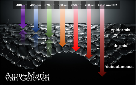
Red light and NIR (Near Infra Red light) have the ability to penetrate varying depths of the skin, resulting in distinct benefits, thus combinations of wavelengths will provide complementary effects.
630 nm Wavelength This wavelength is often used for its skin rejuvenation benefits. It has a relatively shallow penetration depth and is absorbed closer to the surface of the skin primarily affecting the epidermis. 630nm light is associated with increased circulation, reduce inflammation, improved skin tone & texture, aiding in the delivery of nutrients and oxygen to skin cells, and stimulating the production of collagen, leading to improved skin elasticity and a reduction of the appearance of fine lines & wrinkles. 660 nm Wavelength At 660nm, red light can penetrate a little deeper into the skin, reaching the dermis. It is known for its ability to stimulate collagen production, enhance cellular metabolism, and promote anti-inflammatory effects, helping to reduce redness and inflammageing. It also promotes wound healing, making it beneficial for post-surgical or post-trauma skin recovery. 810 nm Wavelength Improve healing & recovery & accelerate wound healing. 830 nm Wavelength Accelerate healing, reduce infection, improve aesthetic outcome following plastic surgery, increase endorfines (mood-enhancing), improve bone repair and growth. 850 nm Wavelength Improve general inflammation body, enhance muscle recovery, improve wound healing, reduced fine lines, wrinkles and hyperpigmentation. Always consult a qualified healthcare professional or dermatologist to determine if and what the most suitable red light therapy approach is for your particular skin condition and rejuvenation goals. Take care! References: Hamblin, Michael R. "Mechanisms and applications of the anti-inflammatory effects of photobiomodulation." AIMS biophysics 4.3 (2017): 337-361. Barolet, Daniel. Regulation of Skin Collagen Metabolism In Vitro Using a Pulsed 660 nm LED Light Source: Clinical Correlation with a Single-Blinded August 2009Journal of Investigative Dermatology 129(12):2751-9 Wunsch A, Matuschka K. (2014). A controlled trial to determine the efficacy of red and near-infrared light treatment in patient satisfaction, reduction of fine lines, wrinkles, skin roughness, and intradermal collagen density increase. Journal of Cosmetic and Laser Therapy, 16(5), 232-237. Avci P, et al. (2013). Low-level laser (light) therapy (LLLT) in skin: stimulating, healing, restoring. Seminars in Cutaneous Medicine and Surgery, 32(1), 41-52. Links to some devices which combine 630 nm and 850 nm: FDA-approved devices ensure safety and regulatory compliance, however the panels are more powerful: Omnilux(tm) Mask (FDA clearance) Very affordable panel (no FDA clearance) Affordable panel (no FDA clearance) |
CategoriesAll Acne Age Clocks Ageing Aquatic Wrinkles Armpits Autophagy Biostimulators Blue Light & HEVIS Circadian Rhythms Cleansing Collagen CoQ10 Cosmetic Intolerance Syndrome Deodorant Dermaplaning Diabetes DNA Damage DNA Repair Dry Skin Epigenetics Evidence Based Skin Care Exfoliation Exosomes Eyes Face Or Feet? Facial Oils Fibroblast Fingertip Units Gendered Ageism Glycation Growth Factors Gua Sha Hair Hair Removal Hallmark Of Aging Healthy Skin Heat Shock Proteins Hormesis Humidity Hyaluron Hyaluronidase Hypo-allergenic Indulging Jade Roller Keratinocytes Licochalcone A Luxury Skin Care Lymphatic Vessel Ageing Malar Oedema Menopause Mitochondrial Dysfunction Mood Boosting Skin Care Neurocosmetics Ox Inflammageing Peptides PH Balance Skin Photo Biomodulation Polynucleotides Proteasome Psoriasis Regeneration Regenerative Treatments Review Safety Scarring Sensitive Skin Skin Care Regimen Skin Flooding Skin Hydration Skin Senescence Skip-Care Sleep Slugging Sunscreen Tanning Under Eye Bags UV Index Vitamin C Vitamin D Well Ageing Skin Care Wound Healing Wrinkles
Archives
December 2024
|
Anne-Marie van Geloven © 2024 All rights reserved
|