Live your best life & take care
12/7/2024 Comments The dark side of vitamin CAlthough Vitamin C in topical applications has many benefits, it also has a dark side; it can be harmful in its oxidised form, temporarily darken the skin and become a pro-oxidant. When vitamin C (ascorbic acid) is exposed to air, light, or heat, it undergoes chemical changes similar to how sugar turns brown when heated. This process doesn't need any special helpers (like enzymes); it just happens because of the conditions around it. Over time, vitamin C breaks down and forms new compounds that have a brown color, much like how sugar becomes caramel. This process is called non-enzymatic oxidation. Oxidized vitamin C can have both beneficial and potentially harmful effects on the skin. 1. ANTIOXIDANT Vitamin C is primarily known for its antioxidant properties, effectively neutralizing reactive oxygen species (ROS) and reducing oxidative stress in the skin. This helps prevent DNA damage and collagen degradation, contributing to anti-aging benefits and improved skin health and beauty [1][2][3]. How vitamin C acts as an antioxidant and undergoes oxidation in your skin Imagine vitamin C as a brave knight patrolling your skin, constantly on guard against harmful invaders called free radicals. These free radicals can damage skin cells, much like how rust can damage metal. Vitamin C, in its role as an antioxidant, sacrifices part of itself (donating an electron) to neutralize these free radicals, preventing them from causing harm. ▌ InInitial defense: When vitamin C donates an electron, it transforms into a less powerful form called the ascorbate radical, similar to a knight losing a piece of armor but still able to fight. ▌ Continued protection: If more free radicals attack, vitamin C can further degrade into dehydroascorbic acid. This form can be regenerated with the help of other antioxidants like glutathione, similar to allies helping the knight repair its armor. ▌ Synergistic effects: Using vitamin C with other antioxidants in skincare products enhances its protective abilities, much like having a team of knights working together for stronger defense. I prefer combining Vitamin C with Licochalcone A for comprehensive skin protection. Vitamin C acts quickly in the skin's outer layer, providing immediate extracellular defense. Meanwhile, Licochalcone A offers long-lasting, intracellular protection against free radicals induced by both UV and High Energy Visible Light, which penetrate deeper into the skin. This synergistic approach ensures a more complete and sustained antioxidant effect. ▌ Final sacrifice: Without support, vitamin C eventually breaks down into other compounds and loses its protective power completely. 2. PRO-OXIDANT At high concentrations, vitamin C can exhibit pro-oxidative properties, generating hydrogen peroxide (H2O2) and leading to increased oxidative stress, particularly when vitamin C interacts with transition metals (Cu and Fe), which can catalyze the formation of harmful radicals [4][5]. This increases the risk of irritation or damage to skin cells. Copper (Cu): Copper compounds can penetrate the skin and participate in redox reactions [6]. Copper can catalyze the oxidation of ascorbate and participate in the Haber-Weiss reaction, generating free radicals [7]. Iron (Fe): Iron can participate in the metal-catalyzed Haber-Weiss reaction, also known as the superoxide-driven Fenton reaction, which produces harmful free radicals [7]. These transition metals can contribute to oxidative stress in the skin through the following mechanisms: ▌ Catalyzing the production of reactive oxygen species (ROS) and reactive nitrogen species (RNS) [8]. ▌Participating in redox cycling, which can generate superoxide anions and hydrogen peroxide [7][8]. ▌ Enhancing lipid peroxidation, protein modification, and DNA damage [8]. While these metals can be harmful in excess, they also play essential roles in normal physiological functions in appropriate amounts. 3. STABILITY & IRRITATION Oxidized vitamin C may lose its effectiveness as an antioxidant and could potentially lead to skin irritation. While fresh vitamin C is beneficial, once it oxidizes, it may not only lose its protective benefits but also contribute to skin stress [9][10]. 4. CONCENTRATION MATTERS The concentration of vitamin C plays a critical role in its effects. At lower (micromolar) concentrations, it protects against oxidative stress; however, at higher (millimolar) concentrations, it can induce cell death due to excessive oxidative stress [5]. Vitamin C is a powerful evidence based antioxidant that provides numerous benefits for skin health, however its oxidized form may not be beneficial for skin health and beauty. It is essential to use either fresh L-Ascorbic Acid or more stable forms of vitamin C in skincare products to maximize benefits while minimizing potential irritation. OTHER RECOMMENDATIONS As vitamin C (especially L-ascorbic acid) oxidizes, it can darken, turning from clear to yellow, then amber, and eventually brown. ▌Use vitamin C serums that have only slightly yellowed and discard products that have turned dark orange or brown. Be aware of signs of oxidation, such as changes in color or smell. ▌Some serums include other ingredients that may contribute to the amber color at purchase. In this case follow the instructions and open jar sign on the packaging and use it within the recommended time frame. ▌ Choose products that combine vitamin C with stabilizing ingredients like glutathione or antioxidant-rich formulas containing vitamin E or Licochalcone A to enhance and prolong antioxidant activity. ▌Store your vitamin C serum properly (cool, dark place. Factors affecting oxidation: Oxygen, metal ions, pH, light, and temperature all influence the rate of vitamin C oxidation. ▌Apply only the recommended amount ▌Although some might recommend to use vitamin C at night as it is less exposed to sunlight, I would rather recommend daytime use for it´s protective benefits, or both, however, this is a personal choice. Well formulated serums containing L-Ascorbic Acid in combination with other antioxidants can maintain efficacy well beyond 24 hours. Reference ▌ Allow it to fully absorb before applying other products or makeup and apply a broad-spectrum sunscreen on top during daytime. TEMPORARILY STAINING Vitamin C effectively brightens skin through multiple mechanisms: it inhibits tyrosinase, the key enzyme in melanin production, and reduces melanin intermediates like dopaquinone. These actions minimize hyperpigmentation and promote a more even skin tone, resulting in a radiant complexion [1][12]. However, vitamin C can also darken the skin temporarily. When vitamin C (especially in the form of L-ascorbic acid) oxidizes, it can produce erythrulose, a compound also found in self-tanners. This reaction can temporarily darken the skin, similar to how a self-tanner works by reacting with proteins in the skin's outer layer through a Maillard reaction, forming melanoidins. The staining can occur on the face, hands, and fingernails, and may even give an orange tint to the hair. It is therefore recommended to wash your hands after application and avoid getting too close to the hairline. L-erythrulose is a primary degradation product of ascorbic acid, and it is formed through the oxidative breakdown of vitamin C, regardless of whether the initial compound is ascorbic acid, dehydroascorbic acid, or 2,3-L-diketogulonate [12]. L-erythrulose is not directly responsible for the amber color of the formula itself. Vitamin C plays a protective role in the skin by acting as an antioxidant, promoting collagen synthesis, and reducing the formation of AGEs [1][13]. It helps maintain skin health by preventing collagen degradation and protecting against UV-induced damage [1][13]. In the rare occasion if you notice any persistent staining or unusual skin reactions, discontinue use and consult a dermatologist. Take care Anne-Marie References [1] Al-Niaimi F, Chiang NYZ. J Clin Aesthet Dermatol. 2017 Jul;10(7):14-17. [2] Khalid A, et al. J Health Rehabil Res. 2024;4(2):1489-1494. [3] Pullar JM, et al. Nutrients. 2017 Aug 12;9(8):866. [4] Kaźmierczak-Barańska J, et al. Nutrients. 2020 May 21;12(5):1501. [5] Chakraborty A, Jana NR. ACS Appl Mater Interfaces. 2017 Dec [6] Hostynek JJ, Maibach HI. Toxicol Mech Methods. 2006;16(5):245-65. [7] Buettner GR, Jurkiewicz BA. Radiat Res. 1996 May;145(5):532-41. [8] Chaudhary P, et al. Front Chem. 2023 May 10;11:1158198. 6;9(48):41807-41817. [9] Jelodar G, et al. Zahedan J Res Med Sci. 2023;25(4):e4037. [10] Podmore ID, et al. Nature. 1998 Apr 9;392(6676):559. [11] De Dormael R, et al. Vitamin C Prevents UV Pigmentation: Meta-analysis. J Clin Aesthet Dermatol. 2019;12(2):E53-E59. [12] Simpson GL, Ortwerth BJ. Biochim Biophys Acta. 2000;1501(1):12-24. [13] Wang K, et al. Role of Vitamin C in Skin Diseases. Front Physiol. 2018;9:819.
Comments
After "deep-diving" into autophagy and impaired autophagy, one of the twelve hallmarks of aging, it makes sense to shine some light on its equally important (however not so famous) partner in cellular housekeeping: the proteasome. It ́s primary function is breaking down proteins that are no longer needed, damaged, or misfolded [1]. Similar to autophagy, it is our body's and skin's very own trash and recycling system, working 24/7 to keep our cells healthy and functioning [2]. The human body is composed of approximately 16-20% protein by weight. This percentage can vary based on factors like age, sex, and overall body composition. Skin, is particularly rich in proteins, about 25-30% of the total protein in the human body is found in the skin and the dry weight of skin is approximately 70% protein. Loss of proteostasis (balance of protein synthesis, folding, and degradation) is one of the twelve hallmarks of aging and the proteasome is an important mechanism within the proteostasis network [3].
THE PROTEASOME The proteasome is a large, barrel-shaped protein complex found in all eukaryotic cells, responsible for the degradation of intracellular proteins [4]. It plays a crucial role in maintaining cellular homeostasis by selectively breaking down short-lived, damaged, or misfolded proteins [5]. The 26S proteasome consists of a 20S core particle and one or two 19S regulatory particles [6]. Proteins targeted for degradation are typically tagged with ubiquitin molecules, which are recognized by the 19S regulatory particle, allowing the protein to be unfolded and fed into the 20S core for proteolysis [7]. The ubiquitination process provides a highly selective mechanism for targeting proteins for degradation in comparison to other systems like lysosomes. Proteasomal degradation is an ATP-dependent process:
This process is crucial for:
▌Maintaining protein quality control [12] ▌Regulating cellular processes by controlling protein levels ▌Recycling amino acids for new protein synthesis The proteasome is involved in numerous vital cellular processes (see illustration), including: ▌Cell cycle regulation ▌Transcriptional control ▌Immune responses ▌Neuronal plasticity Its proper function is essential for cellular health, and dysfunction of the proteasome system has been implicated in various diseases, including neurodegenerative disorders and cancer. The proteostasis network The proteostasis network (PN) is a complex system of cellular machinery that maintains the integrity of the proteome consisting of collaborating systems to ensure proper protein folding, repair damaged proteins and eliminate those beyond repair. ▌Molecular chaperones and co-chaperones ▌The ubiquitin-proteasome system (UPS) ▌Autophagy machinery ▌Translational machinery
PROTEASOME VS AUTOPHAGY
Complementary cleaning and recycling systems While the proteasome primarily handles short-lived and soluble proteins, autophagy is responsible for degrading long-lived proteins, protein aggregates, and even entire organelles [13]. The proteasome plays critical roles in cell cycle control, gene expression, protein quality control, and immune responses, while other systems like autophagy are more involved in bulk degradation and cellular remodeling. The systems are not entirely independent and often work together to maintain cellular health [14]. The ubiquitin-proteasome system (UPS) and autophagy interact through various mechanisms:
PROTEASOME AND EPIGENETICS The proteasome also plays a significant role in epigenetics - the study of heritable changes in gene expression that don't involve changes to the underlying DNA sequence and recognised as one of the hallmarks of aging [19]. The proteasome influences epigenetics through several mechanisms: ▌Histone regulation + modification: The proteasome degrades histones, proteins that package DNA, influencing chromatin structure and gene accessibility [20].. ▌Transcription factor control + regulation: By regulating the levels of transcription factors, the proteasome indirectly affects gene expression patterns [21]. ▌Epigenetic modifier turnover + DNA methylation: The proteasome controls the levels of enzymes that modify histones and DNA, such as histone deacetylases (HDACs) and DNA methyltransferases (DNMTs) [22]. ▌Non-proteolytic functions: Some proteasome subunits have been found to directly interact with chromatin, suggesting a more direct role in gene regulation [23]. These interactions create a complex feedback loop between protein degradation and gene expression, highlighting the proteasome's far-reaching influence on cellular function PROTEASOME AND (SKIN) HEALTH The proteasome is likely present in skin cells and in extracellular fluids associated with skin, such as sweat and plays a vital role in maintaining health and skin quality by regulating the turnover of various proteins. Proteins are fundamental to life for several reasons:
Important proteins in skin and the human body based on their overall impact and prevalence:
Dysfunction of the proteasome in skin cells can lead to various dermatological issues, including ▌accelerated aging of skin cells ▌reduced collagen production and increased breakdown ▌impaired elastin function ▌wrinkles, sagging and loss of elasticity ▌impaired wound healing and barrier function ▌increased susceptibility to UV damage and DNA damage [26] Or more skin conditions like:
PROTEASOME AND CELLULAR SENESCENCE The proteasome plays a crucial role in preventing cellular senescence, a state of permanent cell cycle arrest associated with aging:
PROTEASOME AND IMMUNE FUNCTION The proteasome is integral to immune system function:
Glycosylated proteins Proteins connected to sugar molecules, known as glycosylated proteins, can be targeted by the proteasome: ▌Ubiquitin-Proteasome System (UPS) is capable of degrading many types of glycoproteins [29]. ▌However, hyperglycemia (high blood sugar) can impair proteasome function. Glucose-derived compounds like methylglyoxal (MGO) can modify proteasome subunits, reducing their activity [29]. Amyloids The proteasome's relationship with amyloids (involved in for example Alzheimer's disease) is more complex. The proteasome can degrade some amyloid precursor proteins and smaller amyloid aggregates [30]. However, larger amyloid fibrils often overwhelm or inhibit the proteasome: ▌Amyloid aggregates can clog the entrance to the proteasome's catalytic core. ▌Some amyloids can directly inhibit proteasome activity. INFLUENCERS PROTEASOME ACTIVITY Challenges in protein clearance Several factors can hinder the proteasome's ability to clear modified or aggregated proteins: Glycation: Advanced glycation end products (AGEs) formed in hyperglycemic conditions can modify the proteasome, reducing its activity [29]. Oxidative stress: Often associated with aging and disease, it can damage both proteins and proteasomes [29]. Aging: Proteasome activity generally declines with age, reducing the cell's capacity to clear problematic proteins [30]. The proteasome's activity is sensitive to pH changes: ▌Optimal pH range for proteasome function is typically between 7.5-8.0. ▌Acidic conditions tend to inhibit proteasome activity, while alkaline conditions can enhance it to a certain extent. ▌Skin pH, which is typically slightly acidic (around 4.7-5.75), may influence extracellular proteasome activity. Oxidative stress has complex effects on the proteasome system in skin: ▌Mild oxidation (hormesis) can stimulate proteasomal degradation, while severe oxidation inhibits it ▌Oxidative stress can cause the 26S proteasome to disassemble into its 20S core and 19S regulatory components [25] ▌In skin, oxidative stress from UV radiation or environmental pollutants may affect proteasome function ▌Severely oxidized proteins may form non-degradable aggregates that can bind to and inhibit the proteasome [24] ▌Oxidative stress can reduce cellular ATP levels, affecting the ATP-dependent 26S proteasome function [25] ▌Oxidative stress can alter the association of chaperone proteins like HSP70 with the proteasome, affecting its function and assembly [25] Temperature can significantly impact proteasome function: ▌The optimal temperature for proteasome activity is typically around 37°C (human body temperature) [27] ▌Higher temperatures may initially increase proteasome activity but can eventually lead to denaturation and loss of function. ▌Low temperatures reduce proteasome activity by slowing down enzymatic reactions. ▌Skin, being exposed to environmental temperature changes, may experience fluctuations in proteasome activity. MAINTAIN AND IMPROVE PROTEASOME Several strategies can help maintain and improve proteasomal function: Exercise: Regular physical activity has been shown to enhance proteasome activity. Diet: ▌Protein: Ensuring adequate intake of high-quality proteins provides the building blocks for maintaining a healthy proteome ▌Polyphenols: Found in green tea, berries, and red wine, can stimulate proteasome function. ▌Omega-3 fatty acids: May help maintain proteasome activity and reduce oxidative stress. ▌Sulforaphane (found in broccoli sprouts): Activates Nrf2, which enhances proteasome function. ▌Spermidine: This natural polyamine has been shown to enhance autophagy and improve proteostasis. ▌Curcumin: This compound from turmeric has been shown to enhance proteostasis and have anti-aging effects ▌Caloric restriction or intermittent fasting: May enhance proteasome activity and promote cellular health. Stress management: Chronic stress can impair proteasome function, so stress reduction techniques will be beneficial. Sleep: Crucial for cellular repair and protein homeostasis. Skincare + ingredients: ▌Sun protection: Use broad-spectrum sunscreens to protect skin from photo-damage, which can impair proteasome function. ▌Retinoids: May enhance proteasome activity in skin cells. ▌Peptides: Certain peptides have been shown to stimulate proteasome function. ▌Licochalcone A: Activates Nrf2, which in turn enhances proteasome function. ▌Niacinamide: Supports proteasome function and improves skin barrier health. In-office treatments: ▌Low-level laser therapy: May improve proteasome function in skin cells. ▌Chemical peels: Can stimulate cellular renewal and potentially enhance proteasome activity. MISCELLANEOUS PROTEASOME FACTS ▌Ancient origins: Proteasomes are found in all three domains of life (bacteria, archaea, and eukaryotes), suggesting they evolved over 2 billion years ago. ▌Rapid recyclers: A single proteasome can degrade about 2 million proteins over its lifetime. ▌Circadian rhythm regulation: The proteasome plays a crucial role in maintaining our body's internal clock by degrading clock proteins at specific times. ▌Stress response: Under stress conditions, cells can form large assemblies of proteasomes called "proteasome storage granules" to quickly respond to changing protein degradation needs. The role of the proteasome in protein quality control, cellular regulation, interplay with autophagy, epigenetics, telomeres, cell senescence and more, makes it a key player in maintaining our health and beauty and an interesting target for new strategies to enhance longevity [28], health span and beauty span. Always consult a qualified healthcare professional to determine what the most suitable approach is for your needs and rejuvenation or regeneration goals. Take care! Anne-Marie References: [1] Glickman MH, Ciechanover A. Physiol Rev. 2002;82(2):373-428. [2] Lecker SH, et al. Annu Rev Biochem. 2006;75:629-649. [3] López-Otín C, et al. Cell. 2013;153(6):1194-1217. [4] Tanaka K. Proc Jpn Acad Ser B Phys Biol Sci. 2009;85(1):12-36. [5] Goldberg AL. Nature. 2003;426(6968):895-899. [6] Finley D. Annu Rev Biochem. 2009;78:477-513. [7] Pickart CM, Cohen RE. Nat Rev Mol Cell Biol. 2004;5(3):177-187. [8] Hershko A, Ciechanover A. Annu Rev Biochem. 1998;67:425-479. [9] Thrower JS, et al. EMBO J. 2000;19(1):94-102. [10] Smith DM, et al. Mol Cell. 2005;20(5):687-698. [11] Groll M, et al. Nature. 1997;386(6624):463-471. [12] Balch WE, et al. Science. 2008;319(5865):916-919. [13] Mizushima N, Komatsu M. Cell. 2011;147(4):728-741. [14] Dikic I. Trends Biochem Sci. 2017;42(11):873-886. [15] Ding WX, et al. Am J Pathol. 2007;171(2):513-524. [16] Zhao J, et al. Cell Metab. 2015;21(6):898-911. [17] Pandey UB, et al. Nature. 2007;447(7146):859-863. [18] Korolchuk VI, et al. Mol Cell. 2010;38(1):17-27. [19] Greer EL, Shi Y. Nat Rev Genet. 2012;13(5):343-357. [20] Qian MX, et al. Cell. 2013;153(5):1012-1024. [21] Muratani M, Tansey WP. Nat Rev Mol Cell Biol. 2003;4(3):192-201. [22] Gu B, Lee MG. Mol Cell. 2013;49(6):1134-1146. [23] Geng F, et al. Proc Natl Acad Sci USA. 2012;109(5):1437-1442. [24] Bach SV, et al. Biomol Concepts. 2016;7(4):215-227. doi:10.1515/bmc-2016-0016 [25] Bonea D, et al. BMC Plant Biol. 2021;21:486. doi:10.1186/s12870-021-03234-9 [26] Minoretti P, et al. Cureus. 2024;16(1):e52548. doi:10.7759/cureus.52548 [27] Groll M, et al. Nat Struct Biol. 2005;12(11):1062-1069. doi:10.1038/nsmb1006 [28] Galatidou S, et al. Mol Hum Reprod. 2024;30(7):gaae023. doi:10.1093/molehr/gaae023 [29=41] Queisser MA, et al. Hyperglycemia impairs proteasome function by methylglyoxal. Diabetes. 2010 [28=42] Mao, Y. Structure and Function of the 26S Proteasome. In: Harris, J.R., Marles-Wright, J. Macromolecular Protein Complexes III. Springer, 2021. [29=43] Schipper-Krom, S. Visualizing Proteasome Activity and Intracellular Localization. Front. Mol. Biosci. 6, 2019. [30=44] Lifespan.io. Loss of Proteostasis. Lifespan.io Topics. Accessed 2024.
Autophagy was initially classified under "altered proteostasis" as part of the hallmarks of aging. However as autophagy is involved in various other aspects of aging, such as DNA repair and metabolism, it's now seen as an "integrative hallmark". Autophagy is the cell´s way of cleaning up and recycling it´s own parts to maintain health and efficiency [1] by breaking down various parts of the cell, such as proteins, fats, and small structures called organelles. This breakdown happens in special compartments within the cell called lysosomes, which contain enzymes that can digest these cellular components. Impaired autophagy is a cause of aging and not just a consequence. When the efficiency of autophagy declines, it contributes to the accumulation of damaged cellular components, affecting other hallmarks of aging and the progression of health and beauty (skin health) problems [1][2].
SIMPLIFIED HOW AUTOPHAGY WORKS
▌Initiation: The process begins when a cell is under stress, such as during nutrient deprivation or oxidative stress. ▌Formation of the autophagosome: A double-membrane structure called a phagophore forms and expands to engulf damaged or unnecessary cellular components. ▌Encapsulation: The phagophore completely surrounds the targeted cellular material, forming a sealed vesicle called an autophagosome. ▌Fusion with lysosome: The autophagosome travels through the cell and fuses with a lysosome, forming an autophagolysosome - see picture below ▌Breakdown and recycling: Inside the autophagolysosome, lysosomal enzymes break down the captured cellular material into basic building blocks like amino acids, fatty acids, and nucleotides. ▌Reuse of materials: The broken-down components are released back into the cell's cytoplasm, where they can be reused to build new cellular structures or generate energy. THREE MAIN TYPES OF AUTOPHAGY (illustration) A. Macroautophagy: The most common form, involving the formation of autophagosomes that engulf cellular components and fuse with lysosomes for degradation. B. Chaperone-mediated autophagy: Selective degradation of specific proteins with the help of chaperone proteins. D. Microautophagy: Direct engulfment of cytoplasmic material by lysosomes. Various impairments in these autophagy mechanisms can occur: ▌Autophagosome formation ▌Decreased lysosomal function ▌Impaired fusion of autophagosomes with lysosomes ▌Accumulation of non-degradable material in lysosomes These impairments lead to the accumulation of damaged cellular components, contributing to the aging process. CONSEQUENCES DECLINE AUTOPHAGIC ACTIVITY
GENERAL CAUSES IMPAIRED AUTOPHAGY - HEALTH
Disruption of key regulatory pathways Autophagy is tightly regulated by several molecular pathways, and disruption of these can impair the process: ▌Nutrient sensing pathways: Inhibition of AMPK and SIRT1 or activation of mTOR can suppress autophagy initiation [1][5]. ▌Mutations affecting proteins like ULK1, Atg13, or other autophagy-related genes can disrupt autophagosome formation [5]. ▌Dysregulation of transcription factor TFEB, which controls expression of autophagy and lysosomal genes, can impair the process [1][5]. Defects in autophagosome formation or maturation Problems with the machinery involved in forming or maturing autophagosomes can impair autophagy: ▌Disruption of membrane sources like the ER or mitochondria can affect autophagosome formation [6]. ▌Mutations affecting proteins involved in autophagosome-lysosome fusion, like Dynein, can block completion of autophagy [6]. Lysosomal dysfunction Since lysosomes are crucial for the degradation step of autophagy, lysosomal defects can severely impair the process: ▌Lysosomal storage disorders, like Pompe disease, directly impair the degradative capacity of lysosomes [1]. ▌Accumulation of undegraded material in lysosomes can overwhelm their function over time [1]. Cellular stress and damage Various cellular stressors can both induce and potentially overwhelm autophagy: ▌Oxidative stress and mitochondrial dysfunction can both trigger and potentially impair autophagy if severe [7][8]. ▌Accumulation of protein aggregates, as seen in neurodegenerative diseases, can overwhelm autophagic capacity [6][7]. Metabolic imbalances Disruptions in cellular metabolism can impair autophagy: ▌Chronic exposure to excess nutrients, like in obesity or alcoholic liver disease, can suppress autophagy through mTOR activation [1][5]. ▌Energy deficits can potentially impair autophagy if severe enough to disrupt basic cellular functions [5]. In many cases, impaired autophagy results from a combination of these factors, often creating a vicious cycle where initial dysfunction leads to further cellular stress and damage, progressively worsening autophagic impairment over time [1][7][8]. This is particularly evident in age-related and neurodegenerative diseases, where multiple factors converge to disrupt cellular homeostasis and autophagic function. SKIN AGING Autophagy impairment contributes significantly to skin aging through multiple mechanisms: [9] ▌Reduced collagen and elastin production by fibroblasts ▌Accumulation of damaged ECM components ▌Altered keratinocyte differentiation and reduced barrier function (thinning) ▌Reduced stem cell function and altered cellular metabolism ▌Accumulation of cellular damage and reduced stress resistance Impaired autophagy in fibroblasts and keratinocytes leads to wrinkles and reduced skin elasticity [10][11] and more visible signs of aging skin. SKIN SPECIFIC CAUSES IMPAIRED AUTOPHAGY - BEAUTY Autophagy decline was observed in both intrinsic and extrinsic skin aging [12]. Oxidative stress and environmental factors Skin cells are constantly exposed to environmental stressors that can impair autophagy: ▌Ultraviolet (UV) radiation is a major factor that can disrupt autophagy in skin cells, particularly keratinocytes and melanocytes [13][14]. ▌Reactive oxygen species (ROS) generated from various environmental factors can deactivate key autophagy regulators like Akt and mTORC1, leading to impaired autophagy initiation [15]. Aging and senescence As skin cells age, their autophagic capacity tends to decline: ▌Premature skin aging is associated with decreased autophagy in various skin cell types [13]. ▌Senescence of mesenchymal cells in the dermis is linked to impaired autophagy and contributes to skin aging [14]. Dysregulation of autophagy pathways Several molecular pathways can become dysregulated, leading to impaired autophagy: ▌Mutations or alterations in autophagy-related genes (ATGs) can disrupt the formation of autophagosomes and impair the process [15][16]. ▌Dysfunction of the mTORC1 signaling pathway, a key regulator of autophagy, can lead to autophagy impairment [17]. Cellular energy imbalances Disruptions in cellular metabolism can impair autophagy in skin cells: ▌Low cellular energy levels (high AMP/ATP ratio) can abnormally trigger AMPK activation, disrupting normal autophagy regulation [17]. ▌Nutrient imbalances can affect mTORC1 activity, which is crucial for proper autophagy function [17]. Inflammatory processes Chronic inflammation in the skin can interfere with normal autophagy: ▌Inflammatory skin conditions like psoriasis and atopic dermatitis are associated with impaired autophagy in various skin cell types [16][17]. Lysosomal dysfunction Since lysosomes are crucial for the final stages of autophagy, their dysfunction can severely impair the process: ▌Accumulation of undegraded material in lysosomes, which can occur with aging or in certain skin conditions, can overwhelm lysosomal function and impair autophagy completion [15][14]. ROLE OF UV AND BLUE LIGHT IN AUTOPHAGY IMPAIRMENT IMPLICATIONS FOR SKIN HEALTH AND PHOTOAGING UV Radiation and autophagy: UV exposure has a complex effect on autophagy in skin cells. Acute UV exposure activates autophagy as a protective mechanism. This process helps degrade oxidized lipids and metabolic wastes, potentially slowing photoaging. However, chronic UV exposure leads to autophagy impairment and accelerated skin aging [13]. UV radiation modulates several signaling pathways involved in regulating autophagy: [14] [18] 1. mTOR (mechanistic target of rapamycin): A negative regulator of autophagy 2. AMPK (AMP-activated protein kinase): Promotes autophagy 3. PI3K/Akt pathway: Influences autophagy regulation 4. p53: Plays a role in UV-induced autophagy response UV exposure also affects the expression and activity of autophagy-related genes like Atg5, Atg7, and LC3 [14]. The UV-induced DNA damage and oxidative stress contribute significantly to autophagy dysfunction over time. Blue light and autophagy: ▌Blue light induces approximately 50% of the oxidative stress in skin cells compared to UV. ▌It penetrates deeper into the skin, affecting both epidermal keratinocytes and dermal fibroblasts. ▌Prolonged exposure may lead to autophagy impairment, contributing to premature skin aging and pigmentation issues. Molecular mechanisms and key players: [14] Several molecular mechanisms and key players are involved in the UV-autophagy relationship:
AUTOPHAGY AND DNA REPAIR Autophagy plays a crucial role in maintaining cellular homeostasis and genomic stability, particularly in skin health and DNA repair [19]. When UVB radiation hits our skin, it activates AMPK, which in turn boosts the autophagy process in our cells [18]. This mechanism is essential for repairing various types of DNA damage, including broken DNA strands, small structural changes, and errors that occur during DNA replication [20]. Autophagy positively regulates the recognition of DNA damage by nucleotide excision repair (NER) and enhances the repair of UV-induced lesions, particularly through the removal of oxidized proteins and lipids [21]. By responding to various DNA lesions and regulating multiple aspects of the DNA damage response (DDR), autophagy helps maintain the integrity of our genetic material and promotes overall skin health. IMPACT ON SKIN CELLS The skin, being the largest organ, is significantly affected by impaired autophagy, which impacts various skin cells differently, leading to visible signs of aging such as wrinkles, reduced skin thickness, and pigmentation changes. Ethnic differences in autophagy capacity may influence susceptibility to skin damage [12]. Autophagy has different effects in three categories of skin cells: [13] ▌stem cells: autophagy supports self-renewal and quiescence. Declining autophagy can lead to stem cell loss over time. ▌short-lived differentiating cells: like keratinocytes, autophagy contributes to differentiation processes like cornification but is less impacted by aging. ▌long-lived differentiated cells (hair follicles and sweat glands): autophagy maintains cell survival and function. Decreased autophagy leads to accumulation of damaged components. The roles of autophagy in skin aging are complex and cell type-specific [13]. Keratinocytes Keratinocytes, the primary cell type in the epidermis, rely heavily on autophagy for differentiation and barrier function [16]. Different autophagy proteins showed distinct localization patterns in the epidermis [12]. LC3 and ATG9L1 were enriched in granular layers, while ATG5-ATG12 and ATG16L1 were in basal/spinous layers [12]. Autophagy plays a critical role in keratinocyte cornification, the process by which these cells form the outermost layer of the skin. Autophagy protects keratinocytes against UV-induced DNA damage and inflammation, potentially slowing photoaging [13]. Impaired autophagy in keratinocytes can lead to: ▌Reduced barrier function ▌Increased susceptibility to environmental stressors [14] ▌Altered epidermal differentiation ▌Accumulation of damaged proteins and organelles ▌Increased DNA damage, senescence, and aberrant lipid composition after oxidative stress [14][22]. mTOR inhibition directly promoted keratinocyte differentiation [12]. Fibroblasts Dermal fibroblasts are responsible for producing extracellular matrix (ECM) components, including collagen and elastin. Fibroblast autophagy helps clear lipofuscin (age pigment) and damaged proteins that accumulate with age. Autophagy impairment in fibroblasts can result in: ▌Reduced proteostasis and ECM production (collagen and elastin production) [13] ▌Accumulation of senescent cells and DNA damage [13] ▌Increased matrix metalloproteinase (MMP) activity, leading to ECM degradation ▌Altered cellular metabolism and energy production ▌Accumulation of autophagosomes, resulting in the deterioration of dermal integrity and skin fragility [10][11] These changes contribute to the formation of wrinkles and loss of skin elasticity [14]. Melanocytes Melanocytes, responsible for skin pigmentation, are particularly sensitive to autophagy impairment [13]. Autophagy defects disturb melanosome biogenesis and transport, leading to pigmentation disorders. Autophagy-deficient melanocytes display a senescence-associated secretory phenotype (SASP), contributing to inflammation and pigmentation changes [23]. Declining melanocyte autophagy may contribute to age-related pigmentation changes and hair graying. The consequences of impaired autophagy: ▌Accumulation of damaged melanosomes ▌Altered melanin production and distribution ▌Increased susceptibility to oxidative stress, inflammation and senescence ▌Pigmentation disorders like vitiligo and hyperpigmentation Stem cells Skin stem cells, including those in hair follicles and the interfollicular epidermis, rely on autophagy for maintenance and function. Impaired autophagy in stem cells can lead to: ▌Reduced self-renewal capacity ▌Altered differentiation potential ▌Accumulation of damaged cellular components ▌Premature stem cell exhaustion These effects contribute to reduced skin regeneration and repair capacity with age [14]. Sweat glands and sebaceous glands Autophagy is essential for normal sebum production in sebaceous glands (long-lived cells) and in sweat glands suppresses accumulation of lipofuscin ("age pigment") during aging and maintains gland function [13]. Autophagy plays a crucial role in the function of sweat glands and sebaceous glands. Impairment can result in: ▌Reduced sweat production, affecting thermoregulation ▌Altered sebum composition and production - can affect skin barrier function and contribute to conditions like acne [24] ▌Increased susceptibility to infections and skin disorders Merkel cells Autophagy regulates serotonin signaling in Merkel cells and may impact age-related changes in touch sensation [13]. Hair follicles In hair follicles, (long lived cells) autophagy promotes hair growth [14] and may counteract age-related hair loss when pharmacologically activated [13]. PIGMENTATION Dysregulation of autophagy in melanocytes affects melanin synthesis and transfer, leading to pigmentation disorders [23]. Autophagy activity correlates with skin lightness measurements and plays a role in melanosome degradation in keratinocytes . autophagy proteins like LC3, p62, ATG9L1, ATG5-ATG12 and ATG16L1 were decreased in hyperpigmented skin, while mTORC1 activity was increased in hyperpigmented elbow skin [12]. Autophagy impairment can lead to various pigmentation disorders: [12] ▌Hyperpigmentation: Accumulation of damaged melanosomes and altered melanin distribution ▌Hypopigmentation: Potential link to vitiligo through increased melanocyte sensitivity to oxidative stress ▌Uneven skin tone: Dysregulation of melanin production and transfer to keratinocytes Restoring autophagy (inhibiting mTORC1 with Torin 1) improved both pigmentation (maintaining skin color uniformity) and epidermal differentiation (barrier function) [12] and could be a therapeutic approach for photoaging and hyperpigmentation. PIGMENTATION ISSUES 1. Senile Lentigo (Age Spots): Studies have shown that autophagy declines in hyperpigmented skin areas such as senile lentigocompared to even-toned skin [12]. This decline in autophagy is associated with increased melanin deposition and melanocyte proliferation in the epidermis [13]. The impaired autophagy in these areas also correlates with reduced levels of late epidermal differentiation markers like filaggrin and loricrin [13]. 2. Photoaging: Ultraviolet (UV) radiation, a major cause of photoaging, affects autophagy in skin cells. While UV exposure initially increases autophagy as a protective mechanism, chronic exposure leads to impaired autophagic function. This impairment contributes to the accumulation of damaged cellular components and oxidized proteins, accelerating the photoaging process [14][12]. 3. Xerotic hyperpigmentation: In areas of skin with severe xerosis (dry skin) and hyperpigmentation, an exacerbated decline in autophagy has been observed. This decline is accompanied by severe dehydration and barrier defects, showing correlations with deteriorating skin physiological conditions [13]. The impaired autophagy in these areas contributes to both pigmentation abnormalities and compromised epidermal differentiation. These examples demonstrate that impaired autophagy is associated with various aspects of skin aging, including pigmentation changes, barrier function decline, and altered epidermal differentiation. The decline in autophagic activity appears to be both a result of aging processes and a contributing factor to the progression of skin aging symptoms [12][13][14]. SOLAR ELASTOSIS Solar elastosis is characterized by the accumulation of abnormal elastotic material (broken elastin fibres due to sun damage) in the dermis. While not directly linked to impaired autophagy, the loss of autophagy and/or it's housekeeping partner proteasome could be a contributing factor. 1. Autophagy is crucial for cellular homeostasis: Autophagy is described as "an essential cellular process that maintains balanced cell life" and is responsible for "clearing surplus or damaged cell components notably lipids and proteins" [12]. 2. Impaired autophagy in photoaging: Loss of autophagy leads to both photodamage and the initiation of photoaging in UV exposed skin [12][18]. 3. UV radiation affects autophagy: UV exposure can both stimulate and impair autophagy, depending on the circumstances. For example, repeated UVA radiation negatively affects the autophagy process in fibroblasts due to modifications in lysosomal functioning [25]. 4. Accumulation of damaged components: When autophagy is impaired, there's a reduced ability to clear damaged cellular components. This could include broken down elastin fibres. The proteasome and autophagy work closely together in cleaning up and recycling proteins like elastin. 5. Chronic inflammation: Photoaging is characterized by a chronic inflammatory response, which can be exacerbated by defects in autophagy. In turn, defects in autophagy have also been shown to cause severe inflammatory reaction in the skin [12]. AUTOPHAGY FAT CELLS Autophagy in fat cells, or adipocytes, plays a significant role in regulating adipose tissue biology and metabolism. 1. Role in adipose tissue biology: Autophagy is crucial for maintaining cellular homeostasis in adipose tissue by degrading and recycling cellular components. It influences adipogenic differentiation and affects the size and function of adipose tissue depots [26]. 2. Influence of obesity: In obesity, autophagy is often altered. Adipocytes in obese individuals show increased autophagic activity, which is associated with enhanced lipid mobilization and metabolic activity [27]. This process can be influenced by proinflammatory cytokines, leading to selective degradation of lipid droplet proteins like Perilipin 1 [27]. 3. Adipocyte browning: Autophagy is involved in the browning of white adipose tissue, which is associated with increased energy expenditure and protection against obesity [28]. Suppression of autophagy can block adipogenesis and lipid accumulation, indicating its role in fat storage and metabolism [28]. 4. Response to fasting: During fasting, autophagy is upregulated in adipose tissue to promote fat breakdown and support metabolic processes like ketogenesis [29]. This response involves the regulation of genes that influence autophagic activity. 5. Regulation by mTOR: The mTOR signaling pathway is a major regulator of autophagy in adipocytes. Under conditions of nutrient deprivation or stress, mTOR activity is inhibited, leading to the activation of autophagy [17]. AUTOPHAGY AND INSULIN RESISTANCE Activation of autophagy is beneficial for improving insulin sensitivity without compromising insulin production [30][31]. Impaired autophagy as a cause of insulin resistance 1. Accumulation of cellular debris: When autophagy is impaired, damaged organelles and proteins accumulate in cells, leading to cellular stress and inflammation that can contribute to insulin resistance [32]. 2. ER stress: Autophagy inhibition can cause severe endoplasmic reticulum stress in adipocytes, which can suppress insulin receptor signaling and contribute to peripheral insulin resistance [33]. 3. Mitochondrial dysfunction: Impaired autophagy can lead to accumulation of damaged mitochondria, which can disrupt cellular metabolism and contribute to insulin resistance [32]. 4. Reduced insulin signaling: Knockdown of autophagy genes like Atg7 in adipocytes can reduce insulin-stimulated phosphorylation of insulin receptor subunits and IRS-1, directly impairing insulin signaling [33]. Insulin resistance as a cause of impaired autophagy 1. Hyperinsulinemia: Chronic exposure to high insulin levels, as seen in insulin-resistant states, can suppress autophagy through activation of mTORC1 and inhibition of FoxO1 [30]. 2. Nutrient excess: The excess nutrients associated with obesity and insulin resistance can inhibit autophagy through mTORC1 activation [32][33]. 3. Altered gene expression: Insulin resistance can downregulate the expression of genes encoding major autophagy components, further impairing autophagic function [34]. Bidirectional relationship The relationship between insulin resistance and impaired autophagy often creates a vicious cycle: 1. Initial insulin resistance can lead to suppression of autophagy. 2. Impaired autophagy then exacerbates cellular stress and dysfunction. 3. This cellular dysfunction further worsens insulin resistance. 4. The cycle continues, progressively worsening both conditions [32][33]. Tissue-specific effects The relationship between autophagy and insulin sensitivity can vary depending on the tissue: 1. In insulin-responsive tissues like muscle, liver, and adipose tissue, moderate activation of autophagy can improve insulin sensitivity by reducing cellular stress and inflammation [30][32]. 2. In pancreatic β-cells, however, excessive autophagy can reduce insulin storage and secretion, potentially worsening glucose intolerance despite improved peripheral insulin sensitivity [30]. PREVENTION AND TREATMENT OPTIONS Targeting nutrient-sensing pathways (mTORC1, AMPK, SIRT1) can enhance autophagic activity and mitigate age-related cellular damage [4][35][36] The most efficient and evidence-based methods to improve autophagy are: 1. Intermittent fasting (IF): ▌The 16/8 method (16 hours fasting, 8 hours eating window) is commonly recommended [37][38]. ▌Alternate-day fasting and the 5:2 diet (5 days normal eating, 2 days restricted calories) are also effective [38][39]. ▌Fasting periods of 18-72 hours show increasing benefits for autophagy [37]. Fasting a lot is not recommended for women in their reproductive age, the use of geroprotectors (a few mentioned under point 6) are more suitable. 2. Calorie restriction (CR): [4] ▌Reducing daily calorie intake by 10-40% can trigger autophagy [38]. ▌Long-term calorie restriction increases the expression of autophagy-related genes [40]. 3. Exercise: [4] ▌Both aerobic exercise and resistance training stimulate autophagy [37][41]. ▌Aerobic exercise (lower intensity, longer duration) may be more effective for autophagy than high-intensity exercise [37]. 4. Ketogenic diet: ▌A high-fat, low-carb diet can mimic fasting effects and trigger autophagy [41]. 5. Sleep: ▌Good quality sleep supports autophagy, as it follows the sleep-wake cycle [41]. 6. Specific nutrients and supplements: ▌Spermidine (naturally occurring in our body and food) has been shown to enhance autophagy [40][42] and is on top of the list. ▌Resveratrol, found in red wine and grapes, may induce autophagy [40] (in very high doses), however there are contradicting study outcomes. ▌Curcumin (from turmeric) has shown potential in animal studies [41]. ▌Green tea contains compounds that may support autophagy [40]. ▌GlyNAC - more information below 7. Stress management: ▌Chronic stress can interfere with autophagy, so stress reduction techniques like meditation or yoga may be beneficial [38]. 8. Pharmacological Interventions: ▌Several antidiabetic medicines and other pharmacological agents are being explored to modulate autophagy and slow aging [3][4]. ▌Genetic approaches to upregulate autophagy-related genes (e.g., ATG7, BECN1) are being investigated as potential therapeutic strategies for neurodegenerative diseases [35][43]. 9. Hormetic stress activates autophagy: Hormesis influences and activates autophagy through various mechanisms, contributing to cellular stress resistance and potential health benefits. ▌Hormesis appears to be executed by a variety of physiological cellular processes, including autophagy that cooperatively interact and converge [44]. ▌Hormetic heat shock activates autophagy in human RPE cells [45]. Heat shock factor 1 (HSF1) plays a role in hormetic autophagy activation [46=73]. HHS enhances the expression of fundamental autophagy-associated genes in ARPE-19 cells through the activation of HSF1 [45]. ▌Inhibition of mTOR (mechanistic target of rapamycin) is a key pathway for hormetic autophagy activation. Inhibition of mTOR (specifically dephosphorylation of mTOR complex 1) triggers augmented autophagy [44]. ▌Hormetic autophagy contributes to stress resistance, longevity, and improved proteostasis [46]. 10. Sunscreen: I promote the use of sunscreens, particularly ones with the natural compounds Licochalcone A (powerful anti-oxidant, Nrf2 activator, Glutathione stimulator and MMP1 inhibitor) [47][48][49][50] and Glycerrhetinic Acid (supports DNA repair) [51]. The regular use of sunscreen can decrease the risk of impaired autophagy in skin: ▌Reduction of oxidative stress: By blocking UV rays, sunscreen helps prevent the generation of excessive ROS, which can impair autophagy [18]. ▌Prevention of DNA damage: Sunscreen protects skin cells from UV-induced DNA damage, which can interfere with autophagy-related gene expression [18][21]. ▌Maintenance of cellular homeostasis: By reducing overall UV-induced stress on skin cells, sunscreen helps maintain the balance necessary for proper autophagy function [21]. Several studies have demonstrated the link between UV protection and autophagy preservation. A study published in the Journal of Investigative Dermatology showed that UV radiation can dysregulate autophagy in skin cells, and that protecting against UV exposure can help maintain normal autophagy function [21]. Research published in the International Journal of Molecular Sciences highlighted that sunscreen use can prevent UV-induced damage to autophagy-related proteins and pathways in skin cells [18]. A review in Frontiers in Pharmacology discussed how sunscreen, as part of a comprehensive photoprotection strategy, can help preserve autophagy function in skin by reducing overall UV-induced cellular stress [21]. By using sunscreen regularly, individuals can significantly reduce their risk of impaired autophagy in skin cells, contributing to overall skin health and slowing the photoaging process. 11. Red light therapy: Red light therapy, particularly at a wavelength of 660 nm, has been shown to promote autophagy, the cellular process of cleaning out damaged cells and regenerating healthier ones. Studies indicate that this therapy can enhance autophagy in various contexts, such skin health [57]. Additionally, red light therapy is often used in combination with fasting to further boost cellular repair processes associated with autophagy. Red light activates autophagy in retinal cells: Studies have shown that red light exposure can activate multiple steps of the autophagy process in retinal pigment epithelium (RPE) cells. It increases autophagy-related proteins and promotes the formation of autophagosomes [58]. 12. Polynucleotides: 1. DNA damage response: DNA damage can trigger autophagy as a protective mechanism. Polynucleotides, particularly damaged DNA, can activate autophagy pathways [59]. 2. RNA-mediated regulation: Certain RNA molecules, such as microRNAs and long non-coding RNAs, can modulate autophagy-related gene expression and signaling pathways [59]. 13. Exosomes: Exosomes have a complex relationship with autophagy: 1. Autophagy regulation: Exosomes can carry proteins and RNAs that influence autophagy in recipient cells. For example, some exosomal microRNAs can target autophagy-related genes [59]. 2. Protein content alteration: Autophagy modulators can significantly alter the protein content of phosphatidylserine-positive extracellular vesicles (PS-EVs), including exosomes, produced by cancer cells [59]. 3. Signaling molecules: Exosomes can contain important signaling molecules like SQSTM1 and TGFβ1 pro-protein, which are involved in autophagy regulation [59]. 4. Intercellular communication: Exosomes derived from cells treated with autophagy modulators can influence the metabolism and phenotype of recipient cells [59]. 5. Autophagy-related protein transport: Exosomes can carry autophagy-related proteins like LC3-II, potentially transferring autophagic capabilities between cells [59]. The relationship between exosomes and autophagy is bidirectional. Autophagy can also influence exosome production and content. The specific effects may vary depending on the cell type, physiological context, and the particular polynucleotides or exosomes involved. GLYNAC AND AUTOPHAGY GlyNAC, a combination of glycine and N-acetylcysteine, has shown promising effects on various aspects of cellular health, including autophagy. Glutathione synthesis and oxidative stress GlyNAC supplementation has been shown to improve glutathione (GSH = body's master antioxidant) synthesis and reduce oxidative stress [52][53][54]. GSH is a crucial antioxidant that plays a role in regulating autophagy and DNA repair. By improving GSH levels, GlyNAC may indirectly support autophagic processes [52][53]. Aging hallmarks GlyNAC supplementation has been shown to improve multiple hallmarks of aging, including mitochondrial dysfunction, oxidative stress, and inflammation [52][53][54].[55].These improvements may indirectly support autophagic processes, as these hallmarks are interconnected with autophagy regulation [1][2]. Direct evidence on autophagy While direct evidence of GlyNAC's effect on autophagy is limited, some studies provide insights: 1. In a study on HIV patients, GlyNAC supplementation improved mitophagy markers, suggesting a potential role in enhancing selective autophagy of mitochondria [53]. 2. N-acetylcysteine, a component of GlyNAC, has been shown to induce autophagy in various cellular models, potentially through its antioxidant properties and effects on mTOR signaling [56]. Potential mechanisms The potential mechanisms by which GlyNAC might influence autophagy include: 1. Reduction of oxidative stress, which can promote autophagy induction [52][53][54]. 2. Improvement of mitochondrial function, which is closely linked to mitophagy regulation [7][8][52][53]. 3. Modulation of nutrient-sensing pathways, such as mTOR, which are key regulators of autophagy [53][56]. Future directions While the evidence suggests that GlyNAC supplementation may have beneficial effects on cellular processes related to autophagy, more direct research is needed to fully elucidate its impact on autophagic flux and regulation. By improving autophagy, we're not just investing in our appearance, but in the fundamental processes that keep our body healthy. Always consult a qualified healthcare professional to determine what the most suitable approach is for your needs and rejuvenation or regeneration goals. Take care! Anne-Marie
References:
[1] Aman, Y., et al. (2021). Autophagy in healthy aging and disease. Nature Aging, 1(8), 634-650. [2] Rubinsztein, D. C., et al. (2011). Autophagy and aging. Cell, 146(5), 682-695. [3] Kaushik, S. et al. (2021). Autophagy and the hallmarks of aging. Ageing Research Reviews, 72. [4] Kitada, M., & Koya, D. (2021). Autophagy in metabolic disease and ageing. Nature Reviews Endocrinology, 17, 647 - 661. [5] Ichimiya T et al. Autophagy and Autophagy-Related Diseases: A Review. Int J Mol Sci. 2020 [6] Budini M. et al. Front. Mol. Neurosci. Autophagy and Its Impact on Neurodegenerative Diseases: New Roles for TDP-43 and C9orf72 (2017). [7] Niture S. et al. Int. J. Hepatol. Emerging Roles of Impaired Autophagy in Fatty Liver Disease and Hepatocellular Carcinoma (2021) [8] Edens B.M. et al. Front. Cell. Neurosci. Impaired Autophagy and Defective Mitochondrial Function in Motor Neuron Degeneration (2016) [9] Jeong D et al. The Role of Autophagy in Skin Fibroblasts, Keratinocytes, Melanocytes, and Epidermal Stem Cells. J Invest Dermatol. 2020 [10] Kim H et al. (2018). Autophagy in Human Skin Fibroblasts: Impact of Age. International Journal of Molecular Sciences, 19 [11] Tashiro K. et al. Biochem. Biophys. Res. Commun. Age-related disruption of autophagy in dermal fibroblasts modulates ECM (2014) [12] Wang M et al. Autophagy: Multiple Mechanisms to Protect Skin from Ultraviolet Radiation-Driven Photoaging. Oxid Med Cell Longev. 2019 [13] Murase D. et al. Int. J. Mol. Sci. Autophagy Declines with Premature Skin Aging Altering Skin Pigmentation and Epidermal Diff. (2020). [14] Eckhart Leopold, Tschachler Erwin , Gruber Florian Autophagic Control of Skin Aging Frontiers in Cell and Developmental Biology 2019 [15] Lin Y. et al. Front. Immunol. The multifaceted role of autophagy in skin autoimmune disorders (2024). [16] Kim HJ, Park J, Kim SK, Park H, Kim JE, Lee S. Autophagy: Guardian of Skin Barrier. Biomedicines. 2022 [17] Klapan K, Simon D, Karaulov A, Gomzikova M, Rizvanov A, Yousefi S, Simon HU. Autophagy and Skin Diseases. Front Pharmacol. 2022 [18] Ma J et al. Autophagy plays an essential role in ultraviolet radiation-driven skin photoaging. Front Pharmacol. 2022 [19] Gomes LR, Menck CFM, Leandro GS. Autophagy Roles in the Modulation of DNA Repair Pathways. Int J Mol Sci. 2017 [20] Umar S.A. et al. RSC Adv. Integrating DNA damage response and autophagy in UV-B induced skin photo-damage (2020). [21] Zhong X. et al. Medicine. Role of autophagy in skin photoaging: A narrative review (2024). [22] Song X. et al. Redox Biol. Autophagy deficient keratinocytes show increased DNA damage and senescence after oxidative stress (2016). [23] Ni C. et al. Int. J. Biochem. Cell Biol. Autophagy deficient melanocytes display SASP with oxidized lipid mediators (2016). [24] Rossiter H. et al. Exp. Dermatol. Inactivation of autophagy changes sebaceous gland morphology and function (2018). [25] Gromkowska-Kępka K.J. et al. J. Cosmet. Dermatol. The impact of ultraviolet radiation on skin photoaging: in vitro studies review (2021). [26] Romero M, Zorzano A. Role of autophagy in the regulation of adipose tissue biology. Cell Cycle. 2019 [27] Ju L. et al. Cell Death Dis. Obesity-associated inflammation triggers autophagy-lysosomal response in adipocytes (2019) [28] Seung-Hyun Ro et al. Front. Physiol., 28 January 2019 Autophagy in Adipocyte Browning: Emerging Drug Target for Intervention in Obesity [29] Furthering fat loss in the fasting response - EurekAlert! Peer reviewed publication Osaka University 2022 [30] Yamamoto S et al. Autophagy Differentially Regulates Insulin Production and Insulin Sensitivity. Cell Rep. 2018 [31] Ning Wang et al. Autophagy: Playing an important role in diabetes and its complications, Medicine in Drug Discovery 2024 [32] Frendo-Cumbo S. et al. Front. Cell Dev. Biol. Communication Between Autophagy and Insulin Action (2021). [33] Budi Y.P. et al. PeerJ. Autophagy's role in high-fat diet-induced insulin resistance in mouse adipose tissues (2022). [34] Kezhong Zhang; “NO” to Autophagy: Fat Does the Trick for Diabetes. Diabetes 1 February 2018 [35] Uddin M. et al. Front. Aging Neurosci. Autophagy and Alzheimer's Disease: Mechanisms to Therapeutic Implications (2018). [36] Cheon S. et al. Exp. Neurobiol. Autophagy, Cellular Aging and Age-related Human Diseases (2019) [37] Al-Bari M. et al. Int. J. Mol. Sci. Targeting Autophagy with Natural Products for Cancer Therapy (2021) - dr Erik Berg Youtube 2023 [38] InsideTracker. Autophagy Fasting: What You Should Know Before Starting Your Fast (2024) [39] Life MD Autophagy Fasting: What You Need to Know Before Starting Jeffrey Vacek, DNP, FNP-C 2023 [40] DECODE AGE Autophagy: Definition, Process, causes & Supplements Dec 20, 2023 By Madhulatha Kesam Reddy Naga [41]. MedicineNet How Do You Trigger Autophagy? Medical Author: Dr. Jasmine Shaikh, MD Medical Reviewer: Pallavi Suyog Uttekar, MD [42] Hofer, S.J.et al. Spermidine is essential for fasting-mediated autophagy and longevity. Nat Cell Biol 26, 1571–1584 (2024) [43] Tan C. et al. Neurobiol. Aging. Autophagy in aging and neurodegenerative diseases (2014) [44] Moore MN. Lysosomes, Autophagy, and Hormesis in Cell Physiology, Pathology, and Age-Related Disease. Dose Response. 2020 [45] Amirkavei M et al. Hormetic Heat Shock Enhances Autophagy through HSF1 in Retinal Pigment Epithelium Cells. Cells. 2022 [46] Kumsta C. et al. Nat. Commun. Hormetic heat stress and HSF-1 induce autophagy in C. elegans (2017) [47] Mann T. et al. Photodermatol. Photoimmunol. Photomed. HEVIS induces skin oxidative stress: Protective effects of Licochalcone A (2019) [48] Lim H.W. et al. J. Am. Acad. Dermatol. Impact of visible light on skin health: Antioxidants in skin protection (2022) [49] Ladewig S. et al. EADV Poster. Licochalcone A protects against HEV light-induced ROS and MMP-1 expression in vitro (2018) [50] Kühn J. et al. Exp. Dermatol. Licochalcone A activates Nrf2 and reduces cutaneous oxidative stress in vivo (2014) [51] Hong M. et al. J. Invest. Dermatol. Glycyrrhetinic Acid: Modulator of Skin Pigmentation and DNA-Repair (2009) [52] Kumar P. et al. Clin. Transl. Med. GlyNAC supplementation improves multiple aging-related deficits in older adults (2021) [53] Kumar P. et al. Clin. Transl. Sci. GlyNAC supplementation improves multiple aging-related deficits in older adults (2020) [54] Kumar P. et al. Antioxidants. GlyNAC improves mitochondrial function and insulin resistance in type 2 diabetes (2022) [55].Kumar P. et al. Nutrients. GlyNAC supplementation increases lifespan and corrects aging-related defects in mice (2021) [56] Sun Y. et al. CNS Neurosci. Ther. N-acetylcysteine induces mitochondria-dependent apoptosis in glioma cells (2016) [57] Yang KL et al. In vitro anti-breast cancer studies of LED red light therapy through autophagy. Breast Cancer. 2021 [58] Pinelli R. et al. Antioxidants. Light pulses and phytochemicals promote autophagy to counter oxidative stress in AMD (2023) [59] Hanelova K. et al. Cell Commun. Signal. Autophagy modulators affect signaling molecules in PS+ extracellular vesicles (2023)
Blue light, is also known as high-energy visible (HEV) light and is the most energetic part of the visible light spectrum (380 - 700 nm) with wavelengths ranging from indigo or ultramarine light 420-440 nanometers, blue light 450-495 nanometers to cyan light 480 - 520 nanometers. Blue light has lower energy than ultraviolet (UV) radiation (280–400 nm) and can reach further into the dermis, up to the depth of 1 mm. [1] Sunlight is the primary natural source of blue light. Up to 50% of the damaging oxidative stress in human skin is generated in the VIS spectrum and the other 50% by UV light [2], contributing to premature ageing, ox-inflammageing and hyperpigmentation like age spots.
Blue light from electronic devices The use of electronic devices has led to increased exposure to artificial blue light sources, however the amount of blue light emitted during the conventional use of electronic devices is by far not enough to trigger harmful skin effects. If you sit in front of a monitor uninterrupted for a week at a distance from the screen of approximately 30 cm, this would be the same as the blue light intensity of spending one minute outside on a sunny day in Hamburg Germany at around midday at midsummer. If you hold a smartphone right next to the skin, the intensity does increase, but it would still take approximately 10 hours of uninterrupted use to match the effect on the skin of just one minute of sunlight. The emissions from electronic devices are barely noticeable in comparison to natural blue light directly from the sun and are, thus negligible. However, blue light or HEV light from sunlight can be harmful for skin. Dr Ludger Kolbe Chief Scientist for Photobiology and his team at Beiersdorf AG did pioneering research regarding the harmful effects of HEVIS. [3-4] I would also like to take the opportunity to debunk an important myth at the start of this article as infrared or near infrared light does not induce damaging free radicals (even in high amounts), there is no such thing "infra-ageing" as a result or IR and in fact red light photobiomodulation supports skin rejuvenation. Read more Direct effects of blue light and HEV Light on skin Blue light and HEV light can have both beneficial and detrimental effects on the skin. The most significant direct effects are mediated through their interaction with chromophores, such as flavins, porphyrins, and opsins, which can trigger the overproduction of reactive oxygen species (ROS), reactive nitrogen species (RNS). and hyperpigmentation. Reactive oxygen and nitrogen species cause DNA damage and modulate the immune response. [1] This oxidative stress can lead to: Photo-ageing: Exposure to blue light and HEV light can induce premature skin aging, causing wrinkles, fine lines, and loss of elasticity. Hyperpigmentation: Blue light and HEV light can stimulate melanin production, leading to uneven skin tone and the development of age spots or other forms of hyperpigmentation. DNA damage: The ROS and RNS generated by blue light and HEV light can cause DNA damage, plus potentially increase the risk of skin cancer. Inflammation: The oxidative stress triggered by blue light and HEV light can cause an inflammatory response in the skin, exacerbating conditions like acne, eczema, and psoriasis. Molecular and physiological mechanisms of direct blue light effects on the skin [1]
Indirect effects of blue light and HEV Light on skin Blue light and HEV light can also have indirect effects on the skin by disrupting the body's circadian rhythms. This occurs via both the central mechanism, which involves stimulation of light-sensing receptors located in the retina, and via the peripheral mechanism, which involves direct interaction with skin cells. By disrupting the normal circadian rhythm, blue light can negatively affect the skin's natural overnight repair and regeneration processes. [1] The circadian rhythm has been shown to affect multiple cellular and physiological processes occurring in the skin:
Molecular mechanisms of indirect effects of blue light on the skin [1]
Ideal daytime & nighttime skin care regimen When considering cosmetic interventions, a strategy of daytime protection plus defense and night-time repair may be optimal. The skin's own repair mechanisms, such as base excision repair and nucleotide excision repair, attempt to mitigate blue light induced DNA damage. [12] Daytime protection plus defense Of course prevention and/or reduction of blue light exposure from sunlight is key. Reduce the time spent on electronic devices, especially before bedtime, can help minimize the disruption of circadian rhythms and the indirect effects of blue light and HEV light on the skin. Against premature ageing and hyperpigmentation an evidence based effective approach could be the daily use of tinted broad-spectrum sunscreen preferably containing Licochalcone A (the most effective anti-oxidant reducing damaging free radical activity from both UV and blue light and moreover protects against collagenase MMP-1 expression) strengthening skin's biological defense [4-5-6-7], while iron oxides in colour pigments provide physical protection against blue light. Against hyperpigmentation there are (tinted) sunscreens which on top contain the most potent human tyrosinase inhibitor found in dermatological skin care called Thiamidol® [8-9] and one of the 3 ingredients in the "new Kligman Trio" (NT) [18] and Glycyrrhetinic Acid which supports skin's DNA repair and skin pigmentation [10] and inhibits hyaluronidase activity (HYAL1). Most regular sun filters used in sunscreen don't offer any protection against blue light, however according to the website of BASF the chemical UV filters Tinosorb® A2B and Tinosorb® M can reduce the exposure to blue light. [11] Ectoin or ectoine has shown positive effects against high-energy visible light by decreasing the levels of OPN3 or Opsin-3, a photoreceptor involved in light perception, after HEVL exposure, suggesting role in mitigating light-induced stress on skin cells. Although ectoin does not act as an anti-oxidant or provide a physical barrier, it effectively preserves cellular integrity and function under HEVL stress conditions. [19] However, ectoine exhibits a complex effect on DNA damage, protecting against some forms of radiation-induced damage while potentially enhancing structural changes in DNA under certain conditions. [20] More data would be needed. Scattering and absorption of blue light [5] The penetration depth of visible light is influenced by the reflection, scattering, and absorption mediated not only by the skin’s physical barrier but also by the VL chromophores in the skin and Fitzpatrick skin or photo-type (FST). The primary VL-scatter and absorption molecules in the skin include hemoglobin, melanin, bilirubin, carotene, lipids, and other structures, including cell nuclei and filamentous proteins like keratin and collagen. Melanin and keratins are the primary VL absorbers and scatterers in the epidermis, while hemoglobin is the dominant absorber, and collagen is the major VL scatter in the dermis. Melanin's absorption spectrum ranges from 200 to 900 nm, with the peak absorption varying based on melanin moiety. This means that individuals with darker skin types, which have higher melanin content, are more prone to hyperpigmentation from blue light or VIS due to the greater absorption and scattering of VIS in their skin on top of the previously mentioned higher levels of tyrosinase–DCT complexes leading to increased melanogenesis, leading to both transient and long-lasting pigmentation [13], dependent upon the total dose and exacerbation of melasma especially in individuals with FSTs III to VI. Blue light tanning Recent data demonstrate synergistic effects between VL and UV-A on erythema and pigmentation. VL-induced pigmentation is more potent and more sustained than UVA1-induced pigmentation in darker skin tones.Typically, three mechanisms are involved in the responsive reaction of melanocytes to VL, with increased melanin content: immediate pigment darkening (IPD), persistent pigment darkening (PPD), and delayed tanning (DT). [15] Read more. VL can also exacerbate post inflammatory hyperpigmentation (study with FST IV and V). [16] Blue light therapy While the detrimental effects of blue light and HEV light on the skin have been well-documented, these wavelengths have also shown promise in the treatment of certain skin conditions. In controlled clinical settings, blue light has been used to: Treat Acne: Blue light can reduce the growth of Propionibacterium acnes, the bacteria responsible for acne, and has an anti-inflammatory effect. Manage Psoriasis and Atopic Dermatitis: Blue light has been found to have an anti-inflammatory and antiproliferative effect, making it potentially beneficial for the treatment of these chronic inflammatory skin diseases. Reduce Itch: Some studies have suggested that blue light may help alleviate the severity of itching in certain skin conditions. Vitiligo: Blue light therapy via LEDs can stimulate repigmentation in patients with vitiligo with minimal adverse events, however larger studies are needed. [17] The optimal protocols for blue light therapy are still being developed, and the long-term safety of this treatment modality requires further investigation and should not be initiated without HCP recommendation and monitoring. Overall, the research suggests that prolonged or excessive exposure to high-energy blue light, can have negative long-term effects on skin structure, function, and appearance in all phototypes. As our understanding of the individual variations in skin's response to blue light exposure deepens, the development of personalised or tailored effective solutions become increasingly more tangible. Always consult a qualified healthcare professional or dermatologist to determine what the most suitable approach is for your particular skin condition and rejuvenation goals. Take care! Anne-Marie
References
Mitochondria are the "powerhouses" or "lungs" of our cells and bioenergetic semi-autonomous organelles with their own genomes and genetic systems. [1] They are responsible for generating the energy that fuels a wide range of cellular processes in the skin, including cell signaling, pigmentation, wound healing, barrier integrity [2], metabolism and quality control. [3] Mitochondria exist in each cell of the body and are generally inherited exclusively from the mother. Their primary role is cellular respiration; a process converting the energy in nutrients (like glucose) into a usable form of energy called ATP or Adenosine Triphosphate. Mitochondria are particularly abundant in the skin, reflecting the skin's high metabolic demand. When the functionality of mitochondria is impaired or declines, it impacts skin's vitality, health and beauty. Mitochondrial dysfunction is 1 of the 12 hallmarks of skin ageing.
The skin is particularly susceptible to mitochondrial stress due to its constant exposure to environmental insults, such as UV radiation, pollution, and other oxidative stressors. These factors can damage mitochondrial DNA, leading to increased production of reactive oxygen species (ROS) and disrupting the delicate balance of cellular processes. [4] In aged post-mitotic cells, heavily lipofuscin-loaded lysosomes perform poorly, resulting in the enhanced accumulation of defective mitochondria, which in turn produce more reactive oxygen species causing additional damage (the mitochondrial-lysosomal axis theory). [5] Optimal mitochondrial function is indispensable for sustaining the specialized functions of each cell type, like keratinocyte differentiation, fibroblast ECM production, melanocytes melanin production and distribution, immune cell surveillance, sebocytes and adipocytes. [6] Mitochondrial dysfunction is both directly and indirectly linked to chronological ageing and photo-ageing. [7] As mitochondrial function declines, the skin's ability to regenerate and repair itself is decreased. [2] This results in visible signs of aging, such as wrinkles, loss of elasticity, dryness, uneven pigmentation, melasma, age spots, lipomas, impaired wound healing. [2-4-5-8-9] Mitochondrial dysfunction also has been implicated in skin conditions like acne, eczema, lupus, psoriasis, vitiligo, atopic dermatitis and even skin cancer. [10] Ageing is associated with changes in mitochondrial morphology, including [6] ▌Hyperfusion or increased fragmentation ▌Loss of mitochondrial connectivity [11-7] ▌Decline in the efficiency of oxidative phosphorylation, leading to reduced ATP production ▌Decline mitochondrial membrane potential (ΔΨM) ▌Compromised cellular energy metabolism ▌Reduced mitochondrial turnover (downregulated biogenesis) ▌Impaired mitochondrial quality control such as mitophagy (removal of damaged mitochondria through autophagy) [6] These alterations are related to the increased production of ROS exhibited by mitochondria during ageing, the accumulation of which causes oxidative damage to mitochondrial and cell components contributing to cellular senescence. [12] Good mitochondrial function or metabolism: [7] ▌Redox homeostasis: (the way of reducing oxidative stress) - mitochondrial respiration and ROS production are essential for keratinocyte differentiation ▌ATP production: Adenosine Triphosphate provides energy to drive and support many processes in living cells (and GTP) ▌Respiration: mitochondrial respiration is the most important generator of cellular energy ▌Biogenesis: allows cells to meet increased energy demands, to replace degraded mitochondria and is essential for the adaptation of cells to stress [6] ▌Calcium homeostasis ▌Cellular growth ▌Programmed cell death (apoptosis) reducing cell senescence [13] ▌Mitochondrial protein synthesis: mitochondria typically produce 13 proteins encoded by mitochondrial DNA (mtDNA) Dysfunctional Mitochondria: [7] ▌Oxidative stress ▌Decreased ATP levels ▌Dysfunctional OXPHOS: Oxidative phosphorylation, a metabolic pathway in which enzymes oxidize nutrients to release stored chemical energy in the form of ATP ▌Altered mitochondrial biogenesis ▌Calcium imbalance ▌Cell death Mitochondrial proteins Mitochondria contain >1,100 different proteins (MitoCoP) that often assemble into complexes and supercomplexes such as respiratory complexes and preprotein translocases. The chaperones Heat Shock Proteins HSP60-HSP10 are the most abundant mitochondrial proteins. [3] Small heat shock proteins form a chaperone system that operates in the mitochondrial intermembrane space. Depletion of small heat shock proteins leads to mitochondrial swelling and reduced respiration. [14] Mitochondrial hyperpigmentation Emerging research has shed light on the intricate relationship between mitochondrial dysfunction and the development of hyperpigmentation, a condition characterized by the overproduction and uneven distribution of melanin in the skin. One of the key mechanisms underlying this connection is the role of mitochondria in the regulation of melanogenesis, the process by which melanin is synthesized. Mitochondria are involved in the production of various cofactors and signaling molecules that are essential for the activity of tyrosinase, the rate-limiting enzyme in melanin synthesis. [15] When mitochondrial function is impaired, it can lead to an imbalance in the production and distribution of these cofactors and signaling molecules, ultimately resulting in the overproduction and uneven deposition of melanin in the skin. [15] This can manifest itself as age spots, melasma, and other forms of hyperpigmentation. The link between mitochondrial dysfunction and hyperpigmentation has been further supported by studies on genetic disorders that involve mitochondrial dysfunction, such as mitochondrial DNA depletion syndrome. In these conditions, patients often exhibit a range of pigmentary skin changes, including patchy hyper- and hypopigmentation, as well as reticular pigmentation. [16] Mitochondrial crosstalk and exosomes Mitochondria can crosstalk and move beyond cell boundaries. [17] Mitochondria-derived material might be transferred to neighboring cells in the form of cell-free mitochondria or included in extracellular vesicles [18-19]. This process supports cellular repair and contributes to vital mitochondrial functions. Besides restoring stressed cells and damaged tissues due to mitochondrial dysfunction, intercellular mitochondrial transfer also occurs under physiological and pathological conditions. [20] The transfer of active mitochondria from mesenchymal stem cells (MSCs) has been identified as a repair mechanism for rejuvenating damaged skin fibroblasts. [21] MITOCHONDRIAL SUPPORT Move According Martin Picard phD being physically active is a protective factor against almost everything health related. Exercise stimulates the production of mitochondria as more energy is required. Be hungry sometimes If there is too much supply of energy acquired via food leads to mass shrinking of mitochondria or fragmentation. Don´t over-eat, be calorie neutral and sometimes being calorie deficient is good for mitochondria. Maintain a healthy weight, preferably with a mediterranean diet containing phenolic and polyphenolic compounds (increase mitochondrial function and number) nitrate rich vegetables, soybeans and cacao beans. Mitohormesis In model organisms, lifespan can be improved by compromising mitochondrial function, which induces a hormetic response (“mitohormesis”), provided that this inhibition is partial and occurs early during development. Feel good Feeling good (positivity), especially at night, has a scientifically proven positive effect on mitochondrial health index, it is even a predictive factor. Q10 or Coenzyme Q10 (CoQ10) Q10 is part of the mitochondrial respiration chain and essential for cellular energy production. About 95% of our cellular energy is generated with support of Q10, which is produced by the human body itself. During skin ageing, both the cellular energy production and levels of Q10 are declined. Q10 is a powerful anti-oxidant [22], thus protecting cells from oxidative stress and damage and has proven to be able to "rescue" senescent cells by decreasing elevated senescent markers like p21 levels and β-Galactosidases positive cell numbers (in-vitro). Q10 is bio-active, increasing collagen type I and elastin production. [23] Q10 can be supplemented via nutrition, however also via topical application and is considered an evidence based active ingredient in skin care products. Ubiquinol (reduced form) shows higher bioavailability compared to ubiquinone (oxidized form). [23] Pyrroloquinoline quinone (PQQ) Q10 improves the energy in the mitochondria, however PQQ has shown to increase the number of mitochondria and a redox maestro. I´ve written a full post about this compound, which can be found as skincare ingredient and supplement. Read more about PQQ Glutathione Glutathione is formed in cell's cytoplasm from glutamic acid, cysteine and glycine. It is present in 2 forms: reduced (GSH) and oxidized (GSSG). Reduced GSH is an active anti-oxidant, while the presence of inactive GSSG is increased under oxidative stress. The ratio between GSH and GSSH is considered a measure of oxidative stress. Glutathione participates in redox reactions, acts as co-factor of many anti-oxidant enzymes and is the most important non-enzymatic anti-oxidant, essential for synthesis of proteins and DNA. Low Glutathione results in accelerated ageing and inflammatory skin diseases. Mitochondrial glutathione (mGSH) is the main line of defense for the maintenance of the appropriate mitochondrial redox environment to avoid or repair oxidative modifications leading to mitochondrial dysfunction and cell death. [24] Glutathione can be increased via supplementation via precursors cysteine or N-acetylcysteine (not recommended for pregnant women), a combination of Glycine and NAC (called GlyNAC) part of the popular "power of three" supplementation, or the reduced form of Glutathione itself, or increased via topical active ingredients like Licochalcone A. [25] I´ve written about GlyNAC in my post on autophagy. Nicotinamide NR nicotinamide ribosome which is the precursor of NMN nicotinamide mononucleotide which is the precursor of NAD+ nicotinamide adenine dinucleotide all could have a protective effect on mitochondria. Nicotinamide adenine dinucleotide is present in living organisms as ions NAD+ and NADP+ and in reduced forms NADH and NADPH. NADH is a cofactor of processes inside mitochondria: ▌ATP production ▌Activation of "youth proteins" sirtuins ▌Activation of PARP Poly (ADP-ribose) polymerase, a family of proteins involved in many cellular processes such as DNA repair, genomic stability and programmed cell death ▌Reduction of ROS (free radicals) NAD levels as lowered during ageing. [26] One of the fans of NMN supplementation is Harvard Professor David Sinclair, best known for his work on understanding why we age and how to slow its effects and also featured in my article about hormesis. There are about 14 studies done to date with NMN supplementation in humans, one of which was done by Professor Sinclair. NMN supplementation does raise NAD levels, however there aren't substantial proven health benefits, unless you are unhealthy. Resveratrol Although systemically Resveratrol promotes mitochondrial biogenesis. [27] Other data shows that UVA (14 J/cm(2)) along with resveratrol causes massive oxidative stress in mitochondria. As a consequence of oxidative stress, the mitochondrial membrane potential decreases which results in opening of the mitochondrial pores ultimately leading to apoptosis in human keratinocytes. [28] Magnesium Magnesium supplementation has been shown to improve mitochondrial function by increasing ATP production, decreasing mitochondrial ROS and calcium overload, and repolarizing mitochondrial membrane potential. There are many forms of Magnesium, however Citrate, Malate and Orotate are particularly good for energy. L-Carnitine Placebo-controlled trials have shown positive effects of L-Carnitine supplementation on both pre-frail subjects and elderly men. The effect is possibly mediated by counteracting age-related declining L-carnitine levels which may limit fatty acid oxidation by mitochondria. NEW Ergothioneine (EGT) Ergothioneine (EGT) is a sulfur-containing amino acid derivative known for its antioxidant properties, particularly in mitochondria. It is transported into cells and mitochondria via the OCTN1 transporter, where it helps reduce reactive oxygen species (ROS) and maintain cellular homeostasis [29]. EGT binds to and activates 3-mercaptopyruvate sulfurtransferase (MPST), enhancing mitochondrial respiration and exercise performance [30]. It also protects against oxidative stress and inflammation, potentially benefiting conditions like neurodegenerative diseases [31]. Melatonin Not much talked about when it comes to mitochondria, however should not be ignored as mitochondria can benefit significantly from melatonin supplementation. 1. Antioxidant protection: Melatonin acts as a powerful antioxidant within mitochondria, scavenging free radicals and reducing oxidative damage to mitochondrial DNA and proteins [32][34]. 2. Regulation of mitochondrial homeostasis: Melatonin helps maintain electron flow, efficiency of oxidative phosphorylation, ATP production, and overall bioenergetic function of mitochondria [32][34]. 3. Preservation of respiratory complex activities: Melatonin helps maintain the activities of mitochondrial respiratory complexes, which are crucial for energy production [32][34]. 4. Modulation of calcium influx: Melatonin regulates calcium influx into mitochondria, helping prevent calcium overload which can be damaging [32][34]. 5. Protection of mitochondrial permeability transition: Melatonin helps regulate the opening of the mitochondrial permeability transition pore, which is important for maintaining mitochondrial integrity [32][34]. 6. Enhancement of mitochondrial fusion: Melatonin promotes mitochondrial fusion, which is part of the quality control process for maintaining healthy mitochondria [33]. 7. Promotion of mitophagy: Melatonin enhances the removal of damaged mitochondria through mitophagy, helping maintain a healthy mitochondrial population [33]. 8. Reduction of nitric oxide generation: Melatonin decreases nitric oxide production within mitochondria, which can be damaging in excess [32][34]. 9. Selective uptake by mitochondria: Melatonin is selectively taken up by mitochondrial membranes, allowing it to exert its protective effects directly within these organelles [34]. 10. Support of mitochondrial biogenesis: Some studies suggest melatonin may promote the formation of new mitochondria [33]. The key antioxidants used by mitochondria are Glutathione (GSH), Glutathione peroxidase (GPx), Coenzyme Q10 (CoQ10), Superoxide dismutase (SOD), Melatonin, Vitamin C (ascorbate) and Vitamin E (α-tocopherol). Red light therapy By incorporating red light therapy into your skin care routine, you can help to counteract the damaging effects of mitochondrial dysfunction and support the skin's natural renewal processes. As we continue to explore the 12 hallmarks of ageing, I am confident that we will gain even more valuable insights and develop breakthrough innovations that will improve skin quality, health, beauty and vitality. Always consult a qualified healthcare professional or dermatologist to determine what the most suitable approach is for your particular skin condition and rejuvenation goals. Take care! Anne-Marie References
Many people associate a tan with health, beauty and an active lifestyle. Although a moderate dose of solar radiation is indispensable for our health, unfortunately, there is no such thing as a real "healthy tan" or "healthy sun-kissed glow" as it is always a visible sign of skin damage. Tanning is a response by the skin to exposure to ultraviolet (UV) radiation (and HEV or Blue Light), either from natural sunlight or artificial sources like tanning beds which leads to photo-ageing, pigmentary disorders (like age spots or hyperpigmentation) and immunosuppression, hence skin cancer. When skin is exposed to sunlight: UV rays and high energy visible light (HEV) or also called Blue Light (the most energetic region of HEV), it produces more melanin, a pigment that darkens the skin as a (partial) protective mechanism to prevent further damage. The amount of artificial blue light emitted during the conventional use of electronic devices is not enough to trigger harmful skin effects. (Click here to read more)
MELANIN Melanin is only produced by cells called melanocytes, mostly distributed in the epidermal-dermal junction. Melanocytes contain specialized organelles called melanosomes to store and produce melanin. Melanosomes are transferred from the melanocytes to the neighboring keratinocytes, which are the most abundant cells in the epidermis. One melanin-forming melanocyte surrounded by 36 keratinocytes and a Langerhans cell is called the melano-epidermal unit. [1.2] Melanocytes use the amino acid tyrosine to produce melanin and protect epidermal keratinocytes and dermal fibroblasts from the damaging effects of solar radiation.. [13] The are two melanin pigment classes:
Differences in skin pigmentation do not result from differences in the number of melanocytes in the skin, as one might assume, but from differences in the melanogenic activity (melano-competence), the type of melanin produced in melanosomes (the ratio between eumelanin and pheomelanin differs per Fitzpatrick phototype) and the size, number and packaging of melanosomes, with melanin content of melanosomes ranging from 17.9% to 72.3%. [7] The amount of melanin is never enough for adequate photoprotection, and a "base tan" does not prevent sunburn. Particularly darker phototypes are more sensitive for the damaging effects of Blue Light. Both eumelanin and pheomelanin production are promoted by UV radiation and Blue Light and therefore sunscreens offering a combination of both UV (A + B) protection and Blue Light defense are recommended for all phototypes. TANNING PROCESS The skin's tanning process occurs in four distinct phases: [3]
ROLE OF UVA, UVB AND BLUE LIGHT One of the most important acute effects of UVR is DNA damage. UVA and UVB show different properties regarding their biological effects on the skin. [7] Shorter wavelengths (nm) correspond to higher energy. Infrared does not induce oxidative stress. Read more UVA radiation (320-400 nm) penetrates deeper into the skin and can induce indirect DNA damage by the generation of reactive oxygen species (ROS), leading to premature skin aging. UVA, in contrast to UVB is not filtered by window glass, is able to penetrate deeper into the skin and reach the dermis. They are present constantly, with relatively equal intensity, during all daylight hours throughout the year. It has been estimated that 50% of exposure to UVA occurs in the shade. UVA rays are less intense than UVB, but there are 30 to 50 times more of them. To produce the same erythemal response, approximately 1000 times more UVA dose is needed compared with UVB. [7] The bulbs used in tanning beds emit mostly UVA. UVB radiation (280-320 nm) is less prevalent than UVA, primarily affects the outermost layers of the skin, causing direct DNA damage (more potent than UVA) and triggers inflammatory responses that lead to increased melanin production. UVB radiation fluctuates throughout the day, are at their strongest at noon. and are more cytotoxic and mutagenic than UVA. The action spectrum for UV-induced tanning and erythema are almost identical, but UVA is more efficient in inducing tanning whereas UVB is more efficient in inducing erythema (redness). Dark skin is twice as effective compared to light skin in inhibiting UVB radiation penetration. [7] UVB helps the skin to produce Vitamin D. Blue light (400-500 nm) visible light accounts for 50% of sunlight [11] and can contribute to immediate, delayed, continuous and long-lasting pigmentation by activating melanocyte-specific photoreceptors and increasing melanin synthesis, particularly in individuals with darker (melano-competent) skin types [9], cause DNA damage [10] and generate damaging reactive oxygen species in both the epidermis and the dermis. [12] The effects may last longer than those induced by UVA and UVB radiation. Blue Light can penetrate even deeper than UVA and reach the hypodermis. Blue light therapy is used to target acne causing bacteria and inflammation, however the risks might outweigh the benefits especially in darker phototypes and it might worsen acne marks. EPIDERMIS AND DERMIS Both dermal fibroblasts and epidermal keratinocytes play a crucial role in regulating skin pigmentation and tanning response. [13 15] In comparison to epidermal tanning, dermal tanning is less visible, however more immediate. Dermal fibroblasts secrete various paracrine factors that regulate melanocyte function, survival, and melanin production. Factors like hepatocyte growth factor (HGF), nerve growth factor (NGF), stem cell factor (SCF), and basic fibroblast growth factor (bFGF) stimulate melanogenesis and pigmentation [14 15] Fibroblast senescence and altered secretory profiles in conditions like melasma contribute to abnormal pigmentation by stimulating melanogenesis. [15] Epidermal keratinocytes produce factors like α-melanocyte stimulating hormone (α-MSH) and Wnt1 that activate melanogenic pathways in melanocytes, leading to increased melanin synthesis and transfer to keratinocytes. [15 16]. Keratinocyte-derived exosomes can enhance melanin production by melanocytes. [16] Differences in autophagic activity between various keratinocytes also influences pigmentation. [15] MicroRNAs MicroRNAs are small, non-coding RNA molecules that regulate gene expression by binding to messenger RNA (mRNA) and typically suppressing protein production, for example collagen. They are classified as epigenetic modulators. Several miRNAs have been identified as differentially expressed in aged skin compared to young skin, including: - miR-383, miR-145, miR-34a (upregulated in sun-exposed aged skin) - miR-6879, miR-3648, miR-663b (downregulated in sun-exposed aged skin) [17] Enjoy the sun, however protect your (and your children's) skin from a photo-damaging tan to remain skin health and beauty. Sunless self-tanning products containing dihydroxyacetone (DHA) or Erythrulose provide a safe alternative to achieve a "sun-kissed" glow. You can use after-sun skin care which helps to rehydrate, reduce damage of "sun-stressed" skin and support it's repair. Always consult a qualified healthcare professional or dermatologist to determine what the most suitable approach is for your particular skin condition and rejuvenation goals. Take care! Anne-Marie
References
In skin biology, senescence is a process by which a cell ages and permanently stops dividing but does not die. This is why they are also referred to as "zombie cells". Age-related accumulation of senescent cells is caused by of increased levels of senescence-inducing stressors and/or reduced elimination of senescent cells. Under normal physiological conditions, senescent cells play an important role maintaining cellular homeostasis and inhibiting proliferation of abnormal cells. However, over time, large numbers of zombie cells can build up in the skin and contribute to the overall reduction in skin's regenerative properties, impacting both its beauty and health.
There are 2 forms of cell senescence: Acute senescence: Senescent cells are produced in response to acute stressors to facilitate for example tissue repair, wound healing. They are cleared by our immune system. Chronic senescence: A not programmed process as response to prolonged stress or damage and these senescent cells are not cleared by our immune system, leading to the accumulation of zombie cells impacting our skin health and beauty. It has been suggested that inflammageing is mainly related to senescent cells and their associated SASP (Senescence Associated Secretory Phenotype) which increase in the body with age and contribute to inflammageing. Senescent cells cause inflammageing and inflammageing causes cell senescence. [1] Senescence can be triggered by a number of stress signals to the cell [1]:
Mechanisms of skin cell senescence:
The presence of senescent cells accelerates the ageing process due to their communication with nearby cells through various molecules: [18]
Fibroblast senescence could be the main driver of the skin ageing. [3] The increased number of senescent fibroblasts results in the production of SASPs rich in pro-inflammatory cytokines, including interleukin (IL)-1, IL-6, IL-8, IL-18, matrix metalloproteinases (MMPs), and a variety of other inflammatory chemokines [2] resulting in the breakdown of collagen, loss of elasticity and wrinkle formation. [3] Autophagy in dermal fibroblasts is essential for maintaining skin balance and managing the ageing process, particularly in response to external stressors like UV radiation and particulate matter (PM), by repairing cellular machineries. [4] Insufficient autophagy leads to an exaggerated skin inflammation triggered by inflammasome activation, resulting in accelerated ageing characteristics. When exposed to UVB (in vitro), skin cell types like fibroblasts and keratinocytes show DNA damage and increased senescence markers, such as increased SASPs. [3] Dermal fibroblasts also release insulin-like growth factor (IGF)-1, essential for epidermal cell proliferation and differentiation. [5] IGF-1 signalling in senescent fibroblasts is significantly decreased [6]. Inhibition of the IGF-1 pathway decreases collagen production in the dermis, causing epidermal thinning. Additionally, mitochondrial dysfunction and increased levels of superoxide anions prompt fibroblast ageing, thereby speeding up the skin ageing process. [5] Fibroblasts isolated from photo-aged skin produce a greater amount of pro-melanogenic growth factors. [14] Ageing-associated pigmentation has also been reported to be driven by (UVA-induced) fibroblast senescence. [15-16] Keratinocyte senescence The epidermis shows less impact of senescent keratinocytes due to their quicker turnover in comparison to fibroblasts. Senescent keratinocytes experience reduced ECM production and cell adhesions [8], along with elevated MMP expression in UV-induced senescence [9], and increased SASP levels, including pro-inflammatory cytokines. [10] Airborn particulate matter (PM2.5) can penetrate a disrupted skin barrier. PM2.5-induced ROS leads to epigenetic modification: reduced DNA methyltransferase, elevated DNA demethylase expression, p16INK4a promotor hypomethylation and therewith accelerated keratinocyte senescence. [11] Keratinocytes are the main type of cells that signal the need for melanogenesis. [12] UVR-induced DNA damage in keratinocytes activates melanogenesis. [13] Melanocyte senescence Senescent melanocytes express markers of inflammageing and dysfunctional telomeres. Senescent melanocyte SASPs induce telomere dysfunction and limit the proliferation of the surrounding cells, hence, senescent melanocytes affect and impair basal keratinocyte proliferation and contribute to epidermal atrophy. [17] STRATEGIES TO COMBAT CELL SENESCENCE PREVENTION Sunscreen: Protection against UV radiation combined with blue light defense (Licochalcone A: powerful anti-oxidant, Nrf2-Activator & increasing Glutathione + Colour pigments) and prevention + repair DNA damage (Glycyrrhetinic Acid) INTERVENTION Senotherapeutics can be classified into three development strategies: [25]
Skin care ingredients: [18]
Of course a healthy life-style and diet (consider also intermittent fasting) will support both your body & skin longevity and beauty Prevention and intervention of skin cell senescence offers a promising approach to improve skin health and beauty. Always consult a qualified healthcare professional or dermatologist to determine the most suitable approach for your particular skin condition and rejuvenation goals. Take care! Anne-Marie References
Like epigenetics and exosomes, neurocosmetics represent a revolutionary approach for skin care incorporating neuroscience principles, leveraging the skin-brain connection to improve skin health and beauty. The term itself is a fusion of the words neuroscience and cosmetics. It differs from psychodermatology which like neurocosmetics connects the interaction between mind and skin, but in a different way. Some describe it as how simple sensory stimulation can improve our overall wellbeing and call it "mood beauty", however this doesn't do it justice as neurocosmetics go beyond mood boosting skincare.
DEFINITION NEUROCOSMETICS Dermatologist Professor Laurent Misery back in 2002 described that neurocosmetics are products which are supposed to modulate the neuro-immuno-cutaneous-system (NICS) function at an epidermal level. Skin cells can produce neuromediators, which are mediators for transmission of information between skin, immune and the nervous system. All skin cells express specific receptors for neuromediators and by binding of the neuromediator to its receptor, modulation of cell properties and skin functions are induced like cell differentiation and proliferation (renewal), pigmentation, etc. Hence, keratinocytes, Langerhans cells, melanocytes, endothelial cells, fibroblasts and the other cells of the skin are modulated and controlled by the nerves and in return skin is able to modulate neuronal activity and growth. [1] SKIN-BRAIN CONNECTION In an article from the International Journal of Novel Research and Developments, the skin-brain connection was described as a psychobiological concept that highlights how emotions, stress, and neurotransmitters impact skin health. Indicating that the skin acts as a neuroimmunoendocrine organ, emphasizing its sensitivity to neural signals and stress responses. [4] CUTANEOUS NERVOUS SYSTEM The skin a sophisticated sensory organ that allows you to interact with your environment through touch and feel. It contains a complex network of nerves that send information about sensations like pressure, pain, itch and temperature from the skin through the spinal cord to the brain [9]. The dynamic interactions between the skin and the nervous system is influenced by factors like stress and inflammation, which can impact skin health and ageing. [7] Nerves in the skin: These nerves are like tiny messengers that tell your brain about what your skin is feeling: pressure, heat or pain. Types of nerve fibers: Some are thick and wrapped in a protective coating, which helps them send messages quickly. Others are thin and slow but are very good at sending messages about pain or temperature changes. [3] Sensory receptors: These receptors can tell if something is touching the skin lightly or if there's a lot of pressure. They can also sense if something is hot, cold, or causing pain. [3] Autonomic nervous system: Part of the cutaneous nervous system helps control things that happen in the skin automatically, like sweating to regulate body temperature. [8] Nerve cells: There are about 20 different types of neurons in our skin. [10] The contribution of epidermal keratinocytes to NICS [3]
CUTANEOUS NEURO-AGEING Neuro-ageing is defined as the changes in the nervous system which cause continuous neurodegeneration due to oxidative stress, neuroinflammation or impaired neuromodulation. As skin ages, Aβ-toxin (increased by oxidative stress) accumulates at the nerve endings innervating the tissue, causing disrupted cellular communication, particularly affecting fibroblasts’ ability to produce collagen and extracellular matrix. On top there is a decrease of nerve growth factor (NGF) production, important for the development and maintenance of nerve cells. Different factors can lead to a drop in NGF production, resulting in malfunctioning keratinocytes and reduced lipolytic activity of adipocytes, visibly impacting skin hydration and firmness. [6] Skin nerve fibres are significantly reduced in number following UV irradiation and in ageing skin [5] and therefore neuro-protectors or targetting neurodegeneration can reduce stress manifestations and promote healthy cellular communication for optimal skin function. [3] Although not much is known regarding skin specific or topical neuroprotectors (most research was focussed on the brain), probably potent anti-oxidants, by significantly reducing oxidative stress from UV and blue light and anti-inflammatory ingredients may inhibit skin neuro-ageing and can be neuroprotective especially when combined with sunscreen and strengthening of the skin barrier. NEUROCOSMETIC VARIETY OF ACTIONS
THE FUTURE OF NEUROCOSMETICS The neurocosmetics market is booming, with a projected value of USD 2.69 billion by 2030. [11] The future of neurocosmetics holds promise for innovative ingredients and concepts that harness new neuroscientific insights to revolutionize skin care and sunscreen formulations, to cater to both physical and emotional aspects of skin health and beauty. Take care! Anne-Marie References
Have you ever wondered what those SPF numbers really mean, or how they're determined? From cutting-edge measurement techniques to the truth about water resistance, UV-filters, the world of sunscreen is far more interesting than you might think. Whether you're a beach enthusiast, interested in your skin`s health and beautyspan, or just curious about the science behind your daily skincare routine, this post will shed new light on the powerful protective shield between you and the sun's rays including some useful tips. SPF SPF means Sun Protection Factor. The labelled SPF is not indicating the amount of time you can stay in the sun safely, like for example with SPF 50, it would be 50 minutes, however it indicates how much longer it takes for you to get a sunburn (primarily but not exclusively caused by UVB). Thus with SPF 50, it would take 50 times longer. This is very specific for you and depends on factors like ▌Your phototype ▌UV index, cloudy day or not ▌Season & climate ▌Time of day ▌Latitude & altitude ▌How much product you applied: amount ▌How well you distributed the product: coverage ▌Rubbing off: clothes or touching towelling ▌Sweating ▌Activities like swimming, sauna, etc SIGNIFICANT DIFFERENCES SPF A misconception I would like to address is that the difference between an SPF 30 and SPF 50 of SPF100 is just minor and thus not worth the investment. First, the listed SPF refers predominantly to UVB rays. I will explain UVA protection. SPF 30 blocks 96.7% and SPF 50 97.8%, of UVB rays, this is about 1% difference in “blocking”, and it might seem not a big difference, however SPF50 is 33.3% more effective than SPF 30! We need to look at the % of UVB rays which are still able to damage your precious skin. This moreover translates into a significant difference in immune-suppresion, genomic stability or DNA damage (the root cause for skin cancer and major contributor to premature aging) and inflammation. For example the difference between SPF 100 and 50+ is 45% less DNA damage and 24% less inflammation and thus a significant difference. [1] UVB + UVA Protection ▌SPF 15: Blocks approximately 93.3% of UVB rays Allows about 6.7% of UVB rays to penetrate The minimum UVA protection factor should be 5 ▌SPF 30: Blocks about 96.7% of UVB rays Allows about 3.3% of UVB rays to penetrate The minimum UVA protection factor should be 10 ▌SPF 50: Blocks around 97.8% of UVB rays Allows about 2.2% of UVB rays to penetrate The minimum UVA protection factor should be approximately 16.7 ▌SPF 50+ (measured SPF ≥ 60): Minimum UVA protection factor of 20 ▌SPF 100 (Medical Device): Blocks approximately 99% of UVB rays Allows about 1% of UVB rays to penetrate The minimum UVA protection factor should be approximately 33.3 MEASUREMENT SPF SPF (Sun Protection Factor) measurement involves several methods, each with its own advantages and pitfalls. In vivo method (ISO 24444) ISO 24444 is the international standard for the in vivo determination of the Sun Protection Factor (SPF) of sunscreen products. This standard specifies a method for evaluating how well a sunscreen protects human skin against erythema, which is the reddening of the skin caused by UV radiation exposure. ▌In vivo testing: The SPF is determined by testing on human subjects. A controlled amount of sunscreen is applied to the skin, and the test involves measuring the Minimal Erythema Dose (MED) with and without sunscreen. The SPF is calculated as the ratio of these doses. ▌Procedure: The test involves exposing treated and untreated skin areas to UV radiation using a solar simulator. The MED is determined by observing the point at which slight but visible reddening occurs on the skin after exposure. ▌SPF Calculation: The SPF value is calculated as an arithmetic mean of all valid individual SPF values obtained from all test subjects. ▌Global Adoption: ISO 24444 has been widely adopted in nearly 60 countries, including those in Europe, Australia, New Zealand, Japan, and several others, ensuring a harmonized approach to SPF testing across different regions. ▌Advantages: Provides real-world data on sunscreen performance. ▌Disadvantages: Requires exposure of human subjects to UV radiation and sunburn (unethical). Can be time-consuming and expensive. Results may vary due to individual skin differences. In Vitro Spectrophotometric Method ▌Process: Uses a spectrophotometer to measure UV transmission through a thin film of sunscreen applied to a substrate. ▌Measurement: Calculates SPF based on the absorption spectrum. ▌Advantages: Rapid, cost-effective, and doesn't require human subjects. ▌Disadvantages: May not accurately represent real-world conditions. Results can be affected by the substrate used and application technique. Double Plate Method (DPM), also known as the Cosmetics Europe In vitro method Is a technique under development as ISO 23675. The Double Plate Method offers a promising alternative for sunscreen testing by eliminating the need for human subjects and providing a more standardized approach to measuring SPF. It is expected to be officially published as an international standard in early 2025. ▌Dual plate system: Utilizes two types of PMMA plates—moulded and sandblasted—to simulate the skin's surface. The combination of these plates helps overcome limitations related to the affinity of different sunscreen formulations for a single type of plate. ▌Automated spreading: The sunscreen is applied to the plates using a robot, ensuring consistent application that mimics human application but with improved reproducibility. ▌UV exposure: The plates are exposed to UV radiation with a spectrum similar to that used in the in vivo ISO 24444 method, allowing for assessment of the sunscreen's photostability and effectiveness. ▌Measurement: Initial absorbance is measured before UV exposure, and final absorbance is measured post-exposure. These measurements are used to calculate the in vitro SPF. ▌Validation and standardization: The method is currently in the validation process by ISO experts and aims to provide accurate, repeatable, and reproducible SPF predictions. Hybrid Diffuse Reflectance Spectroscopy (HDRS) Hybrid Diffuse Reflectance Spectroscopy (HDRS) is newer technique and associated with the ISO 23698 standard. This method is being developed as a non-invasive alternative to traditional SPF measurement methods like ISO 24444, which involves in vivo testing on human skin using UV radiation to provoke an erythemal response. ▌Non-Invasive: HDRS does not require UV exposure that causes erythema (skin reddening), thus addressing ethical concerns associated with traditional SPF testing methods. ▌Hybrid approach: Combines in vivo diffuse reflectance spectroscopy on the skin with in vitro transmission measurements of sunscreen products. This allows for comprehensive assessment without causing physical harm to test subjects[5]. ▌Comprehensive assessment: Provides a hybrid spectrum that evaluates both UVB and UVA protection, correlating closely with traditional in vivo SPF and in vitro UVA protection factor (UPF) test results[3]. ▌Ethical and safe: Eliminates the need for UV-induced skin reactions, making it a more ethical testing method. ▌Efficient: Reduces the time required for testing compared to traditional methods. ▌Reliable: Demonstrated good correlation with established standards like ISO 24444 and ISO 24443, making it a viable alternative for sunscreen testing. The HDRS method is currently at the Final Draft International Standard (FDIS) stage, indicating it is close to becoming an official ISO standard, expected to be published in early 2025. Researchers and regulatory bodies continue to work on improving these methods to ensure more accurate and reliable SPF measurements across different sunscreen formulations. UVA PROTECTION A higher SPF value generally correlate with higher UVA protection, especially in regions requiring the 1:3 UVAPF-to-SPF ratio for broad-spectrum labeling. It is called the UVA-COLIPA ratio as defined in ISO 24443 or Critical Optical Radiation Absorption (CORA). CORA is a measure used to assess the UVA protection of sunscreen products. According to European regulations, the UVA protection factor of a sunscreen must be at least one-third of its labeled SPF value. This ensures that sunscreen products provide a minimal and balanced level of protection against both UVA and UVB radiation. UVA protection in sunscreens is sometimes not listed but disclosed on the product by a black circle with UVA in it, or listed and measured using different systems across various continents: Europe The UVAPF is not per se disclosed on the product.Look for the black circle with UVA written in it. ▌PPD (Persistent Pigment Darkening): Measures UVA protection directly. ▌UVAPF (UVA Protection Factor): Must be at least 1/3 of the labeled SPF value. ▌Critical Wavelength: At least 370 nm for broad-spectrum protection. Asia (particularly Japan and Korea) PA System: Derived from PPD measurements. ▌PA+ (PPD 2-4) ▌PA++ (PPD 4-8) ▌PA+++ (PPD 8-16) ▌PA++++ (PPD 16 or higher) United States ▌Broad Spectrum: Indicates UVA protection, but no specific rating system. ▌Critical wavelength of at least 370 nm required for broad-spectrum labeling. Australia ▌Broad Spectrum: Similar to US, requires UVA protection to be at least 1/3 of the labeled SPF like in Europe Measurement methods ▌In vivo PPD Test: Measures skin darkening after UVA exposure. ▌Critical Wavelength: Determines the wavelength below which 90% of UV absorption occurs. ▌In vitro PMMA Plate Method: Used for measuring UVAPF-to-SPF ratio in Europe. HOW SUN-FILTERS WORK UVA FILTER ▌absorption maximum between 320 and 400 nm UVB FILTER ▌absorption maximum between 290 and 320 nm BROADSPECTRUM FILTER ▌absorption throughout the UV spectrum from 290 to 400 nm MINERAL VS CHEMICAL The terms "mineral" and "chemical" filters in sunscreens are often considered inaccurate because they do not accurately reflect the chemical nature of the ingredients used. Instead, the terms "organic" and "inorganic" are more precise: Why the terms matter 1. Chemical nature: The term "chemical" suggests synthetic or artificial, which can be misleading since both organic and inorganic filters involve chemical processes. "Organic" refers to carbon-containing compounds, while "inorganic" refers to mineral-based compounds without carbon. 2. Mechanism of action: The terms "physical" and "chemical" imply different mechanisms of action (reflection vs. absorption), but both types of filters can absorb UV radiation. 3. Consumer perception: Using accurate terminology helps consumers make informed choices based on their preferences for natural or synthetic ingredients and their environmental impact. CHEMICAL OR ORGANIC FILTERS ▌Composition: These are carbon-based compounds designed to absorb UV radiation. They include aromatic compounds with carbonyl groups, such as cinnamates and benzophenones. ▌Mechanism: Organic filters absorb UV radiation and undergo a reaction, releasing the absorbed energy as heat or light of a lower-energy longer wavelength such as infrared radiation (i.e., heat). ▌Examples: Avobenzone, octocrylene, and oxybenzone and ecamsule are common organic filters. ▌Stability: Most newer organic filters are photostable, meaning they don´t stop working after absorbing too much UV light. However, avobenzone and octinoxate are photo-unstable and are therefore often combined with other filters. Butyl Methoxydibenzoylmethane, (avobenzone), provides excellent protection across the entire UVA range, including UVA1 (340-400 nm) and UVA2 (320-340 nm). This makes it the global gold standard for UVA protection. ▌Advantages: Chemical filters have a high “staying power”, meaning they don´t clump and stay in an even layer on the skin, often have lighter pleasant textures and offer high UVA protection. ▌Act to block ultraviolet radiation, which is light with wavelengths shorter than visible light ▌UVA1 (300-400) also called long UVA ▌UVA2 (315-340) ▌UVB (290-315) radiation ▌UVC (100-290) nm - not relevant .PHYSICAL OR INORGANIC FILTERS ▌Composition: These are mineral-based compounds, typically metal oxides like titanium dioxide (TiO2) and zinc oxide (ZnO). ▌Mechanism: Inorganic filters primarily reflect and scatter (actually also into the skin) UV radiation but can also absorb it due to their semiconducting properties. Act to block ultraviolet radiation which is light with wavelengths shorter than visible light. ▌Advantages: They offer broad-spectrum protection, are photostable, less likely to cause irritation. ▌Disadvantages: might leave a white cast, are sometimes cosmetically less elegant (greasy and thick) or less suitable for darker phototypes, and tend to clump together on your skin, even though you might not notice this. You need quite a large amount of zinc oxide to absorb a relatively small amount of UV and the risk is rather high that you don´t use enough. ▌Dermatologists in the US were recommending mineral sunscreens, because in the US the sunfilters approved by the FDA are restricted and to reach the UVA1 protection level, had to contain either avobenzone as organic filter or zinc oxide as inorganic filter. Although zinc oxide has lower UVA-PF, it was considered to have less irritation potential and was therefore preferred. Note: Avobenzone is an excellent filter found in sunscreens suitable and tested on sensitive skin, however it is always recommended to ask for a sample and try before you buy. ▌Experimental studies have shown that when particle sizes are very small, as in micronized sunscreens, the mechanism of action is similar to that of chemical filters. Some say that only 5-10% of the mode of action is “reflection and scattering” and the rest is comparable to chemical filters. WATERRESISTANT – WATERPROOF - SWEATPROOF In Europe and other regions, the terms "water-resistant," "waterproof," and "sweatproof" on sunscreen labels have specific meanings and regulations. The ISO standard for measuring water resistance in sunscreens is ISO 16217. This standard outlines the procedure for evaluating water resistance by comparing the Sun Protection Factor (SPF) before and after water immersion. According to the guidelines: 1. A sunscreen can be labeled as "water-resistant" if it retains at least 50% of its SPF value after 40 minutes (2 x 20 minutes) of water immersion compared to the initial SPF value before immersion. 2. For "very water-resistant" claims, the product must maintain its effectiveness after 80 minutes (4 x 20 minutes) of water immersion. ▌Measurement method: The sunscreen is applied to the skin and immersed in water according to a strict ISO-protocol for the claimed duration. Afterwards, the SPF is measured to ensure it remains effective. ▌Disadvantages: ▌Variability: Differences in application thickness and skin type can affect results. ▌Environmental factors: Chlorine, saltwater, and physical activity can impact sunscreen effectiveness, hence the testing method does not reflect real world. While there are regional differences in how water resistance is labeled and regulated, no sunscreen can be truly waterproof or sweatproof. Consumers should look for "water-resistant" labels and reapply sunscreen regularly (every 2 hours) and preferably after swimming, sweating or toweling to maintain protection. Europe Water-resistant ▌Regulations: European regulations do not allow claims of "waterproof" or "sweatproof" due to the potential for misleading consumers. United States Water-resistant ▌Definition: Similar to Europe, U.S. regulations allow sunscreens to be labeled as "water-resistant" for either 40 or 80 minutes. ▌Regulations: The FDA prohibits the use of "waterproof" and "sweatproof" on labels since 2011, requiring clear indications of how long the product remains effective in wet conditions. Australia Water-resistant ▌Definition: Australian regulations are strict, allowing water-resistant claims only if the sunscreen maintains its SPF after immersion in water for up to 4 hours. ▌Measurement method: Similar testing methods are used as in Europe and the U.S., with rigorous standards set by the Therapeutic Goods Administration (TGA). SAFETY CONCERNS & MYTHS Nanoparticles: Zinc oxide and titanium must be ground into tiny particles to avoid forming a “white cast”. This can be either micro-particles (100-250o nm) or even smaller than 100 nm (nanoparticles). Even these smallest particles don´t penetrate beyond the stratum corneum and are considered safe. They might penetrate deeper and cause reactions when applied on damaged skin, for example just after an aesthetic procedure like peeling, fractional laser etc. Endocrine disruption: Claims about hormone disruption are largely based on animal studies with unrealistically high doses. Human studies have not shown significant risks, which was confirmed after careful re-evaluation by regulatory bodies. The only filter to avoid is 4-Methylbenzylidene Camphor, also known as 4-MBC or Enzacamene, is a chemical sunscreen agent used primarily as a UVB filter. 4-MBC has been banned in the European Union due to concerns about its safety or lacking proper safety data. Systemic absorption: While some sunscreen ingredients can be absorbed into the bloodstream, the levels are considered too low to cause harm. Larger companies and probably some smaller ones too, have serious safety departments who will make toxicology calculations taking lifetime exposure of the ingredient(s) and formula into consideration. They are in constant exchange with regulatory bodies and both exist to keep you safe. There is zero tolerance for systemic or side effect of skincare or sunscreens. Free radical formation: Some filters in sunscreens react with UV and form free radicals, thus cause oxidative stress. Intelligent sunscreen formulations contain anti-oxidants to neutralize free radicals from UV, Blue Light and potentially UV-filters. My personal favorite is Licochalcone A, because it it is the most potent anti-oxidant to neutralize free radical activity from both UV and High Energy Visible Light. Moreover, it can work as both first line defence (extracellular) and second line defense (intracellular), backed up by science. DIY sunscreens: Crafting sunscreens at home can lead to uneven distribution of the filters if ingredients are not well mixed, too low concentrations of filters and thus inadequate protection. Serious sunscreen brands put their products through a long development process including SPF, UVA, microbiology, stability, safety and tolerability testing, product in use studies with hundreds of volunteers and clinical studies under supervision of a dermatologist. The potential skin damage from insufficient SPF far outweighs any cost savings, for both aesthetic and health reasons. Sunscreens cause skin cancer: They don´t and there is ample scientific evidence to support this. I do want to re-emphasise to apply sunscreen in the recommended amount and ensure adequate coverage to be well protected. Reapply every 2 hours, especially after swimming, perspiring or towelling. Sunlight is inherently healthy: While some sun exposure is absolutely beneficial, excessive exposure is a known carcinogen and will make your skin age faster or cause hyperpigmentation. Do I need to remind you of famous pictures of a woman with leather-like looking very tanned wrinkled skin and the truck driver with severe solar elastosis on the side of his face exposed to sunlight? Sun is fun, however please be safe. Read more. I must apply sunscreen every day: In case of skin cancer prevention I would consider Australia a reliable benchmark. The Cancer Council Australia and the Australian Radiation Protection and Nuclear Safety Agency (ARPANSA) recommend using sunscreen on days when the UV Index forecast is 3 or higher. However, if you want to be safe and significantly decrease the risk of skin cancer, prevent premature aging and/or hyperpigmentation, daily use of sunscreen in face (or other unexposed areas) is highly recommended even with a lower UV Index, especially when using medication, skincare or undergo treatments making your skin more prone to sun-damage. Too much or too often is almost not possible when it comes to sunscreen use. TikTok trends and celebrity recommendations: Use common sense and what works well for them, might not work for you. "Scary sunscreen stories" seem to go viral at the moment and I wish the same people with huge following like Dr. Andrew David Huberman (associate professor of neurobiology and ophthalmology at Stanford University School of Medicine), or Gary Brecka (human biologist and biohacker) would instead of creating and spreading sunscreen myths focus on proper evidence based education on sunscreen use and skin cancer prevention. Ocean safe and sustainable formulas/products: The term "reef-safe" has become a buzzword in the sunscreen industry. Ocean or reef safe formula´s are usually formulated without microplastics (UNEP definition), with biodegradable polymers and improved filter-systems complying with regulations like the ones in Hawaii and Palau, are more sustainable formula´s in preferably in ditto packaging. Sustainability is extremely complicated, involving the whole supply-chain from ingredient sourcing, production, packaging (primary and secondary), transportation to recyclability and even marketing materials. I consider every step towards preserving our marine life and environment in general a significant one. TIPS 1. Select the right sunscreen: It's crucial to choose a sunscreen that suits your skin type, purpose and one you enjoy using. Opt for a higher SPF than you think you need, as you often apply less than the recommended amount to reach the labelled SPF on the product. The findings of this study suggest that at the start of the workday proper application of 2 mg/cm2 of SPF50+ (which is 60 or higher) sunscreen will degrade to an SPF level of less than 30 at 4 hours after application. Read more Take this into consideration when buying your sunscreen, you don’t reapply before your lunch break and go outside in the sun for a walk. Big disclaimer is that matters might be worse than reflected, as in some areas your sunscreen will have worn off completely and coverage is important for protection. A useful tip is to apply sunscreen twice; studies show that double application helps achieve the labeled SPF more reliably. Of course you can double up with a daycare containing SPF and a sunscreen. 2. Apply sunscreen properly: The most important of all tips. Take the time to apply sunscreen thoroughly about 15-20 minutes prior to going outside. Coverage and even distribution of the correct amount are key. The majority of sunscreens can be used after your daily moisturiser or serum and before (gently applied) make-up. Not all ingredients might go well together. Tinted products containing iron oxides offer additional protection against UV and High Energy Visible Light, however make-up with SFP is not sufficient as you will probably not apply enough of it to reach the listed SPF without looking cakey. 3. Be aware of Blue Light: Although not mentioned in this post, blue light from sunlight can harm your skin. It's important to be informed about its effects, particularly darker phototypes. Read more. 4. Rethink tanning: There is no such thing as a healthy tan (except maybe a spray tan). A tan indicates skin damage. It's essential to recognize this and take protective measures. Read more. 5. Consider DNA damage: DNA damage from UV exposure is serious, though the skin can repair itself to an extent, there are ways to prevent damage (sunscreen) and support this repair process. Read more. 6. Prioritise SPF: Using (expensive) rejuvenating serums or creams is futile without daily sunscreen protection. Sunscreen is the foundation of any effective skincare routine. Moisturisers with a high SPF will offer the same UV protection as sunscreen, because SPF is regulated. The same amount as sunscreen is recommended to be applied and reapplied: 2mg per cm2. Calculate about 1 gram for face, 1 gram for the neck, 1 gram for décolletage, 1 gram for the back of 1 hand, 2 grams for your scalp and 2 grams per forearm. The precise amount depends on your skin surface. 7. Eye safety: Some filters may cause irritation when they migrate into the eye area. This is very annoying. You can avoid migration of the product by applying a little bit of translucent powder, a trick used by make-up artists to “set” foundation and concealer, however this works well for sunscreen too. Wear sunglasses for extra protection of the delicate eye area. Although some might recommend the use of mineral filters in the eye area, I am hesitant to make such a recommendation as mineral filters are more prone to migrate and clump than chemical filters. 8. Shiny greasy skin: Some sunscreens might make your skin look greasy or shiny. Moreover, skin´s sebum production is increased during daytime: circadian rhythms. There are special sunscreens for oily skin types with mattifying pigments and even sebum regulating technology. For example L-Carnitine has shown to reduce sebum production by 48%. Careful blotting, the use of a translucent or even better a powder with iron oxide containing colour pigments also help to mattify. Always consult a qualified healthcare professional to determine what the most suitable approach is for your skin health and beauty. Sun is fun! Take care. Anne-Marie Reference [1] van Bodegraven et. al. Redefine photoprotection: Sun protection beyond sunburn. Experimental Dermatology, 2024
Skin complaints related to visual display terminals (VDTs) such as smartphones, tablets, and computers are on the rise. Visually it's a rosacea-like dermatitis (erythema, oedema, papules or pustules), and can be accompanied by itch, pain and smarting (1). Some only have subjective symptoms and no visible skin problems.
It was thought that this skin condition might be caused by prolonged and repeated exposure to blue light emitted by electronic devices. The term "blue light" refers to the high-energy visible (HEV) light spectrum, which is present in the blue region of the visible light spectrum. As we are spending more and more time with or in front of screens from computers, tablets, or smartphones, there are increasing concerns about the harmful impact of blue light on skin. When referring to these light sources, we talk about artificial blue light. Dr Ludger Kolbe (Chief Scientist Photobiology) tested the radiation onto the skin emitted by different smartphones and tablets from various distances. Findings: the amount of blue light emitted during the conventional use of electronic devices is by far not enough to trigger harmful skin effects. If you sit in front of a monitor uninterrupted for a week at a distance from the screen of approximately 30 cm, this would be the same as the blue light intensity of spending one minute outside on a sunny day in Hamburg Germany at around midday at midsummer. If you hold a smartphone right next to the skin, the intensity does increase, but it would still take approximately 10 hours of uninterrupted use to match the effect on the skin of just one minute of sunlight as described. The emissions from electronic devices are barely noticeable in comparison to natural blue light directly from the sun and are, thus, negligible. The same does not apply for natural HEVIS, which does harm the skin. So is HEVIS from a screen is not causes screen dermatitis, what does? Overall, there is an overlap with skin complaints in buildings with ‘climate control’ problems (2), which may explain why screen dermatitis is relatively more common in atopic patients. The same authors found psychosocial conditions and high work-related stress to be indicators for developing VDT-related facial skin problems. Berg et al. (3) found that these patients frequently have sensitive skin: they are so-called ‘stingers’, reacting with stinging or itching when lactic acid (5%) (LAST-test or Lactic Acid Sting Test). In a large literature study, screen dermatitis was found to show many similarities with skin damaged by UV light or ionizing radiation (4). Ionizing radiation consisting of particles, rays with sufficient energy to cause ionization in the medium through which it passes. Examples are heat or light from the sun, microwaves from an oven, X rays from an X-ray tube and gamma rays from radioactive elements. In this large literature study, not only clinical but also immuno-histological manifestations were evaluated. Most striking was the increased number of mast cells in screen dermatitis, containing histamine. The latter is known to be released when mast cells are exposed to UV light and may be responsible for symptoms of itch, pain, oedema and erythema in screen dermatitis. Furthermore, Langerhans' cells in the epidermis were significantly decreased or virtually absent in screen dermatitis as well as in skin damaged by UV light or ionizing radiation. On the other hand, levels of some neuropeptides were determined, and although several differences were found with normal skin, no single marker was 100% able to distinguish between healthy skin and screen dermatitis (1). It is unclear whether VDTs leak electric or magnetic fields that affect our cells (4). In keeping with these findings are the conclusions of a case-referent study, stating that screen dermatitis most likely is the result of non-specific or irritant factors in subjects with sensitive skin (2). In conclusion: It is highly recommended to use SPF and protect the skin from natural HEVIS (with for example product containing Licochalcone A). However protection against artificial blue light won't likely prevent or improve screen dermatitis symptoms. Always consult your dermatologist for a proper diagnose and treatment. Take care. References:
2/23/2023 Comments Cosmetic Intolerance Syndrome
Dermatologists and pharmacy-assistants or pharmacists are regularly confronted with consumers or patients who have tired about a 100 skin care products and "react" to almost all of them. This could be seen as sensitive or hyper-sensitive skin: self-reported facial presence of different sensory perceptions including tingling, stinging, burning, tingling, pain and pruritis (itch). Sensitive skin was first described by Maibach in 1987 under the name of Cosmetic Intolerance Syndrome. Irritant contact dermatitis (ICD) is caused by the non–immune-modulated irritation of the skin by a substance, leading to skin changes. Allergic contact dermatitis (ACD) is a delayed reaction in which a foreign substance comes into contact with the skin; skin changes occur after re-exposure to the substance. Sounds dramatic, and it is. Buying products to find out that you can not use them is a waste of money, disappointing and if you don't know the cause it is utterly frustrating or problematic. Symptoms can appear local systemic, occur immediately or sometimes with a delay of several hours or days and range from mild to very severe in case of a serious allergic reaction (even life threatening anaphylaxis) and may impact quality of life and/or sleep.
An easy way to avoid wasting your money on a new product is to ask for a sample. It is the most important reason why they exist. Usually with a few applications you can tell if you like the product and your skin likes it too. To find out specifically why your skin is (hyper-)reactive it is smart to go to your dermatologist and ask him/her/they for a test and advice after doing some home-work. You can prior to the appointment make a report as detailed as possible. Checklist for the appointment for a dermatologist / allergologist should contain: symptoms with timing, affected areas (face, scalp, body), co-medication, (family) history of skin diseases (for example atopic dermatitis, psoriasis, rosacea), occupational hazards (for example hair dressers exposed to chemicals), dietary changes, stress, menstrual cycle, use of washing detergents / fabric softeners, sun exposure (photosensitivity) or environmental triggers or changes (like mega-city pollution or skiing by low temperatures or wind), wear of (tight) clothes and anything you deem relevant like recently undergone (aesthetic) treatments. Of course cross-check the INCI's or ingredient lists of the products you use if there are common ingredient(s) listed. Most common triggers in cosmetics are easy to find: latex, dyes, metals and fragrance. Some ingredients are beneficial for skin, but your skin may need slowly adjust to them, like tretinoid or retinol. Some ingredients may be beneficial for some, however not good for your skin when used on a daily base like alpha-hydroxy-acids in high concentrations. I know that many mention preservatives as a potential trigger. Some certainly can be. However my experience (about 9 years of clinical tolerability and safety tests with modern dermatological skin care) is that almost all users tolerate products containing evidence based modern preservatives really well (rarely any side effects) and benefit from them as they reduce the risk of contaminations by unwanted growth of microflora in a product. Hence, they are there for your safety. Large companies have thousands of people working in R&D every day and know what they are doing and are there to provide you with the best they can offer, based on the latest regulations, insights and science. Moreover, they would not market products risking high complaint rates or more dermatological skin care without clinical proof of good / very good tolerability (even in sensitive skin). I therefor like to challenge the negative reputation preservatives in general have. We all remember that parabens were "killed" by reputation and because companies want to produce products consumers enjoy and love, they started formulating all their products without them. Truth is that most parabens were and are very safe based on ample evidence. In this case a family of beneficial preservatives suffered significantly because of 1 or 2 bad family-members. We don't formulate with parabens or known potential allergens. A very active and skilled safety department is taking care of that. Don't completely rely on the word hypo-allergenic. It only means that the risk of a reaction to one of the ingredients is reduced and does not provide a 100% guarantee. Don't think that "everything natural" is always good for you. For example latex (a well known potential allergen) comes from nature. Click the button below for a full list of potential allergens in skin care provided by the FDA. There are preservative, fragrance and dye free options available in skin care. Although "free off" claims are frowned upon by the industry or sometimes not even legal. Usually these "minimal" products come in specific safe packaging (mostly pumps) to avoid contamination, often also protecting them from air and light to avoid oxidation of certain ingredients in the formula. There are evidence based dermo-cosmetic products which actively sooth and calm the skin with ingredients like Symsitive®, reduce related redness or inflammation with licochalcone A or cool or reduce itch with methoxy-propanediol or polidocanol or a combination. Sometimes the solution is as simple as the problem. Sensitive (hyper) reactive skin can be caused by barrier impairment, which allows irritants to penetrate the skin, sensors in the skin to be exposed and water to evaporate from the skin.....hence often confused or overlapping with dry skin as the symptoms are identical. If your skin barrier is the problem, the solution is to avoid over-exfoliation or harsh cleansing, maintain the skin's healthy pH (around 5) and use products containing ingredients supporting barrier repair like urea and/or dexpanthenol. These are considered the gold standard as written in this position paper by Prof. M. Augustin et al 2019. In my opinion all skin is "sensitive", and it should be. It is our skin's function to protect us by sensing heat, cold, touch, pressure and pain for example. However when the skin does not tolerate "normal conditions", hence is overreacting to (common) ingredients in skin care, it is telling you "something is off" and it might be Cosmetic Intolerance Syndrome. Listen to your skin. Don't forget to ask for a sample and advice from the pharmacist when buying a new product or go to your dermatologist/allergologist to get a better understanding of the problem, so they can provide a tailored solution for you. Take care
The fibroblast is one of the most important cells involved in ageing skin. You can find it in the lower layer of the epidermis and the dermis. It has many functions, one of which is the production of key components like hyaluron (filling + hydration), collagen (strength + structure) and elastin (flexibility + stretch). It particularly has to work hard to replenish hyaluronic acid or hyaluron as this filling component only has a half-life in the skin of several hours up to a day. Good quality collagen can last 15 years and elastin up to 70 years. It is also believed to be involved in the clean-up of dysfunctional components, like for example broken elastin, which is visible photodamage-damage and called solar elastosis. Fibroblast senescence (agedness) does also increase the risk of age spots. In proper ageing skin management, the fibroblast is a key target-cell.
Many aesthetic in-office treatments like ultrasound, radio-frequency, chemical peelings, laser etc. are based on causing controlled damage to the skin provoking wound-healing. This is the base of their rejuvenating or aesthetic impact. The number of new fibroblasts (myofibroblasts) is increased during the wound-healing process. Some injectables, like for example hyaluron-fillers cause the fibroblasts at the injection site to stretch and bio-stimulate collagen production. There are specific bio-stimulating injectable treatments. The most popular ones are Sculptra®, Radiesse®, Ellanse®, and a new one which combines hyaluron-filling and bio-stimulation is HArmonyCa®. As we age the fibroblast is undergoing some changes because of intrinsic and extrinsic factors. It loses it’s production power, it flattens, loses mechanical tension and therewith the ability to interact with other cells in the skin. It is becoming “tired and deaf”. My hypothesis was that injecting large droplets of hyaluron into the dermis might cause the fibroblast to become “lazy” via a negative feedback mechanism: when something is present in abundance, the fibroblast might not be stimulated enough to work hard to replenish it. This is not yet scientifically proven. It is important to keep the fibroblast in good shape and biologically active. We can stimulate it’s biological activity with skincare containing bio-stimulators, or ingredients which activate the production of important skin components by the fibroblast. On the other side we need to protect the cell from damage. Bio-stimulating active ingredients in skincare which have shown to particularly stimulate the fibroblast* are for example:
Protection from photo-damage we can achieve with a combination of sunscreen and anti-oxidants, more specifically Licochalcone A. Licochalcone A has a proven broad ability to protect the skin from damaging free-radicals or oxidative stress from UVA, UVB and HEVIS (High Energy Visible Light) affecting keratinocytes and fibroblasts. I am not yet aware of skincare ingredients which increase the number of (new) fibroblasts, like the semi or minimal invasive in-office treatments. It’s an interesting field to explore if this is possible without injury, inflammation or irritation. However, you probably get "more bang for your buck" by starting a a skincare routine with focus on bio-stimulation and protection of the fibroblast pre- and post minimal and semi invasive aesthetic treatments. This could be something we will proof with a clinical study. Take care *in vitro 2/18/2023 Comments Skincare peri and post menopause
Our life expectance is increasing and the average age when menopause occurs didn't change much in the last decade. This is why more women will have to care for post menopause skin for a longer time. During and after menopause our skin will go through some changes and might even become problematic. In this blog post I will have a closer look into these changes.
Change During the start of menopause, also called peri-menopause, women will notice some changes to their skin. This is because estrogen levels start to decline (-35% between age 35-50) and as estrogen level decline, androgen level proportionately become more dominant. As a result, the majority of women experience drier skin. Or when the hormone levels are differently balanced they may get a more oily skin or develop acne tarda (adult acne), because the oil gland activity is increased. Another problem is that the skin's pH level will increase, which will impact skin health, barrier and microflora or microbiome. A higher pH value may result in problematic skin. Loss of biological activity Around this period the metabolic biological activity in the skin will decrease faster than in our 20s or 30s. The production of important components like hyaluronic acid (filling + hydration), collagen (strength + structure) and elastin (flexibility + stretch) by fibroblasts (a very important skin cell) isn't sufficient, while the speed of their degradation is inclining because the skin's natural resilience against damaging free radical activity is reduced and the activity of degradation enzymes, like hyaluronidase, collagenase and elastase is elevated. Therewith the presence of those important skin components is declining 30% in the first years. This leads to more advanced signs of ageing skin and an overall loss of skin quality: skin firmness, skin surface eveness, skin tone eveness and glow (Goldie, Clin Cosmet Invest Dermatol, 2021). Solution Skin ageing is a multifaceted continuous biological degenerative process, with an impact on overall skin quality, self perceived attractiveness, confidence and comfort (Quality of Life). The optimal solution should improve all 4 emergent perceptual categories or EPG's of skin quality (an important component of human attractiveness) as mentioned above. This can be achieved by supporting skin's own resilience against the inclined loss by degradation (reduce free radical and enzymatic activity) and increase skin's own biological activity, hence skin's own production of hyaluron, collagen and elastin with bio-active ingredients or bio-stimulators and inhibit human tyrosinase activity (reduce age spots). I will explain the 4 key actions below: 1. Bio-stimulators Some evidence based bio-actives we can find in skincare are:
2. Enzyme inhibitors Some ingredients in skincare which inhibit enzymatic degradation are:
3. Anti-oxidants Damaging free radical activity is increased in mature skin and ROS (Radical Oxidative Species) increase degradation of all components, enzymatic degradation and human tyrosinase activity, a powerful cocktail of anti-oxidants is a "must-have". The combination of fresh activated L-Ascorbic-Acid (primary defence with instant neutralisation of extra-cellular free radicals) and Licochalcone A (secondary defence with long-lasting intracellular stress protection is a valuable addition in any day or nighttime skincare regimen. Licochalcone A is moreover one of the most powerful anti-oxidants (if not the most powerful one) proven to reduce (deep) oxidative stress from High Energy Visible Light or HEVIS. As we know, free radicals from HEVIS damage the important skin-cell called the fibroblast and increase the risk of age spots. 4. Human tyrosinase inhibition A relatively new, effective and safe ingredient in skincare which was tested on inhibiting human tyrosinase is Thiamidol. Other ingredients in skincare were tested on mushrooms (Hornyak, Journal of Investigative Dermatology 2018 & Mann et al. 2018) and are not potent in reducing human tyrosinase activity. It took 10 years of pioneering research (dr Ludger Kolbe) and comparing 50.000 actives to patent and market it. In the mean time Thiamidol is loved and recommended globally by many dermatologists and evidence based with 35+ studies including >2000 participants with all Fitzpatrick phototypes. Every AM routine should at least have a skincare product with SPF of 15 or higher. An improvement of skin quality leads to an improvement of quality of life (van Geloven et al. EADV 2022). Hope this was helpful. Take care
Vitamin C is a "must have" skin care ingredient our skin needs at any age.
One of the best researched skin care ingredients and proven to be very beneficial for skin is Vitamin C. Our skin uses Vitamin C as an anti-oxidant and the dermal fibroblasts need Vitamin C for the production of collagen. Two very good reasons to add this ingredients into your daily skincare routine whether you are twenty or eighty. Moreover, our skin depends on us for the needed supply, as our skin is not able to produce Vitamin C itself. We can either include enough Vitamin C in our diet or apply Vitamin C topically there where we need it the most. Usually this is the skin which is exposed to (sunlight) as this increases damaging free radical activity in our skin. An active form of vitamin C can reduce the free radical activity, which we call anti-oxidative effect. There are 4 things to consider when buying a skincare product containing Vitamin C:
Day or night? Some recommend to use Vitamin C during the night, as the active form of Vitamin C will oxidize in daylight. Hence, your skin can benefit from the Vitamin C longer during the night. I would recommend Vitamin C to be used during daytime (thus added to your morning routine), as we need protection from damaging free radicals the most during daytime and the oxidization of Vitamin C is actually a sign that the ingredient is doing it’s job! It’s even better to add Vitamin C both to your day & night time skincare routine. Is L-Ascorbid Acid enough? Vitamin C is counteracting free radicals from UV light. However, UV is not the only damaging light form as there is also High Energy Visible Light or abbreviated HEVIS. This penetrates even deeper into the skin where also the dermal fibroblasts reside. The dermal fibroblasts are our collagen and hyaluronic acid producing cells and a key target in an effective anti-ageing skincare strategy. Lichochalcone A (Licorice-root extract) has proven to be the most potent anti-oxidant to protect the dermal fibroblasts and neutralize free radicals from HEVIS. Moreover, Lichocalcone A increases Glutathione, which is a skin’s own anti-oxidant. Licorice-root extract is an anti-ageing hero. Summary The combination of Vitamin C and Lichocalcone A will protect our skin and dermal fibroblasts from free radical damage by UV and HEVIS and will provide superior biological cell protection in comparison to Vitamin C only. For me this is a good reason to use a product containing both ingredients as a first step after my cleansing routine in the morning. If you have sensitive eyes, I recommend to use an eye care prior, which will form a barrier to help to prevent the low pH Vitamin C product to migrate into the eye area. Afterwards you can use the other products of your skincare routine. I would like to put emphasis on using a SPF of 30 or higher during the day. This will not only help to protect your skin, but also support the anti-oxidative benefits and make them last longer. Hope this was helpful. Take care! 7/22/2018 Comments Skin care with something blue
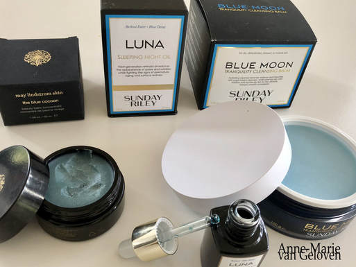
Lately I was trying out several skin care products with a very similar smell, which I actually started to appreciate during my evening skin care routine.
Usually, an overpowering fragrance in a product puts me off, however I consider this one soothing. The (in my opinion) pleasant odour comes from an ingredient called Tanacetum Annuum or Blue Tansy (Moroccan Blue Chamomile - not to be confused with Tanacetum Vulgare) and is found as the signature ingredient in some more luxury "Blue" products like May Lindrom's beauty balm concentrate called "The Blue Cocoon", Sunday Riley's tranquility cleansing balm called "Blue Moon" (Blue Tansy Leaf oil) and her sleeping night oil called "Luna". All products are relatively "oily" and you only need the littlest amount. Blue Tansy is "calming", as it supposed to have anti-inflammatory and anti-allergenic, anti-histaminic and anti-fungal properties. Tanacetum Annuum is an essential oil with a very dark blue collar due to chamazulene. The aromatic description is sweet, warm fruity, with subtle floral, camphorous and herbaceous undertones. It's most often mixed in with other oils or ingredients to dilute it, as the recommendation is not to use concentrations above 5%. Although it has anti-inflammatory properties, some might have intolerance for it as it contains camphor, which can cause sensitivity. Therefore, I would not recommend to use multiple products containing Blue Tansy in conjunction. Pure Blue Tansy oil is not easy to get hold of, thus an expensive ingredient. If I was asked choose one product, I would pick Sunday Riley Luna sleeping night oil which also contains Retinol. Luna is easy to use and incorporate in a night time regimen, is less expensive when compared to May Lindrom's "The Blue Cocoon", very popular amongst "beauty guru's" and receives many positive reviews. Alternatively, there are other evidence based skin care ingredients with proven anti-inflammatory properties, for example Arctiin (anti-inflammageing, stimulates hyaluronic acid and collagen production) and Licochalcone (also powerful anti-oxidant). They don't have the blue colour or "calming" odour, which some may find offensive. Hope you enjoy healthy skin & take care. |
CategoriesAll Acne Age Clocks Ageing Aquatic Wrinkles Armpits Autophagy Biostimulators Blue Light & HEVIS Circadian Rhythms Cleansing Collagen CoQ10 Cosmetic Intolerance Syndrome Deodorant Dermaplaning Diabetes DNA Damage DNA Repair Dry Skin Epigenetics Evidence Based Skin Care Exfoliation Exosomes Eyes Face Or Feet? Facial Oils Fibroblast Fingertip Units Gendered Ageism Glycation Growth Factors Gua Sha Hair Hair Removal Hallmark Of Aging Healthy Skin Heat Shock Proteins Hormesis Humidity Hyaluron Hyaluronidase Hypo-allergenic Indulging Jade Roller Keratinocytes Licochalcone A Luxury Skin Care Lymphatic Vessel Ageing Malar Oedema Menopause Mitochondrial Dysfunction Mood Boosting Skin Care Neurocosmetics Ox Inflammageing Peptides PH Balance Skin Photo Biomodulation Polynucleotides Proteasome Psoriasis Regeneration Regenerative Treatments Review Safety Scarring Sensitive Skin Skin Care Regimen Skin Flooding Skin Hydration Skin Senescence Skip-Care Sleep Slugging Sunscreen Tanning Under Eye Bags UV Index Vitamin C Vitamin D Well Ageing Skin Care Wound Healing Wrinkles
Archives
December 2024
|
Anne-Marie van Geloven © 2024 All rights reserved
|