Live your best life & take care
![]() Lipid peroxidation is a chemical process involving the oxidative degradation of lipids, particularly polyunsaturated fatty acids (PUFAs). This process occurs naturally during aging and can be induced by external factors such as UV radiation, pollution, and cigarette smoke when free radicals, especially reactive oxygen species (ROS), interact with lipids in cell membranes. The initial interaction generates lipid radicals and lipid peroxides, which can then react with additional oxidizing agents, creating a cascading chain reaction that leads to oxidative stress and significant cellular damage [1]. While sebum (the oily substance produced by sebaceous glands) primarily consists of saturated and monounsaturated fatty acids, it also contains smaller quantities of polyunsaturated fatty acids. Lipid peroxidation plays a role in various skin conditions, including acne and signs of photoaging [2][3], rosacea, psoriasis and eczema. ![]() OILY SKIN AND LIPID PEROXIDATION Oily skin types are particularly vulnerable to lipid peroxidation due to higher sebum (lipid) production and concentration at the skin surface. Sebum, an oily substance produced by sebaceous glands, is composed of various lipids, including triglycerides, free fatty acids, and squalene. When exposed to environmental stressors such as UV radiation and pollution [3], sebum components can undergo oxidation, leading to: 1. Altered composition: Oxidation changes the sebum's composition, resulting in the formation of comedogenic substances that can clog pores and initiate inflammatory processes associated with acne [4]. 2. Squalene oxidation: Squalene, a major component of sebum, is highly susceptible to oxidation. When oxidized, it generates harmful byproducts that contribute to inflammation and skin conditions like acne [4][5]. 3. Increased membrane permeability: Oxidation alters the fluidity and integrity of the skin's lipid barrier, making it more permeable and susceptible to further oxidative damage [6][7]. 4. Changed sebum consistency: The products of lipid peroxidation can accumulate and change the composition of sebum, leading to thicker consistency and potential blockage of hair follicles, which is a precursor to acne formation [5]. INFLAMMATORY RESPONSES AND ANTIOXIDANT CAPACITY Lipid peroxidation triggers inflammatory responses in the skin. The oxidized lipids can act as signaling molecules that promote inflammation, leading to conditions such as acne vulgaris. Increased levels of lipid peroxides correlate with inflammatory lesions in acne patients [4][6]. Oily skin types often experience an imbalance in antioxidant defenses. Deficiencies in antioxidants like vitamins E and C are common in those suffering from acne, which can exacerbate oxidative stress and promote lipid peroxidation [2]. Although sebum contains some antioxidants (like vitamin E), excessive oxidative stress from environmental factors can overwhelm these defenses, leading to increased lipid peroxidation and subsequent skin damage. FERROPTOSIS AND SKIN HEALTH Lipid peroxidation often initiates the process of ferroptosis. It occurs when the cell's antioxidant defenses, particularly the glutathione peroxidase 4 (GPX4) enzyme, fail to neutralize lipid peroxides. When lipids are damaged by oxidative stress, they generate lipid peroxides, which can accumulate and become toxic to cells. When lipid peroxides reach critical levels, they trigger ferroptosis, leading to ferroptosis a form of regulated cell death. PREVENTING LIPID PEROXIDATION Its clear that managing lipid pre-oxidation will be beneficial, especially for oily skin and to reduce its impact, you can try: 1. Antioxidant-rich skincare: Incorporating topical products containing antioxidants such as vitamins C and E, and Licochalcone A can help neutralize free radicals and prevent oxidative damage to lipids [2]. Licochalcone A has been shown to inhibit lipid peroxidation in keratinocytes (the main cell type in the epidermis) induced by UV radiation [8], and in human dermal fibroblasts exposed to hydrogen peroxide [9]. 2. Melatonin: The hormone melatonin, is widely present in various tissues including the skin and regulates circadian rhythms and promotes sleep. Melatonin can penetrate membranes and mitigate lipid peroxidation and protein oxidation, as well as oxidative damage to the mitochondria and DNA caused by UVR [10]. 3. Daily cleansing and regular exfoliation: Using non-abrasive exfoliants like salicylic acid or glycolic acid and daily cleansing can help remove oxidized lipids and pollution particles or dirt from the skin's surface, preventing clogged pores and reducing the risk of acne formation [4]. Try to avoid overcleansing or overexfolliation, not because the skin “compensates” by increasing the sebum production (which is a myth), however because you might damage the skin barrier. 4. Sun protection: Exposure to UV light generates reactive oxygen species (ROS), which can initiate lipid peroxidation in sebum. This process leads to the formation of lipid hydroperoxides and other oxidized lipids that can disrupt skin barrier function and promote inflammation [5]. Applying broad-spectrum sunscreen daily protects against UV-induced oxidative stress which contributes to lipid peroxidation [3]. Sunscreens containing antioxidants (like Licochalcone A) can provide additional protection. 5. Healthy diet: Consuming a diet rich in omega-3 fatty acids, fruits, and vegetables can enhance the skin's antioxidant defenses and reduce inflammation associated with lipid peroxidation [2]. 6. Avoiding environmental stressors: Reducing exposure to pollutants and smoking can minimize oxidative stress on the skin. ![]() 7. Sebum regulating topical ingredients: There are several evidence based skin care ingredients. I will highlight one called L-Carnitine. L-Carnitine is a skin’s own amino acid derivative is produced from the amino acids lysine and methionine. It supports sebum regulation through several mechanisms: ▌Increased β-oxidation (“fat burning”): L-carnitine significantly augments β-oxidation in human sebocytes, which is the process by which fatty acids are broken down [11]. This leads to a decrease in intracellular lipid content. ▌Sebum secretion reduction: Topical application of a 2% L-carnitine formulation for 3 weeks significantly decreased the sebum secretion [11]. ▌Bio-availability: Topically applied L-carnitine is bioavailable and can reach the dermis, allowing it to interact with sebaceous glands [11]. 8. Mattifying pigments: Mattifying pigments can be beneficial for oily skin, as they help absorb excess sebum and reduce shine. These pigments typically include ingredients such as clays, minerals, starches, polymers, charcoals, and silicas. 9. Blotting papers are thin, absorbent sheets designed to absorb thus remove excess oil from the skin's surface, providing a temporary matte appearance [12]. 10. Powder: Powder with ingredients like silica or talc, can absorb some oil and provide a matte finish. However, I would not recommend to apply a powder several times per day, because you are building up a “dirt layer” on top of the skin. Always consult a qualified healthcare professional to determine what the most suitable approach is for your skin health and beauty. Take care Anne-Marie References: [1] Ayala A, Muñoz MF, Argüelles S. Lipid peroxidation: malondialdehyde and 4-hydroxy-2-nonenal. Oxid Med Cell Longev. 2014;2014:360438. doi:10.1155/2014/360438. [2] Briganti S, Picardo M. Antioxidant activity and lipid peroxidation in skin diseases. J Eur Acad Dermatol Venereol. 2003;17(6):663-9. doi:10.1046/j.1468-3083.2003.00751.x. [3] Niki E. Lipid oxidation in skin. Free Radic Res. 2015;49(7):827-34. doi:10.3109/10715762.2014.976213. [4] Bowe WP, Logan AC. Lipid peroxidation in acne vulgaris. Lipids Health Dis. 2010;9:141. doi:10.1186/1476-511X-9-141. [5] Picardo M, Ottaviani M, Camera E, Mastrofrancesco A. Sebaceous gland lipids. Dermatoendocrinol. 2009;1(2):68-71. doi:10.4161/derm.1.2.8472. [6] Yadav DK, Kumar S, Choi EH, et al. Dynamics of oxidized skin lipid bilayer and ROS permeability. Sci Rep. 2019;9:4496. doi:10.1038/s41598-019-40913-y. [7] Ludovici M, Kozul N, Materazzi S, et al. Sebaceous gland density and stratum corneum lipidome. Sci Rep. 2018;8:11500. doi:10.1038/s41598-018-29742-7. [8] Kim HK, Kim JH, Lee SJ, Lee HS. Licochalcone A inhibits lipid peroxidation in keratinocytes from UV radiation. J Invest Dermatol. 2005;125(5):1009-16. doi:10.1111/j.0022-202X.2005.23845.x. [9] Huang J, Wang Z, Liu Y. Licochalcone A protects human dermal fibroblasts from hydrogen peroxide damage. Mol Med Rep. 2013;7(6):1977-82. doi:10.3892/mmr.2013.1397. [10] Bocheva G et al. Protective Role of Melatonin and Its Metabolites in Skin Aging. Int. J. Mol. Sci. 2022;23:1238. [11] Peirano RI, et al. Topically applied L-carnitine effectively reduces sebum secretion in human skin. J Cosmet Dermatol. 2012;11(11):30-36. [12] Wu PC, et al. Blotting paper as a disposable tool for sampling chemical residues from skin surface. J Food Drug Anal. 2019;27(2):610-613.
Comments
![]()
After "deep-diving" into autophagy and impaired autophagy, one of the twelve hallmarks of aging, it makes sense to shine some light on its equally important (however not so famous) partner in cellular housekeeping: the proteasome. It ́s primary function is breaking down proteins that are no longer needed, damaged, or misfolded [1]. Similar to autophagy, it is our body's and skin's very own trash and recycling system, working 24/7 to keep our cells healthy and functioning [2]. The human body is composed of approximately 16-20% protein by weight. This percentage can vary based on factors like age, sex, and overall body composition. Skin, is particularly rich in proteins, about 25-30% of the total protein in the human body is found in the skin and the dry weight of skin is approximately 70% protein. Loss of proteostasis (balance of protein synthesis, folding, and degradation) is one of the twelve hallmarks of aging and the proteasome is an important mechanism within the proteostasis network [3].
THE PROTEASOME The proteasome is a large, barrel-shaped protein complex found in all eukaryotic cells, responsible for the degradation of intracellular proteins [4]. It plays a crucial role in maintaining cellular homeostasis by selectively breaking down short-lived, damaged, or misfolded proteins [5]. The 26S proteasome consists of a 20S core particle and one or two 19S regulatory particles [6]. Proteins targeted for degradation are typically tagged with ubiquitin molecules, which are recognized by the 19S regulatory particle, allowing the protein to be unfolded and fed into the 20S core for proteolysis [7]. The ubiquitination process provides a highly selective mechanism for targeting proteins for degradation in comparison to other systems like lysosomes. Proteasomal degradation is an ATP-dependent process:
![]()
This process is crucial for:
▌Maintaining protein quality control [12] ▌Regulating cellular processes by controlling protein levels ▌Recycling amino acids for new protein synthesis The proteasome is involved in numerous vital cellular processes (see illustration), including: ▌Cell cycle regulation ▌Transcriptional control ▌Immune responses ▌Neuronal plasticity Its proper function is essential for cellular health, and dysfunction of the proteasome system has been implicated in various diseases, including neurodegenerative disorders and cancer. The proteostasis network The proteostasis network (PN) is a complex system of cellular machinery that maintains the integrity of the proteome consisting of collaborating systems to ensure proper protein folding, repair damaged proteins and eliminate those beyond repair. ▌Molecular chaperones and co-chaperones ▌The ubiquitin-proteasome system (UPS) ▌Autophagy machinery ▌Translational machinery ![]()
PROTEASOME VS AUTOPHAGY
Complementary cleaning and recycling systems While the proteasome primarily handles short-lived and soluble proteins, autophagy is responsible for degrading long-lived proteins, protein aggregates, and even entire organelles [13]. The proteasome plays critical roles in cell cycle control, gene expression, protein quality control, and immune responses, while other systems like autophagy are more involved in bulk degradation and cellular remodeling. The systems are not entirely independent and often work together to maintain cellular health [14]. The ubiquitin-proteasome system (UPS) and autophagy interact through various mechanisms:
PROTEASOME AND EPIGENETICS The proteasome also plays a significant role in epigenetics - the study of heritable changes in gene expression that don't involve changes to the underlying DNA sequence and recognised as one of the hallmarks of aging [19]. The proteasome influences epigenetics through several mechanisms: ▌Histone regulation + modification: The proteasome degrades histones, proteins that package DNA, influencing chromatin structure and gene accessibility [20].. ▌Transcription factor control + regulation: By regulating the levels of transcription factors, the proteasome indirectly affects gene expression patterns [21]. ▌Epigenetic modifier turnover + DNA methylation: The proteasome controls the levels of enzymes that modify histones and DNA, such as histone deacetylases (HDACs) and DNA methyltransferases (DNMTs) [22]. ▌Non-proteolytic functions: Some proteasome subunits have been found to directly interact with chromatin, suggesting a more direct role in gene regulation [23]. These interactions create a complex feedback loop between protein degradation and gene expression, highlighting the proteasome's far-reaching influence on cellular function PROTEASOME AND (SKIN) HEALTH The proteasome is likely present in skin cells and in extracellular fluids associated with skin, such as sweat and plays a vital role in maintaining health and skin quality by regulating the turnover of various proteins. Proteins are fundamental to life for several reasons:
Important proteins in skin and the human body based on their overall impact and prevalence:
Dysfunction of the proteasome in skin cells can lead to various dermatological issues, including ▌accelerated aging of skin cells ▌reduced collagen production and increased breakdown ▌impaired elastin function ▌wrinkles, sagging and loss of elasticity ▌impaired wound healing and barrier function ▌increased susceptibility to UV damage and DNA damage [26] Or more skin conditions like:
PROTEASOME AND CELLULAR SENESCENCE The proteasome plays a crucial role in preventing cellular senescence, a state of permanent cell cycle arrest associated with aging:
PROTEASOME AND IMMUNE FUNCTION The proteasome is integral to immune system function:
Glycosylated proteins Proteins connected to sugar molecules, known as glycosylated proteins, can be targeted by the proteasome: ▌Ubiquitin-Proteasome System (UPS) is capable of degrading many types of glycoproteins [29]. ▌However, hyperglycemia (high blood sugar) can impair proteasome function. Glucose-derived compounds like methylglyoxal (MGO) can modify proteasome subunits, reducing their activity [29]. Amyloids The proteasome's relationship with amyloids (involved in for example Alzheimer's disease) is more complex. The proteasome can degrade some amyloid precursor proteins and smaller amyloid aggregates [30]. However, larger amyloid fibrils often overwhelm or inhibit the proteasome: ▌Amyloid aggregates can clog the entrance to the proteasome's catalytic core. ▌Some amyloids can directly inhibit proteasome activity. INFLUENCERS PROTEASOME ACTIVITY Challenges in protein clearance Several factors can hinder the proteasome's ability to clear modified or aggregated proteins: Glycation: Advanced glycation end products (AGEs) formed in hyperglycemic conditions can modify the proteasome, reducing its activity [29]. Oxidative stress: Often associated with aging and disease, it can damage both proteins and proteasomes [29]. Aging: Proteasome activity generally declines with age, reducing the cell's capacity to clear problematic proteins [30]. The proteasome's activity is sensitive to pH changes: ▌Optimal pH range for proteasome function is typically between 7.5-8.0. ▌Acidic conditions tend to inhibit proteasome activity, while alkaline conditions can enhance it to a certain extent. ▌Skin pH, which is typically slightly acidic (around 4.7-5.75), may influence extracellular proteasome activity. Oxidative stress has complex effects on the proteasome system in skin: ▌Mild oxidation (hormesis) can stimulate proteasomal degradation, while severe oxidation inhibits it ▌Oxidative stress can cause the 26S proteasome to disassemble into its 20S core and 19S regulatory components [25] ▌In skin, oxidative stress from UV radiation or environmental pollutants may affect proteasome function ▌Severely oxidized proteins may form non-degradable aggregates that can bind to and inhibit the proteasome [24] ▌Oxidative stress can reduce cellular ATP levels, affecting the ATP-dependent 26S proteasome function [25] ▌Oxidative stress can alter the association of chaperone proteins like HSP70 with the proteasome, affecting its function and assembly [25] Temperature can significantly impact proteasome function: ▌The optimal temperature for proteasome activity is typically around 37°C (human body temperature) [27] ▌Higher temperatures may initially increase proteasome activity but can eventually lead to denaturation and loss of function. ▌Low temperatures reduce proteasome activity by slowing down enzymatic reactions. ▌Skin, being exposed to environmental temperature changes, may experience fluctuations in proteasome activity. MAINTAIN AND IMPROVE PROTEASOME Several strategies can help maintain and improve proteasomal function: Exercise: Regular physical activity has been shown to enhance proteasome activity. Diet: ▌Protein: Ensuring adequate intake of high-quality proteins provides the building blocks for maintaining a healthy proteome ▌Polyphenols: Found in green tea, berries, and red wine, can stimulate proteasome function. ▌Omega-3 fatty acids: May help maintain proteasome activity and reduce oxidative stress. ▌Sulforaphane (found in broccoli sprouts): Activates Nrf2, which enhances proteasome function. ▌Spermidine: This natural polyamine has been shown to enhance autophagy and improve proteostasis. ▌Curcumin: This compound from turmeric has been shown to enhance proteostasis and have anti-aging effects ▌Caloric restriction or intermittent fasting: May enhance proteasome activity and promote cellular health. Stress management: Chronic stress can impair proteasome function, so stress reduction techniques will be beneficial. Sleep: Crucial for cellular repair and protein homeostasis. Skincare + ingredients: ▌Sun protection: Use broad-spectrum sunscreens to protect skin from photo-damage, which can impair proteasome function. ▌Retinoids: May enhance proteasome activity in skin cells. ▌Peptides: Certain peptides have been shown to stimulate proteasome function. ▌Licochalcone A: Activates Nrf2, which in turn enhances proteasome function. ▌Niacinamide: Supports proteasome function and improves skin barrier health. In-office treatments: ▌Low-level laser therapy: May improve proteasome function in skin cells. ▌Chemical peels: Can stimulate cellular renewal and potentially enhance proteasome activity. MISCELLANEOUS PROTEASOME FACTS ▌Ancient origins: Proteasomes are found in all three domains of life (bacteria, archaea, and eukaryotes), suggesting they evolved over 2 billion years ago. ▌Rapid recyclers: A single proteasome can degrade about 2 million proteins over its lifetime. ▌Circadian rhythm regulation: The proteasome plays a crucial role in maintaining our body's internal clock by degrading clock proteins at specific times. ▌Stress response: Under stress conditions, cells can form large assemblies of proteasomes called "proteasome storage granules" to quickly respond to changing protein degradation needs. The role of the proteasome in protein quality control, cellular regulation, interplay with autophagy, epigenetics, telomeres, cell senescence and more, makes it a key player in maintaining our health and beauty and an interesting target for new strategies to enhance longevity [28], health span and beauty span. Always consult a qualified healthcare professional to determine what the most suitable approach is for your needs and rejuvenation or regeneration goals. Take care! Anne-Marie References: [1] Glickman MH, Ciechanover A. Physiol Rev. 2002;82(2):373-428. [2] Lecker SH, et al. Annu Rev Biochem. 2006;75:629-649. [3] López-Otín C, et al. Cell. 2013;153(6):1194-1217. [4] Tanaka K. Proc Jpn Acad Ser B Phys Biol Sci. 2009;85(1):12-36. [5] Goldberg AL. Nature. 2003;426(6968):895-899. [6] Finley D. Annu Rev Biochem. 2009;78:477-513. [7] Pickart CM, Cohen RE. Nat Rev Mol Cell Biol. 2004;5(3):177-187. [8] Hershko A, Ciechanover A. Annu Rev Biochem. 1998;67:425-479. [9] Thrower JS, et al. EMBO J. 2000;19(1):94-102. [10] Smith DM, et al. Mol Cell. 2005;20(5):687-698. [11] Groll M, et al. Nature. 1997;386(6624):463-471. [12] Balch WE, et al. Science. 2008;319(5865):916-919. [13] Mizushima N, Komatsu M. Cell. 2011;147(4):728-741. [14] Dikic I. Trends Biochem Sci. 2017;42(11):873-886. [15] Ding WX, et al. Am J Pathol. 2007;171(2):513-524. [16] Zhao J, et al. Cell Metab. 2015;21(6):898-911. [17] Pandey UB, et al. Nature. 2007;447(7146):859-863. [18] Korolchuk VI, et al. Mol Cell. 2010;38(1):17-27. [19] Greer EL, Shi Y. Nat Rev Genet. 2012;13(5):343-357. [20] Qian MX, et al. Cell. 2013;153(5):1012-1024. [21] Muratani M, Tansey WP. Nat Rev Mol Cell Biol. 2003;4(3):192-201. [22] Gu B, Lee MG. Mol Cell. 2013;49(6):1134-1146. [23] Geng F, et al. Proc Natl Acad Sci USA. 2012;109(5):1437-1442. [24] Bach SV, et al. Biomol Concepts. 2016;7(4):215-227. doi:10.1515/bmc-2016-0016 [25] Bonea D, et al. BMC Plant Biol. 2021;21:486. doi:10.1186/s12870-021-03234-9 [26] Minoretti P, et al. Cureus. 2024;16(1):e52548. doi:10.7759/cureus.52548 [27] Groll M, et al. Nat Struct Biol. 2005;12(11):1062-1069. doi:10.1038/nsmb1006 [28] Galatidou S, et al. Mol Hum Reprod. 2024;30(7):gaae023. doi:10.1093/molehr/gaae023 [29=41] Queisser MA, et al. Hyperglycemia impairs proteasome function by methylglyoxal. Diabetes. 2010 [28=42] Mao, Y. Structure and Function of the 26S Proteasome. In: Harris, J.R., Marles-Wright, J. Macromolecular Protein Complexes III. Springer, 2021. [29=43] Schipper-Krom, S. Visualizing Proteasome Activity and Intracellular Localization. Front. Mol. Biosci. 6, 2019. [30=44] Lifespan.io. Loss of Proteostasis. Lifespan.io Topics. Accessed 2024. ![]()
Mitochondria are the "powerhouses" or "lungs" of our cells and bioenergetic semi-autonomous organelles with their own genomes and genetic systems. [1] They are responsible for generating the energy that fuels a wide range of cellular processes in the skin, including cell signaling, pigmentation, wound healing, barrier integrity [2], metabolism and quality control. [3] Mitochondria exist in each cell of the body and are generally inherited exclusively from the mother. Their primary role is cellular respiration; a process converting the energy in nutrients (like glucose) into a usable form of energy called ATP or Adenosine Triphosphate. Mitochondria are particularly abundant in the skin, reflecting the skin's high metabolic demand. When the functionality of mitochondria is impaired or declines, it impacts skin's vitality, health and beauty. Mitochondrial dysfunction is 1 of the 12 hallmarks of skin ageing.
The skin is particularly susceptible to mitochondrial stress due to its constant exposure to environmental insults, such as UV radiation, pollution, and other oxidative stressors. These factors can damage mitochondrial DNA, leading to increased production of reactive oxygen species (ROS) and disrupting the delicate balance of cellular processes. [4] In aged post-mitotic cells, heavily lipofuscin-loaded lysosomes perform poorly, resulting in the enhanced accumulation of defective mitochondria, which in turn produce more reactive oxygen species causing additional damage (the mitochondrial-lysosomal axis theory). [5] Optimal mitochondrial function is indispensable for sustaining the specialized functions of each cell type, like keratinocyte differentiation, fibroblast ECM production, melanocytes melanin production and distribution, immune cell surveillance, sebocytes and adipocytes. [6] Mitochondrial dysfunction is both directly and indirectly linked to chronological ageing and photo-ageing. [7] As mitochondrial function declines, the skin's ability to regenerate and repair itself is decreased. [2] This results in visible signs of aging, such as wrinkles, loss of elasticity, dryness, uneven pigmentation, melasma, age spots, lipomas, impaired wound healing. [2-4-5-8-9] Mitochondrial dysfunction also has been implicated in skin conditions like acne, eczema, lupus, psoriasis, vitiligo, atopic dermatitis and even skin cancer. [10] Ageing is associated with changes in mitochondrial morphology, including [6] ▌Hyperfusion or increased fragmentation ▌Loss of mitochondrial connectivity [11-7] ▌Decline in the efficiency of oxidative phosphorylation, leading to reduced ATP production ▌Decline mitochondrial membrane potential (ΔΨM) ▌Compromised cellular energy metabolism ▌Reduced mitochondrial turnover (downregulated biogenesis) ▌Impaired mitochondrial quality control such as mitophagy (removal of damaged mitochondria through autophagy) [6] These alterations are related to the increased production of ROS exhibited by mitochondria during ageing, the accumulation of which causes oxidative damage to mitochondrial and cell components contributing to cellular senescence. [12] Good mitochondrial function or metabolism: [7] ▌Redox homeostasis: (the way of reducing oxidative stress) - mitochondrial respiration and ROS production are essential for keratinocyte differentiation ▌ATP production: Adenosine Triphosphate provides energy to drive and support many processes in living cells (and GTP) ▌Respiration: mitochondrial respiration is the most important generator of cellular energy ▌Biogenesis: allows cells to meet increased energy demands, to replace degraded mitochondria and is essential for the adaptation of cells to stress [6] ▌Calcium homeostasis ▌Cellular growth ▌Programmed cell death (apoptosis) reducing cell senescence [13] ▌Mitochondrial protein synthesis: mitochondria typically produce 13 proteins encoded by mitochondrial DNA (mtDNA) Dysfunctional Mitochondria: [7] ▌Oxidative stress ▌Decreased ATP levels ▌Dysfunctional OXPHOS: Oxidative phosphorylation, a metabolic pathway in which enzymes oxidize nutrients to release stored chemical energy in the form of ATP ▌Altered mitochondrial biogenesis ▌Calcium imbalance ▌Cell death Mitochondrial proteins Mitochondria contain >1,100 different proteins (MitoCoP) that often assemble into complexes and supercomplexes such as respiratory complexes and preprotein translocases. The chaperones Heat Shock Proteins HSP60-HSP10 are the most abundant mitochondrial proteins. [3] Small heat shock proteins form a chaperone system that operates in the mitochondrial intermembrane space. Depletion of small heat shock proteins leads to mitochondrial swelling and reduced respiration. [14] Mitochondrial hyperpigmentation Emerging research has shed light on the intricate relationship between mitochondrial dysfunction and the development of hyperpigmentation, a condition characterized by the overproduction and uneven distribution of melanin in the skin. One of the key mechanisms underlying this connection is the role of mitochondria in the regulation of melanogenesis, the process by which melanin is synthesized. Mitochondria are involved in the production of various cofactors and signaling molecules that are essential for the activity of tyrosinase, the rate-limiting enzyme in melanin synthesis. [15] When mitochondrial function is impaired, it can lead to an imbalance in the production and distribution of these cofactors and signaling molecules, ultimately resulting in the overproduction and uneven deposition of melanin in the skin. [15] This can manifest itself as age spots, melasma, and other forms of hyperpigmentation. The link between mitochondrial dysfunction and hyperpigmentation has been further supported by studies on genetic disorders that involve mitochondrial dysfunction, such as mitochondrial DNA depletion syndrome. In these conditions, patients often exhibit a range of pigmentary skin changes, including patchy hyper- and hypopigmentation, as well as reticular pigmentation. [16] Mitochondrial crosstalk and exosomes Mitochondria can crosstalk and move beyond cell boundaries. [17] Mitochondria-derived material might be transferred to neighboring cells in the form of cell-free mitochondria or included in extracellular vesicles [18-19]. This process supports cellular repair and contributes to vital mitochondrial functions. Besides restoring stressed cells and damaged tissues due to mitochondrial dysfunction, intercellular mitochondrial transfer also occurs under physiological and pathological conditions. [20] The transfer of active mitochondria from mesenchymal stem cells (MSCs) has been identified as a repair mechanism for rejuvenating damaged skin fibroblasts. [21] MITOCHONDRIAL SUPPORT Move According Martin Picard phD being physically active is a protective factor against almost everything health related. Exercise stimulates the production of mitochondria as more energy is required. Be hungry sometimes If there is too much supply of energy acquired via food leads to mass shrinking of mitochondria or fragmentation. Don´t over-eat, be calorie neutral and sometimes being calorie deficient is good for mitochondria. Maintain a healthy weight, preferably with a mediterranean diet containing phenolic and polyphenolic compounds (increase mitochondrial function and number) nitrate rich vegetables, soybeans and cacao beans. Mitohormesis In model organisms, lifespan can be improved by compromising mitochondrial function, which induces a hormetic response (“mitohormesis”), provided that this inhibition is partial and occurs early during development. Feel good Feeling good (positivity), especially at night, has a scientifically proven positive effect on mitochondrial health index, it is even a predictive factor. Q10 or Coenzyme Q10 (CoQ10) Q10 is part of the mitochondrial respiration chain and essential for cellular energy production. About 95% of our cellular energy is generated with support of Q10, which is produced by the human body itself. During skin ageing, both the cellular energy production and levels of Q10 are declined. Q10 is a powerful anti-oxidant [22], thus protecting cells from oxidative stress and damage and has proven to be able to "rescue" senescent cells by decreasing elevated senescent markers like p21 levels and β-Galactosidases positive cell numbers (in-vitro). Q10 is bio-active, increasing collagen type I and elastin production. [23] Q10 can be supplemented via nutrition, however also via topical application and is considered an evidence based active ingredient in skin care products. Ubiquinol (reduced form) shows higher bioavailability compared to ubiquinone (oxidized form). [23] Pyrroloquinoline quinone (PQQ) Q10 improves the energy in the mitochondria, however PQQ has shown to increase the number of mitochondria and a redox maestro. I´ve written a full post about this compound, which can be found as skincare ingredient and supplement. Read more about PQQ Glutathione Glutathione is formed in cell's cytoplasm from glutamic acid, cysteine and glycine. It is present in 2 forms: reduced (GSH) and oxidized (GSSG). Reduced GSH is an active anti-oxidant, while the presence of inactive GSSG is increased under oxidative stress. The ratio between GSH and GSSH is considered a measure of oxidative stress. Glutathione participates in redox reactions, acts as co-factor of many anti-oxidant enzymes and is the most important non-enzymatic anti-oxidant, essential for synthesis of proteins and DNA. Low Glutathione results in accelerated ageing and inflammatory skin diseases. Mitochondrial glutathione (mGSH) is the main line of defense for the maintenance of the appropriate mitochondrial redox environment to avoid or repair oxidative modifications leading to mitochondrial dysfunction and cell death. [24] Glutathione can be increased via supplementation via precursors cysteine or N-acetylcysteine (not recommended for pregnant women), a combination of Glycine and NAC (called GlyNAC) part of the popular "power of three" supplementation, or the reduced form of Glutathione itself, or increased via topical active ingredients like Licochalcone A. [25] I´ve written about GlyNAC in my post on autophagy. Nicotinamide NR nicotinamide ribosome which is the precursor of NMN nicotinamide mononucleotide which is the precursor of NAD+ nicotinamide adenine dinucleotide all could have a protective effect on mitochondria. Nicotinamide adenine dinucleotide is present in living organisms as ions NAD+ and NADP+ and in reduced forms NADH and NADPH. NADH is a cofactor of processes inside mitochondria: ▌ATP production ▌Activation of "youth proteins" sirtuins ▌Activation of PARP Poly (ADP-ribose) polymerase, a family of proteins involved in many cellular processes such as DNA repair, genomic stability and programmed cell death ▌Reduction of ROS (free radicals) NAD levels as lowered during ageing. [26] One of the fans of NMN supplementation is Harvard Professor David Sinclair, best known for his work on understanding why we age and how to slow its effects and also featured in my article about hormesis. There are about 14 studies done to date with NMN supplementation in humans, one of which was done by Professor Sinclair. NMN supplementation does raise NAD levels, however there aren't substantial proven health benefits, unless you are unhealthy. Resveratrol Although systemically Resveratrol promotes mitochondrial biogenesis. [27] Other data shows that UVA (14 J/cm(2)) along with resveratrol causes massive oxidative stress in mitochondria. As a consequence of oxidative stress, the mitochondrial membrane potential decreases which results in opening of the mitochondrial pores ultimately leading to apoptosis in human keratinocytes. [28] Magnesium Magnesium supplementation has been shown to improve mitochondrial function by increasing ATP production, decreasing mitochondrial ROS and calcium overload, and repolarizing mitochondrial membrane potential. There are many forms of Magnesium, however Citrate, Malate and Orotate are particularly good for energy. L-Carnitine Placebo-controlled trials have shown positive effects of L-Carnitine supplementation on both pre-frail subjects and elderly men. The effect is possibly mediated by counteracting age-related declining L-carnitine levels which may limit fatty acid oxidation by mitochondria. NEW Ergothioneine (EGT) Ergothioneine (EGT) is a sulfur-containing amino acid derivative known for its antioxidant properties, particularly in mitochondria. It is transported into cells and mitochondria via the OCTN1 transporter, where it helps reduce reactive oxygen species (ROS) and maintain cellular homeostasis [29]. EGT binds to and activates 3-mercaptopyruvate sulfurtransferase (MPST), enhancing mitochondrial respiration and exercise performance [30]. It also protects against oxidative stress and inflammation, potentially benefiting conditions like neurodegenerative diseases [31]. Melatonin Not much talked about when it comes to mitochondria, however should not be ignored as mitochondria can benefit significantly from melatonin supplementation. 1. Antioxidant protection: Melatonin acts as a powerful antioxidant within mitochondria, scavenging free radicals and reducing oxidative damage to mitochondrial DNA and proteins [32][34]. 2. Regulation of mitochondrial homeostasis: Melatonin helps maintain electron flow, efficiency of oxidative phosphorylation, ATP production, and overall bioenergetic function of mitochondria [32][34]. 3. Preservation of respiratory complex activities: Melatonin helps maintain the activities of mitochondrial respiratory complexes, which are crucial for energy production [32][34]. 4. Modulation of calcium influx: Melatonin regulates calcium influx into mitochondria, helping prevent calcium overload which can be damaging [32][34]. 5. Protection of mitochondrial permeability transition: Melatonin helps regulate the opening of the mitochondrial permeability transition pore, which is important for maintaining mitochondrial integrity [32][34]. 6. Enhancement of mitochondrial fusion: Melatonin promotes mitochondrial fusion, which is part of the quality control process for maintaining healthy mitochondria [33]. 7. Promotion of mitophagy: Melatonin enhances the removal of damaged mitochondria through mitophagy, helping maintain a healthy mitochondrial population [33]. 8. Reduction of nitric oxide generation: Melatonin decreases nitric oxide production within mitochondria, which can be damaging in excess [32][34]. 9. Selective uptake by mitochondria: Melatonin is selectively taken up by mitochondrial membranes, allowing it to exert its protective effects directly within these organelles [34]. 10. Support of mitochondrial biogenesis: Some studies suggest melatonin may promote the formation of new mitochondria [33]. The key antioxidants used by mitochondria are Glutathione (GSH), Glutathione peroxidase (GPx), Coenzyme Q10 (CoQ10), Superoxide dismutase (SOD), Melatonin, Vitamin C (ascorbate) and Vitamin E (α-tocopherol). Red light therapy By incorporating red light therapy into your skin care routine, you can help to counteract the damaging effects of mitochondrial dysfunction and support the skin's natural renewal processes. As we continue to explore the 12 hallmarks of ageing, I am confident that we will gain even more valuable insights and develop breakthrough innovations that will improve skin quality, health, beauty and vitality. Always consult a qualified healthcare professional or dermatologist to determine what the most suitable approach is for your particular skin condition and rejuvenation goals. Take care! Anne-Marie References
3/20/2024 Comments Telomeres: tiny caps with big impact![]()
Our DNA is as like precious book of life filled with information and instructions, with telomeres acting like the protective covers. Just as book covers get worn over time, our telomeres naturally shorten as we age. This shortening is like a biological clock, ticking away with each cell division.
Telomere shortening is considered one of the twelve key hallmarks of aging. Those hallmarks all play an important role in longevity, health-span, and skin quality, thus both health and beauty. Telomeres are the protective end-caps of chromosomes, similar to the plastic caps at the end of shoelaces. They maintain genomic stability and prevent chromosomal damage. Telomeres become slightly shorter each time a cell divides, and over time they become so short that the cell is no longer able to successfully divide. They shorten more rapidly in dermal fibroblasts compared to epidermal keratinocytes, hence there are significant differences amongst our cells. Telomeres in skin cells may be particularly susceptible to accelerated shortening because of both proliferation and DNA-damaging agents such as reactive oxygen species and sun exposure. [16]. When a cell is no longer able to divide due to telomere shortening, this can lead to
![]()
This consequently affects both health and beauty
FACTORS INFLUENCING TELOMERE SHORTENING Sleep quality Poor sleep quality significantly impacts telomere length:
INTERVENTIONS FOR TELOMERE PRESERVATION 1. Possible strategies to preserve telomere length
Telomerase is an enzyme that plays a crucial role in maintaining the length of telomeres and skin cell function. Telomerase is a ribonucleoprotein enzyme, meaning it contains both protein (TERT plus dyskerin) and RNA components (TER or TERC). Its primary function is to add repetitive DNA sequences (telomeres) to the ends of chromosomes, preventing them from shortening during cell division. Telomerase is active in embryonic stem cells, some adult stem cells, cancer cells, certain skin cells, specifically:
Poor sleep quality is associated with shorter telomere length. Studies have found significant associations between shortened telomere length and poor sleep quality and quantity, including obstructive sleep apnea [17]. Not feeling well rested in the morning was significantly associated with shorter telomere length in older adults [18]. Sleep loss and poor sleep quality may activate DNA damage responses and cellular senescence pathways [17]. Poor sleep can increase oxidative stress and inflammation, which may accelerate telomere shortening [17]. Disruption of circadian rhythms due to poor sleep may negatively impact telomere maintenance [17]. Improving sleep quality through lifestyle changes and sleep hygiene practices may help preserve telomere length. [19]
A study showed that diet, exercise, stress management, and social support could increase telomere length by approximately 10% over five years [20].
Adopt a plant-rich diet, such as the Mediterranean diet, which includes whole grains, nuts, seeds, green tea, legumes, fresh fruits (berries), vegetables (leafy greens), omega-3 fatty acids from sources like flaxseed and fish oil or fatty fish and foods rich in folate. This diet is rich in antioxidants and anti-inflammatory properties that help maintain telomere length [21]. 5. Fasting Fasting, especially intermittent fasting, has attracted interest for its potential impact on health, including telomere preservation. Multiple studies have shown that intermittent fasting (IF) and other fasting regimens can reduce markers of oxidative stress and inflammation. Research on animals has demonstrated that caloric restriction and intermittent fasting can boost telomerase activity and enhance telomere maintenance in specific tissues. A human study by Cheng et al. (2019) found a correlation between intermittent fasting and longer telomeres, by reducing PKA activity and IGF1 levels, which are crucial for regulating telomerase function. A study showed that 36 hours of fasting induced changes in DNA methylation and another one histone modifications, hence fasting has the potential to induce epigenetic changes. Important note: Be careful with a time-restricted eating schedule (often seen as a form of intermittent fasting, where you eat all meals within an 8 hour time-frame), especially women in menopause or people with a pre-existing heart condition. The American Heart Association presented data indicating that people with a pre-existing heart condition have a 91% higher risk of of death of a heart disease when following the time-restricted eating schedule with an 8 hour window, compared to those who eat within a 12-16 hours window. However, several experts have criticised the data, which aren´t published in a peer reviewed journal. When considering fasting, or a time-restricted eating schedule, especially for a longer period, talk to a qualified HCP first. 6. Exercise
EMERGING TECHNOLOGIES IN TELOMERE-TARGETING SKINCARE Small RNAs in skincare Small RNAs play a significant role in the effectiveness of telomere-targeting skincare by influencing skin regeneration and cellular processes. Recent research has highlighted their potential in enhancing wound healing and reducing scarring, which are critical aspects of maintaining healthy skin. Small RNAs, such as microRNAs, are involved in regulating gene expression related to skin aging and and show potential in telomere maintenance [29]. They can modulate the expression of genes that control cellular senescence, oxidative stress response, and inflammation, all of which are crucial for preserving telomere integrity and function [30].
RNAi technology in development RNAi-based skincare approaches could target genes involved in telomere maintenance or have effects on markers related to telomere biology:
RNA-based telomere extension is a method developed at Stanford University and uses modified RNA to extend telomeres in cultured human cells, allowing cells to divide more times than untreated cells [35]. IN OFFICE DERMATOLOGICAL TREATMENTS Aesthetic, regenerative treatments that support skin quality may indirectly support telomere preservation.
Telomere shortening questionable as stand-alone hallmark [36] Telomere length (TL) has long been considered one of the best biomarkers of aging. However, recent research indicates TL alone can only provide a rough estimate of aging rate and is not a strong predictor of age-related diseases and mortality. Other markers like immune parameters and epigenetic age may be better predictors of health status and disease risk. TL remains informative when used alongside other aging biomarkers like homeostatic dysregulation indices, frailty index, and epigenetic clocks. TL meets some criteria for an ideal aging biomarker (minimally invasive, repeatable, testable in animals and humans) but its predictive power for lifespan and disease is questionable. There is inconsistency in epidemiological studies on TL's association with aging processes and diseases. This has led to debate about TL's reliability as an aging biomarker. It's unclear if telomere shortening reflects a "mitotic clock" or is more a marker of cumulative stress exposure. TL is still widely used in aging research but there are ongoing questions about its usefulness as a standalone biomarker of biological age. As research in regenerative medicine advances, we're seeing promising developments in therapies targeting telomere biology for longevity, health and beauty. While telomere research is exciting, it's important to remember that it's just one part of a comprehensive approach to aging, and future treatments will likely combine multiple strategies to target preferably all 12 hallmarks for the best results. Always consult a qualified healthcare professional or dermatologist to determine what the most suitable approach is for you. . Take care! Anne-Marie
References
[1] Martin, H., Doumic, M., Teixeira, M.T. et al. Telomere shortening causes distinct cell division regimes during replicative senescence in Saccharomyces cerevisiae. Cell Biosci11, 180 (2021) [2] M. Borghesan, W.M.H. Hoogaars, M. Varela-Eirin, N. Talma, M. Demaria, A Senescence-Centric View of Aging: Implications for Longevity and Disease, Trends in Cell Biology, Volume 30, Issue 10, 2020, Pages 777-791, ISSN 0962-8924, [3] McHugh D, Gil J. Senescence and aging: Causes, consequences, and therapeutic avenues. J Cell Biol. 2018 Jan 2;217(1):65-77. [4] Oeseburg, H., de Boer, R.A., van Gilst, W.H. et al. Telomere biology in healthy aging and disease. Pflugers Arch - Eur J Physiol 459, 259–268 (2010) [5] Catarina M Henriques, Miguel Godinho Ferreira, Consequences of telomere shortening during lifespan, Current Opinion in Cell Biology, Volume 24, Issue 6, 2012 [6] Henriques CM, Ferreira MG. Consequences of telomere shortening during lifespan. Curr Opin Cell Biol. 2012 [7] Chaib, S., Tchkonia, T. & Kirkland, J.L. Cellular senescence and senolytics: the path to the clinic. Nat Med 28, 1556–1568 (2022) [8] Lei Zhang et al. Cellular senescence: a key therapeutic target in aging and diseases JCI The Journal of Clinical Investigation 2022 [9] Muraki K, Nyhan K, Han L, Murnane JP. Mechanisms of telomere loss and their consequences for chromosome instability. Front Oncol. 2012 Oct 4;2:135. [10] Marlies Schellnegger et al. Aging, 25 January 2024 Sec. Healthy Longevity Volume 5 - 2024 Unlocking longevity: the role of telomeres and it´s targeting interventions [11] Bär C, Blasco MA. Telomeres and telomerase as therapeutic targets to prevent and treat age-related diseases. F1000Res. 2016 Jan 20;5:F1000 Faculty Rev-89. [12] Kasiani C. Myers et al. Blood (2022) 140 (Supplement 1): 1895–1896. Gene therapies November 15 2022 Successful Ex Vivo Telomere Elongation with EXG-001 in a patients with Dyskeratosis Congenital Kasiani C. Myers et al. [13] Falckenhayn C, Winnefeld M, Lyko F, Grönniger E. et al. Identification of dihydromyricetin as a natural DNA methylation inhibitor with rejuvenating activity in human skin. Front Aging. 2024 Mar 4;4:1258184 [14] Minoretti P, Emanuele E. Clinically Actionable Topical Strategies for Addressing the Hallmarks of Skin Aging: A Primer for Aesthetic Medicine Practitioners. Cureus. 2024 Jan 19;16(1):e52548 [15] Guterres, A.N., Villanueva, J. Targeting telomerase for cancer therapy. Oncogene 39, 5811–5824 (2020). [16] Buckingham EM, Klingelhutz AJ. The role of telomeres in the ageing of human skin. Exp Dermatol. 2011 Apr;20(4):297-302. [17] Debbie Sabot, Rhianna Lovegrove, Peta Stapleton, The association between sleep quality and telomere length: A systematic literature review, Brain, Behavior, & Immunity - Health, Volume 28, 2023, 100577, ISSN 2666-3546 [18] Iloabuchi, Chibuzo et al. Association of sleep quality with telomere length, a marker of cellular aging: A retrospective cohort study of older adults in the United States Sleep Health: Journal of the National Sleep Foundation, Volume 6, Issue 4, 513 – 521 [19] Rossiello, F., Jurk, D., Passos, J.F. et al. Telomere dysfunction in ageing and age-related diseases. Nat Cell Biol 24, 135–147 (2022) [20] Elisabeth Fernandez Research September 16 2013 Lifestyle changes may lengthen telomeres, A measure of cell aging. Diet, Meditation, Exercise can improve key element of Immune cell aging, UCSF Scientist report [21] Martínez P, Blasco MA. Telomere-driven diseases and telomere-targeting therapies. J Cell Biol. 2017 Apr 3;216(4):875-887. [22] Guo, J., Huang, X., Dou, L. et al. Aging and aging-related diseases: from molecular mechanisms to interventions and treatments. Sig Transduct Target Ther 7, 391 (2022). [23] Hachmo Y, Hadanny A, Abu Hamed R, Daniel-Kotovsky M, Catalogna M, Fishlev G, Lang E, Polak N, Doenyas K, Friedman M, Zemel Y, Bechor Y, Efrati S. Hyperbaric oxygen therapy increases telomere length and decreases immunosenescence in isolated blood cells: a prospective trial. Aging (Albany NY). 2020 Nov 18;12(22):22445-22456 [24] Gutlapalli SD, Kondapaneni V, Toulassi IA, Poudel S, Zeb M, Choudhari J, Cancarevic I. The Effects of Resveratrol on Telomeres and Post Myocardial Infarction Remodeling. Cureus. 2020 Nov 14;12(11):e11482. [25] Widgerow AD, Ziegler ME, Garruto JA, Bell M. Effects of a Topical Anti-aging Formulation on Skin Aging Biomarkers. J Clin Aesthet Dermatol. 2022 Aug;15(8):E53-E60. PMID: 36061477; PMCID: PMC9436220. [26] Alt, C.; Tsapekos, M.; Perez, D.; Klode, J.; Stoffels, I. An Open-Label Clinical Trial Analyzing the Efficacy of a Novel Telomere-Protecting Antiaging Face Cream. Cosmetics 2022, 9, 95. [27] Cosmetics & Toiletries Telomere protection: Act on the origin of youth, June 3th 2015 Sederma [28] Yu Y, Zhou L, Yang Y, Liu Y. Cycloastragenol: An exciting novel candidate for age-associated diseases. Exp Ther Med. 2018 Sep;16(3):2175-2182. [29] Gerasymchuk M, Cherkasova V, Kovalchuk O, Kovalchuk I. The Role of microRNAs in Organismal and Skin Aging. Int J Mol Sci. 2020 Jul 25;21(15):5281. [30] Jacczak B, Rubiś B, Totoń E. Potential of Naturally Derived Compounds in Telomerase and Telomere Modulation in Skin Senescence and Aging. International Journal of Molecular Sciences. 2021; 22(12):6381. [31] Roig-Genoves, J.V., García-Giménez, J.L. & Mena-Molla, S. A miRNA-based epigenetic molecular clock for biological skin-age prediction. Arch Dermatol Res 316, 326 (2024). [32] Eline Desmet, Stefanie Bracke, Katrien Forier, Lien Taevernier, Marc C.A. Stuart, Bart De Spiegeleer, Koen Raemdonck, Mireille Van Gele, Jo Lambert, An elastic liposomal formulation for RNAi-based topical treatment of skin disorders: Proof-of-concept in the treatment of psoriasis, International Journal of Pharmaceutics, Volume 500, Issues 1–2, 2016, Pages 268-274, ISSN 0378-5173 [33] Oger E, Mur L, Lebleu A, Bergeron L, Gondran C, Cucumel K. Plant Small RNAs: A New Technology for Skin Care. J Cosmet Sci. 2019 May/Jun;70(3):115-126. PMID: 31398100. [34] Vimisha Dharamdasani, Abhirup Mandal, Qin M. Qi, Isabella Suzuki, Maria Vitória Lopes Badra Bentley, Samir Mitragotri, Topical delivery of siRNA into skin using ionic liquids, Journal of Controlled Release, Volume 323, 2020, Pages 475-482, ISSN 0168-3659 [35] Krista Conger January 2015 Stanford Medicine News Center Telomere extension turns back aging clock in cultured human cells, study finds [36] Alexander Vaiserman, Dmytro Krasnienkov Telemore length as marker of biological age: state-of-the-art, open issues and future perspectives Front. [37] Martínez P, Blasco MA. Telomere-driven diseases and telomere-targeting therapies. J Cell Biol. 2017 Apr 3;216(4):875-887 ![]()
Glycation is one of the basic root causes of endogeneous (intrinsic) skin ageing and a very challenging one or almost impossible one to reverse. Glycation is an ageing reaction which begins in early life, developing clinical symptoms at around 30, and progressively accumulates in tissues and skin due to the glycated collagens that are difficult to be decomposed. Glycation occurs naturally in the body when sugars react with proteins and lipids to form advanced glycation end products (AGEs). AGEs can be exogenously ingested (through food consumption), inhaled via tobacco or endogenously produced and formed both intracellularly and extracellularly. AGE modifications lead to dermal stiffening, diminished contractile capacity of dermal fibroblasts, lack of elasticity in the connective tissues, contribute to hyperpigmentation and a yellowish skin appearance. The formation of AGEs is amplified through exogenous factors, e.g., ultraviolet radiation.
AGEs cause changes in the skin through 3 processes:
One study published in the Journal of Investigative Dermatology found that levels of AGEs were higher in the skin of older individuals compared to younger ones. The study also showed that there was a correlation between the level of AGEs and the severity of skin ageing. This suggests that inhibiting the production or accumulation of AGEs in the skin is a potential target for anti-ageing interventions or skin ageing management. AGEs are complex and heterogeneous, more than a dozen AGEs have been detected (however not all) in tissues and can be divided into three categories according to their biochemical properties. AGEs are formed through four pathways:
GLYCATION INHIBITION IS KEY AGEs can be crosslinked through side chains to form a substance of very high molecular weight, which is not easily degraded. The consequences from skin glycation are irreversible. This makes prevention or inhibition of the process the best potential strategy to maintain skin health and ageing skin management. One way to do this is by altering the diet to reduce the intake of sugars and carbohydrates, which are known to contribute to glycation. Several studies have found that reducing sugar intake can result in significant improvements in skin health, including reducing wrinkles and improving skin texture. ![]()
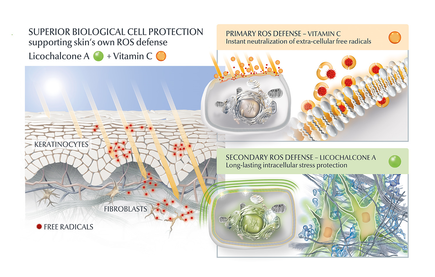
AGE inhibitors
Another potential strategy is the use of topical agents that inhibit the formation or accumulation of AGEs in the skin. One study published in the Journal of Cosmetic Science found that a cream containing carnosine, a peptide that inhibits glycation, improved skin elasticity and reduced the appearance of wrinkles in individuals with ageing skin. Skincare containing NAHP or Acetyl Hydroxyproline inhibits the formation of AGEs significantly (in vitro), most likely through a mechanism where NAHP competes with the proteins for the sugar. Finally, NAHP sacrifices itself in place of the proteins and gets (at least partially) glycated. NAHP also prevents loss of cellular contractile forces in a glycated in vitro dermis model and counteracts the diminished cell-matrix interaction that is caused by glyoxal-induced AGE formation. Take-aways from a study published in the International Journal of Cosmetic Science [2]: 1. NAHP significantly and dose-dependently inhibited the formation of advanced glycation end-products (AGEs) in a protein solution. 2. NAHP dose-dependently inhibited the formation of N-(carboxymethyl)lysine (CML), a specific AGE. 3. In fibroblast-populated collagen lattices, NAHP prevented the glycation-induced disturbance of fibroblast contractile capacity. 4. Ex vivo application of NAHP to skin explants decreased AGE fluorescence compared to glucose-treated samples. Anti-Oxidants I would suggest to combine those ingredients with an ingredient like Licochalcone A. Numerous high ranked publications support that Licochalcone A protects cells from oxidative stress mediated by e.g. UV and HEVIS (blue light) induced reactive oxidative species (ROS). Due to the activation and nuclear translocation of the transcription factor NrF2, the expression of anti-inflammatory, antioxidant and detoxifying enzymes are induced. These enzymes protect the skin cells (like keratinocytes and fibroblasts) from ROS-induced damage, like lipid peroxidation and DNA as well as protein damage. If Licochalcone A is combined with L-Ascorbic Acid, (the most active form of Vitamin C), it supporting skin's own collagen production, provides superior biological cell protection amongst other relevant benefits. Vitamin C (and E) has shown to inhibit protein glycation [4]. SPRAY TAN A study in Redox Biology indicates that sunless tanning with dihydroxyacetone (DHA) causes glycation and potential DNA damage in the epidermis [1].: 1. Glycation: DHA exposure led to the formation of advanced glycation end products (AGEs) in epidermal cells, confirmed by mass spectrometric detection of N-ε-(carboxyethyl)-l-lysine (CEL). 2. DNA damage: DHA induced a cellular stress response, including activation of stress-related genes and phosphoprotein signaling. While not directly measuring DNA damage, these responses are often associated with cellular stress that can lead to DNA damage. 3. Significance: The effects were observed at low millimolar concentrations of DHA, relevant to topical application. The stress response was rapid and pronounced, occurring within minutes of exposure. 4. Location: The effects were primarily observed in epidermal cells and reconstructs, with no mention of dermal effects. This study suggests that sunless tanning with DHA may not be as safe as previously thought, warranting further investigation into its long-term effects on skin health [1], however no need to panic as this negative effect is only seen in the top layer of the skin. DOUBLE TROUBLE: GLYCATION + UVB [3] The combination of GO-AGEs and UVB exposure has a more pronounced effect on skin inflammation and oxidative stress than either factor alone. This suggests a synergistic relationship between glycation and UV exposure in accelerating skin aging processes. 1. GO-AGEs combined with UVB irradiation significantly increased the secretion of pro-inflammatory cytokines (IL-1β, IL-6, IL-8) in skin cells compared to either GO-AGEs or UVB alone. 2. The GO-AGEs + UVB treatment group showed a more than three-fold increase in IL-6 and IL-8 mRNA levels compared to other groups. 3. GO-AGEs + UVB treatment induced significantly higher release of nitric oxide (NO) compared to other groups. 4. The combination of GO-AGEs and UVB enhanced reactive oxygen species (ROS) release, creating about 1.5 times more oxidative stress compared to control and other groups. 5. Cell viability was notably affected in the GO-AGEs, UVB, and GO-AGEs + UVB treatment groups compared to the control group. These findings are significant as they demonstrate that the combination of GO-AGEs and UVB exposure has a more pronounced effect on skin inflammation and oxidative stress than either factor alone. This suggests a synergistic relationship between glycation and UV exposure in accelerating skin aging processes. Use sunscreen daily. . GLYCATION AND SKIN HEALTH Acne In addition to its role in ageing, glycation in the skin has also been linked to a range of skin health problems. One study published in the Journal of Cosmetic Dermatology found that the level of AGEs in the skin was significantly higher in individuals with acne than in those without acne. The study also showed that treating acne with a topical antibiotic significantly reduced the levels of AGEs in the skin. Atopic Dermatitis Another study published in the Journal of Investigative Dermatology found that individuals with atopic dermatitis had higher levels of AGEs in their skin than healthy individuals. This suggests that glycation may play a role in the development of inflammatory skin conditions. Diabetes + Woundhealing The correlation between high sugar levels and skin ageing can be seen in diabetic patients, where one-third of this population has skin complications. A prominent feature of ageing human skin is the fragmentation of collagen fibers, which severely damages the structural integrity and mechanical properties of the skin. Elevated levels of MMP-1 and MMP-2 and higher crosslinked collagen in the dermis of diabetic skin lead to the accumulation of fragmented and crosslinked collagen, thereby impairing the structural integrity and mechanical properties of dermal collagen in diabetes. Collagen crosslinking makes it impossible for them to easily repair, resulting in reduced skin elasticity and wrinkles. Keratinocytes and fibroblasts are the main cells involved in wound healing, but due to the high glucose (HG) microenvironment in diabetics, the functional state of these cells is impaired, thereby accelerating cellular senescence (programmed cell death). Conclusion We can't completely stop the glycation process, therefore it's important that we inhibit it from a young age onwards, hence monitor the sugar intake of our children, use daily SPF and invest in good dermo-cosmetic products containing ingredients like NAHP and powerful anti-oxidants like L-Ascorbid Acid (Vitamin C is needed for the production of collagen) and Licochalcone A (also anti-inflammatory). Preventing signs of ageing, specifically caused by glycation is most effective. If your skin shows (advanced) signs of ageing, you can get visible improvement using skin component (hyaluron, collagen and elastin) bio-stimulating ingredients like Retinol, Bakuchiol, Arctiin, Creatine or Glycine Saponin. Consult your dermatologist if you wish to improve your skin's appearance or skin health issues. Take care Anne-Marie References [1] Perer J, Jandova J, Fimbres J, Jennings EQ, Galligan JJ, Hua A, Wondrak GT. The sunless tanning agent dihydroxyacetone induces stress response gene expression and signaling in cultured human keratinocytes and reconstructed epidermis. Redox Biol. 2020 Sep;36:101594. [2] Knoblich C, et al. N‐acetyl‐L‐hydroxyproline – A potent skin anti‐ageing active preventing advanced glycation end‐product formation in vitro and ex vivo. Int J Cosmet Sci. 2023;1-10. doi:10.1111/ics.12930 [3] Sultana, R., Parveen, A., Kang, MC. et al. Glyoxal-derived advanced glycation end products (GO-AGEs) with UVB critically induce skin inflammaging: in vitro and in silico approaches. Sci Rep 14, 1843 (2024). https://doi.org/10.1038/s41598-024-52037-z [3] Sadowska-Bartosz I, Bartosz G. Prevention of protein glycation by natural compounds. Molecules. 2015 Feb 16;20(2):3309-34. doi: 10.3390/molecules20023309. PMID: 25690291; PMCID: PMC6272653. Special thanks: Ph.D. dr Julia M. Weise Manager Biological Testing & Dorothea Schweiger Lab Manager Facial Skin Biology Beiersdorf HQ Hamburg ![]()
Psoriasis occurs in many different forms and levels of severity. The first signs may appear between the age of 15 and 35 and 75% of patients are diagnosed before the age of 46 according to the World Health Organisation. As there is no cure for the disease, the highest prevalence is seen in a more mature age group age 50-69. Many of the treatments which are part of the standard treatment guidelines for psoriasis cause as a side effect premature ageing skin. For example PUVA, next to being an effective treatment, does cause (severe) photo-damage. Many patients will undergo such treatments on and off or continuously throughout their life.
Although the primary goal of dermatology is to improve the functional attributes of the skin (health) and lessen the tremendous burden psoriasis may cause, ultimately one aims to improve the skin's physical attributes (appearance). Ageing skin is a biological degenerative process which influences the activity of collagen and hyaluronic acid producing cells (mainly fibroblasts) and leads to a decrease of skin components like collagen, hyaluron and functional elastin. Effective anti-aging skincare can support the protection of those cells and skin components (anti-oxidants and SPF) and thus slow down the fastened degenerative process. Some active ingredients (for example biologically active Glycine Saponin and Arctiin) have proven to effectively stimulate fibroblasts in the production of collagen and hyaluronic acid and thus replenish dermal components. Loss of those components and photo-damage eventually lead to visible signs of ageing. Although ageing skin is natural, premature ageing skin isn't necessary. Most patients will probably already use a moisturising facial skincare product. It makes sense to recommend anti-ageing skincare instead to be used in conjunction with treatments which as a side effect cause premature ageing skin, particularly for exposed areas like the face. As psoriasis oftentimes doesn't occur in the face (except in the hairline), anti-ageing skincare will pose a low to no risk to aggravate facial skin and there are anti-ageing skincare products available which have proven to be suitable for psoriasis patients. Take care. |
CategoriesAll Acne Age Clocks Ageing Aquatic Wrinkles Armpits Autophagy Biostimulators Blue Light & HEVIS Circadian Rhythms Cleansing Collagen CoQ10 Cosmetic Intolerance Syndrome Deodorant Dermaplaning Diabetes DNA Damage DNA Repair Dry Skin Epigenetics Evidence Based Skin Care Exfoliation Exosomes Eyes Face Or Feet? Facial Oils Ferroptosis Fibroblast Fingertip Units Gendered Ageism Glycation Growth Factors Gua Sha Hair Hair Removal Hallmark Of Aging Healthy Skin Heat Shock Proteins Hormesis Humidity Hyaluron Hyaluronidase Hypo-allergenic Indulging Jade Roller Keratinocytes Licochalcone A Lipid Peroxidation Luxury Skin Care Lymphatic Vessel Ageing Malar Oedema Menopause Mitochondrial Dysfunction Mood Boosting Skin Care Neurocosmetics Oily Skin Ox Inflammageing Peptides PH Balance Skin Photo Biomodulation Polynucleotides Proteasome Psoriasis Regeneration Regenerative Treatments Review Safety Scarring Sensitive Skin Skin Care Regimen Skin Flooding Skin Hydration Skin Senescence Skip-Care Sleep Slugging Sunscreen Tanning Under Eye Bags UV Index Vitamin C Vitamin D Well Ageing Skin Care Wound Healing Wrinkles
Archives
January 2025
|
Proudly powered by Weebly
Anne-Marie van Geloven © 2024 All rights reserved
|