Live your best life & take care
Collagen is a vital component of the skin's extracellular matrix, providing essential structural support and elasticity. Collagen-stimulating treatments, skincare products, and supplements have gained popularity for their effectiveness in gradual prejuvenation and rejuvenation approaches. These methods can help maintain skin health and combat signs of aging when used appropriately. However, it's important to note that excessive collagen stimulation can potentially lead to adverse effects, including fibrosis and skin stiffness, which may be detrimental to overall skin health and beauty. Therefore, a balanced and informed approach to collagen stimulation is crucial for achieving optimal results while minimizing potential risks. TYPES OF COLLAGEN AND THEIR ROLES 1. Type I collagen: Predominantly found in skin, tendons, and bones, providing tensile strength. 2. Type III collagen: Often found alongside Type I, contributing to skin elasticity and firmness. While these types are beneficial for youthful skin, excessive production can lead to fibrotic tissue formation and stiffness [1]. More about collagen types click here EXCESSIVE COLLAGEN STIMULATION Excessive collagen production, particularly type I collagen, can contribute to fibrosis and scarring in pathological conditions: 1. In hypertrophic scars, there is an overproduction of primarily type III collagen, which is later replaced by type I collagen. These scars contain "an overload of primarily type III collagen oriented parallel to the epidermal surface with multiple nodules containing myofibroblasts, large extracellular collagen filaments and abundant acidic mucopolysaccharides" [2]. 2. Many rejuvenating in-office treatments (for example energy based devices)are based on "controlled damage and repair”, thus wound healing. During wound healing, abnormal extracellular matrix (ECM) reconstruction, particularly abnormal collagen remodelling, leads to the formation of hypertrophic scars. In these scars, "thin collagen fibres with increased synthesis and crosslinks result in raised scars" [2]. 3. The relative ratio of type III to type I collagen is reduced in pathological scars compared to unscarred adult dermis. Additionally, hydroxylation of type I collagen was found to be significantly higher in keloids, leading to excessive collagen cross-linking [3]. IN-OFFICE TREATMENTS AND COLLAGEN STIMULATION These treatments aim to maintain or restore natural collagen production rather than overstimulate it to unnatural levels. Some examples are: 1. Exosomes and Polynucleotides: Aim to stimulate healthy collagen production but require careful application. 2. Radiofrequency and Ultrasound: Use heat to remodel collagen. While generally safe, a study by Zelickson et al. [4] reported that excessive heating during RF treatments could potentially lead to collagen denaturation and subsequent fibrosis if not properly controlled. 3. Microneedling: Promotes collagen production but risks scarring if not performed properly. A review by Iriarte et al. [5] noted that while microneedling is generally safe, excessive or improper use could potentially lead to scarring or hyperpigmentation. 4. Laser treatments: Excessive use of ablative lasers can potentially lead to scarring and fibrosis. A study by Hantash et al. [6] found that ablative fractional resurfacing can induce dermal remodeling and new collagen formation, but also noted that improper use could lead to adverse effects. It's important to emphasize that these potential adverse effects are typically associated with improper use, overtreatment, or individual susceptibility rather than being inherent risks of the treatments themselves when performed correctly. More research is needed to fully understand the long-term effects of repeated collagen stimulation treatments on skin structure and function. POTENTIAL RISKS ▌Excessive collagen production: Can lead to fibrosis, characterized by stiff, non-functional tissue: increased extracellular matrix deposition, with collagen being the main component, leading to a drastic reduction of tissue functionality [7]. In skin, this can result in reduced elasticity and increased stiffness. ▌Imbalance in collagen types: Overproduction of certain collagen types can lead to reduced skin elasticity and increased stiffness. The ratio of type I to type III collagen naturally increases with age, which is associated with changes in skin tension, elasticity, and healing [7]. RECOMMENDATIONS FOR SAFE USE ▌ Prejuvenation: Focus on treatments (performed by a professional) that promote balanced collagen production without overstimulation. The effect of a collagen-stimulating procedure is a gradual process and can take up to 12 weeks or longer before a final result. This gradual improvement is due to the time required for the body to produce new collagen in response to the stimulation. Laser treatments, for example, can trigger collagen synthesis deep within the skin, with effects continuing for several months post-treatment [8]. Leave sufficient time in between procedures. Support your skin with a skincare routine tailored to your skintype, goals and use of daily sunscreen. Be very rigorous when it comes to the use of home devices or treatments. Many of them are not well researched or might cause damage when not properly used or performed. ▌Rejuvenation: Opt for treatments or a combination of treatments that complement each other, working in different layers of the skin in different ways. Don't expect a "one-day transformation". Rebuilding collagen takes time and a consistent approach. The skin is not able to replenish what it lost over a period of many years in just a few days [9]. Support in-office collagen stimulating treatments with a good skincare regimen, daily use of sunscreen, healthy lifestyle and diet or supplementation if necessary [10]11]. The effectiveness of combining different treatments for skin rejuvenation has been demonstrated in clinical studies. For instance, a study published in the Journal of Clinical and Aesthetic Dermatology showed that a combination of microneedling and platelet-rich plasma significantly improved skin texture and collagen production compared to microneedling alone [12]. The importance of a consistent skincare regimen and sun protection in maintaining collagen levels has been well-documented. A review in the Archives of Dermatological Research highlighted that daily use of broad-spectrum sunscreen can prevent collagen degradation caused by UV radiation [13]. While collagen stimulation is beneficial for skin prejuvenation, "banking" or rejuvenation, it is crucial to balance its production to avoid the formation of fibrotic tissue and maintain healthy skin elasticity. Further research is needed to optimize treatment protocols and minimize risks associated with excessive collagen stimulation. Always consult a qualified healthcare professional to determine the most suitable approach for your skin goals, health, and beauty. Take care Anne-Marie References: [1] Wang Kang , Wen Dongsheng , Xu Xuewen , Zhao Rui , Jiang Feipeng , Yuan Shengqin , Zhang Yifan , Gao Ya , Li Qingfeng Extracellular matrix stiffness—The central cue for skin fibrosis Frontiers in Molecular Biosciences 2023 DOI=10.3389/fmolb.2023.1132353 [2] Meirte J, Moortgat P, Anthonissen M, Maertens K, Lafaire C, De Cuyper L, Hubens G, Van Daele U. Short-term effects of vacuum massage on epidermal and dermal thickness and density in burn scars: an experimental study. Burns Trauma. 2016 Jul 8;4:27. doi: 10.1186/s41038-016-0052-x. PMID: 27574695; PMCID: PMC4964043. [3] Zhou Claire Jing , Guo Yuan Mini review on collagens in normal skin and pathological scars: current understanding and future perspective Frontiers in Medicine 2024 [4] Zelickson, B. D., Kist, D., Bernstein, E., Brown, D. B., Ksenzenko, S., Burns, J., ... & Kilmer, S. (2004). Histological and ultrastructural evaluation of the effects of a radiofrequency‐based nonablative dermal remodeling device: a pilot study. Archives of Dermatology, 140(2), 204-209. [5] Iriarte, C., Awosika, O., Rengifo-Pardo, M., & Ehrlich, A. (2017). Review of applications of microneedling in dermatology. Clinical, Cosmetic and Investigational Dermatology, 10, 289-298. [6] Hantash, B. M., Bedi, V. P., Kapadia, B., Rahman, Z., Jiang, K., Tanner, H., ... & Zachary, C. B. (2007). In vivo histological evaluation of a novel ablative fractional resurfacing device. Lasers in Surgery and Medicine, 39(2), 96-107. [7] Wang, C., Rong, Y., Ning, F., & Zhang, G. (2011). The content and ratio of type I and III collagen in skin differ with age and injury. African Journal of Biotechnology, 10(13), 2524-2529. https://doi.org/10.5897/AJB10.1999 [8] Alam, M., Hughart, R., Champlain, A., Geisler, A., Paghdal, K., Whiting, D., Hammel, J. A., Maisel, A., Rapcan, M. J., West, D. P., & Poon, E. (2018). Effect of Platelet-Rich Plasma Injection for Rejuvenation of Photoaged Facial Skin: A Randomized Clinical Trial. JAMA Dermatology, 154(12), 1447-1452. https://doi.org/10.1001/jamadermatol.2018.3977 [9] Ganceviciene, R., Liakou, A. I., Theodoridis, A., Makrantonaki, E., & Zouboulis, C. C. (2012). Skin anti-aging strategies. Dermato-endocrinology, 4(3), 308-319. https://doi.org/10.4161/derm.22804 [10] Katta, R., & Desai, S. P. (2014). Diet and dermatology: the role of dietary intervention in skin disease. The Journal of clinical and aesthetic dermatology, 7(7), 46-51. [11] Addor, F. A. S. (2017). Antioxidants in dermatology. Anais brasileiros de dermatologia, 92, 356-362. https://doi.org/10.1590/abd1806-4841.20175697 [12] Asif, M., Kanodia, S., & Singh, K. (2016). Combined autologous platelet-rich plasma with microneedling verses microneedling with distilled water in the treatment of atrophic acne scars: a concurrent split-face study. Journal of Cosmetic Dermatology, 15(4), 434-443. https://doi.org/10.1111/jocd.12207 [13] Battie, C., & Verschoore, M. (2012). Cutaneous solar ultraviolet exposure and clinical aspects of photodamage. Indian Journal of Dermatology, Venereology, and Leprology, 78, S9-S14. https://doi.org/10.4103/0378-6323.97351
Comments
The UV Index (UVI) is a valuable tool for assessing the strength of ultraviolet (UV) radiation from the sun at any given location and time. The UVI values are determined using the STAR (System for Transfer of Atmospheric Radiation) model. This model takes into account various atmospheric conditions to estimate UV radiation levels. The values provided reflect typical conditions for each location and serve as reference points. Actual UV Index readings can vary due to local factors, such as temporary changes in ozone levels and other atmospheric conditions. The values range from 0 to 11+, serving as a standardized guide for sun protection measures. This helps us understand the potential for skin damage based on UV exposure levels. They are specified for the 21st of each month across different regions. Higher UVI values indicate a greater risk of harm, particularly concerning sunburn, DNA damage, premature skin aging and hyperpigmentation [1][2]. HIGHEST AND LOWEST UV INDEX VALUES Highest UV Index The highest recorded UV Index values can reach 12 or more, especially in regions near the equator, high-altitude areas, and places with low ozone levels. The Atacama Desert in Chile has documented peaks as high as 20, highlighting the extreme UV exposure possible in certain environments [2]. Lowest UV Index The lowest values are typically observed at night or during winter months in polar regions, where solar angles are significantly reduced, often resulting in readings close to zero [2][3]. GEOGRAPHIC INFLUENCES ON UV LEVELS UV exposure varies widely across different geographical regions and withing the regions: ▌Europe: Generally experiences moderate UV levels due to higher latitudes and frequent cloud cover [4]. ▌Asia: Significant variability; tropical areas encounter high UV levels while northern regions have lower indices [2]. ▌Australia: Known for high UV exposure, particularly during summer months, due to its proximity to the equator and clearer skies. ▌USA: Southern states typically report higher UV indices compared to their northern counterparts. ▌Latin America: High UV indices are prevalent near the equator, while southern regions like Argentina experience lower values [2][3]. ▌Altitude: Higher altitudes receive more intense UV radiation due to a thinner atmosphere [2]. ▌Reflection: Beaches can experience increased UV levels due to sunlight reflecting off water and sand [3]. ▌Northern vs. Southern hemisphere: The Southern hemisphere generally has higher UV levels attributed to less atmospheric pollution and ozone depletion [2]. ▌Equatorial regions: These areas maintain consistently high UV indices throughout the year due to direct sunlight [2][3]. INDOOR vs OUTDOOR UV EXPOSURE The UV Index indoors is significantly lower than outdoor levels on a sunny day. This is primarily due to the filtering effect of window glass, which blocks most UVB radiation. On a clear day, outdoor UV levels can reach up to 8,000 µW/cm², while indoor levels near a window may be as low as 250 µW/cm², dropping further with distance from the window. The indoor UVI reduction is primarily due to the filtering effect of glass windows, which block most UVB (320–400 nm) radiation while allowing some UVA (320–400 nm) rays to penetrate and can still contribute to premature skin aging, hyperpigmentation and DNA damage. Blue Light (400–495 nm): Part of visible light spectrum; penetrates glass easily. High energy Visible Light is responsible for 50% of the free radical activity [5] and like UV radiations contributes to premature skin aging, hyperpigmentation and DNA damage. Factors influencing indoor UV exposure include window size, orientation, and surrounding obstructions like trees. Direct and indirect exposure ▌Direct exposure occurs when sunlight directly enters through windows. ▌Indirect (Diffuse) exposure results from sunlight scattering off surfaces or atmospheric particles. While diffuse exposure is reduced by walls and roofs, it can still penetrate through windows [3]. Factors affecting indoor exposure 1. Window glass: Standard glass blocks most UVB but allows some UVA and High energy Visible Light through. 2. Sky view: More visible sky from indoors increases diffuse UV exposure. 3. Distance from windows: The intensity of UV radiation decreases with distance from windows due to the inverse square law [3]. 4. Window orientation and size: Larger windows facing south (in the Northern Hemisphere) or north (in the Southern Hemisphere) allow more sunlight into indoor spaces [3]. 5. Scattering (indirect – diffuse exposure) CHANGING UVI OVER TIME There is scientific evidence indicating that the UV Index (UVI) is influenced by various environmental factors, including changes in ozone levels and climate conditions, which can affect UV radiation exposure over time. 1. UV radiation: A study by Fountoulakis et al. (2020) analyzed long-term changes in UV-B radiation and found that variations in UV levels are primarily driven by changes in aerosols and total ozone, with significant regional differences observed. The study indicates that while some areas have experienced increases in UV-B irradiance, others have shown decreases, particularly during summer months in polar regions due to improvements in ozone levels [6]. 2. Impact of ozone depletion: Research has shown that the decline of stratospheric ozone has historically led to increased UV radiation at certain wavelengths. For instance, a study by Bais et al. (2011) projected that UV irradiance would likely return to its 1980 levels by the early 21st century at northern mid-latitudes and high latitudes, suggesting ozone recovery influences UV radiation levels [7].While standard windows block most harmful UVB rays, damaging UVA and blue light (or HEVIS) can still penetrate indoors, affecting skin´s beauty and health. Awareness of these factors and UV Index enables you to take appropriate protective measures against harmful effects of sunlight even indoors while considering the benefits of controlled exposure for vitamin D synthesis [3]. Take care Anne-Marie References [1] Federal Office for Radiation Protection (BfS). (n.d.). What is the UV Index? Retrieved December 7, 2024, from bfs.de/EN/topics/opt/uv/index/introduction/introduction_node.html [2] Fioletov V, Kerr JB, Fergusson A. The UV index: definition, distribution, and factors affecting it. Can J Public Health. 2010;101(4):I5-9. doi: 10.1007/BF03405303. [3] Heckman CJ, Liang K, Riley M. Awareness and impact of the UV index: A systematic review of international research. Prev Med. 2019;123:71-83. doi: 10.1016/j.ypmed.2019.03.004. [4] World Health Organization. (n.d.). Radiation: The UV index. Retrieved December 7, 2024, from who.int/news-room/questions-and-answers/item/radiation-the-ultraviolet-(uv)-index [5] Albrecht S et al. Effects on detection of radical formation in skin due to solar irradiation measured by EPR spectroscopy. Methods. 2016;109:44-54. [6] Fountoulakis I et al. Long-term changes in UV-B radiation. Atmos Chem Phys. 2020;20(5):3075-3091. [7] Bais AF et al. Projections of UV radiation changes in the 21st century: impact of ozone recovery and cloud effects. Atmos Chem Phys. 2011;11(20):7533-7545. doi: 10.5194/acp-11-7533-2011 [8] Eleftheratos K et al. Ozone, DNA-active UV radiation, and cloud changes due to enhanced greenhouse gas concentrations. Atmos Chem Phys. 2022;22:12827–12855. doi: 10.5194/acp-22-12827-2022 12/7/2024 Comments The dark side of vitamin CAlthough Vitamin C in topical applications has many benefits, it also has a dark side; it can be harmful in its oxidised form, temporarily darken the skin and become a pro-oxidant. When vitamin C (ascorbic acid) is exposed to air, light, or heat, it undergoes chemical changes similar to how sugar turns brown when heated. This process doesn't need any special helpers (like enzymes); it just happens because of the conditions around it. Over time, vitamin C breaks down and forms new compounds that have a brown color, much like how sugar becomes caramel. This process is called non-enzymatic oxidation. Oxidized vitamin C can have both beneficial and potentially harmful effects on the skin. 1. ANTIOXIDANT Vitamin C is primarily known for its antioxidant properties, effectively neutralizing reactive oxygen species (ROS) and reducing oxidative stress in the skin. This helps prevent DNA damage and collagen degradation, contributing to anti-aging benefits and improved skin health and beauty [1][2][3]. How vitamin C acts as an antioxidant and undergoes oxidation in your skin Imagine vitamin C as a brave knight patrolling your skin, constantly on guard against harmful invaders called free radicals. These free radicals can damage skin cells, much like how rust can damage metal. Vitamin C, in its role as an antioxidant, sacrifices part of itself (donating an electron) to neutralize these free radicals, preventing them from causing harm. ▌ InInitial defense: When vitamin C donates an electron, it transforms into a less powerful form called the ascorbate radical, similar to a knight losing a piece of armor but still able to fight. ▌ Continued protection: If more free radicals attack, vitamin C can further degrade into dehydroascorbic acid. This form can be regenerated with the help of other antioxidants like glutathione, similar to allies helping the knight repair its armor. ▌ Synergistic effects: Using vitamin C with other antioxidants in skincare products enhances its protective abilities, much like having a team of knights working together for stronger defense. I prefer combining Vitamin C with Licochalcone A for comprehensive skin protection. Vitamin C acts quickly in the skin's outer layer, providing immediate extracellular defense. Meanwhile, Licochalcone A offers long-lasting, intracellular protection against free radicals induced by both UV and High Energy Visible Light, which penetrate deeper into the skin. This synergistic approach ensures a more complete and sustained antioxidant effect. ▌ Final sacrifice: Without support, vitamin C eventually breaks down into other compounds and loses its protective power completely. 2. PRO-OXIDANT At high concentrations, vitamin C can exhibit pro-oxidative properties, generating hydrogen peroxide (H2O2) and leading to increased oxidative stress, particularly when vitamin C interacts with transition metals (Cu and Fe), which can catalyze the formation of harmful radicals [4][5]. This increases the risk of irritation or damage to skin cells. Copper (Cu): Copper compounds can penetrate the skin and participate in redox reactions [6]. Copper can catalyze the oxidation of ascorbate and participate in the Haber-Weiss reaction, generating free radicals [7]. Iron (Fe): Iron can participate in the metal-catalyzed Haber-Weiss reaction, also known as the superoxide-driven Fenton reaction, which produces harmful free radicals [7]. These transition metals can contribute to oxidative stress in the skin through the following mechanisms: ▌ Catalyzing the production of reactive oxygen species (ROS) and reactive nitrogen species (RNS) [8]. ▌Participating in redox cycling, which can generate superoxide anions and hydrogen peroxide [7][8]. ▌ Enhancing lipid peroxidation, protein modification, and DNA damage [8]. While these metals can be harmful in excess, they also play essential roles in normal physiological functions in appropriate amounts. 3. STABILITY & IRRITATION Oxidized vitamin C may lose its effectiveness as an antioxidant and could potentially lead to skin irritation. While fresh vitamin C is beneficial, once it oxidizes, it may not only lose its protective benefits but also contribute to skin stress [9][10]. 4. CONCENTRATION MATTERS The concentration of vitamin C plays a critical role in its effects. At lower (micromolar) concentrations, it protects against oxidative stress; however, at higher (millimolar) concentrations, it can induce cell death due to excessive oxidative stress [5]. Vitamin C is a powerful evidence based antioxidant that provides numerous benefits for skin health, however its oxidized form may not be beneficial for skin health and beauty. It is essential to use either fresh L-Ascorbic Acid or more stable forms of vitamin C in skincare products to maximize benefits while minimizing potential irritation. OTHER RECOMMENDATIONS As vitamin C (especially L-ascorbic acid) oxidizes, it can darken, turning from clear to yellow, then amber, and eventually brown. ▌Use vitamin C serums that have only slightly yellowed and discard products that have turned dark orange or brown. Be aware of signs of oxidation, such as changes in color or smell. ▌Some serums include other ingredients that may contribute to the amber color at purchase. In this case follow the instructions and open jar sign on the packaging and use it within the recommended time frame. ▌ Choose products that combine vitamin C with stabilizing ingredients like glutathione or antioxidant-rich formulas containing vitamin E or Licochalcone A to enhance and prolong antioxidant activity. ▌Store your vitamin C serum properly (cool, dark place. Factors affecting oxidation: Oxygen, metal ions, pH, light, and temperature all influence the rate of vitamin C oxidation. ▌Apply only the recommended amount ▌Although some might recommend to use vitamin C at night as it is less exposed to sunlight, I would rather recommend daytime use for it´s protective benefits, or both, however, this is a personal choice. Well formulated serums containing L-Ascorbic Acid in combination with other antioxidants can maintain efficacy well beyond 24 hours. Reference ▌ Allow it to fully absorb before applying other products or makeup and apply a broad-spectrum sunscreen on top during daytime. TEMPORARILY STAINING Vitamin C effectively brightens skin through multiple mechanisms: it inhibits tyrosinase, the key enzyme in melanin production, and reduces melanin intermediates like dopaquinone. These actions minimize hyperpigmentation and promote a more even skin tone, resulting in a radiant complexion [1][12]. However, vitamin C can also darken the skin temporarily. When vitamin C (especially in the form of L-ascorbic acid) oxidizes, it can produce erythrulose, a compound also found in self-tanners. This reaction can temporarily darken the skin, similar to how a self-tanner works by reacting with proteins in the skin's outer layer through a Maillard reaction, forming melanoidins. The staining can occur on the face, hands, and fingernails, and may even give an orange tint to the hair. It is therefore recommended to wash your hands after application and avoid getting too close to the hairline. L-erythrulose is a primary degradation product of ascorbic acid, and it is formed through the oxidative breakdown of vitamin C, regardless of whether the initial compound is ascorbic acid, dehydroascorbic acid, or 2,3-L-diketogulonate [12]. L-erythrulose is not directly responsible for the amber color of the formula itself. Vitamin C plays a protective role in the skin by acting as an antioxidant, promoting collagen synthesis, and reducing the formation of AGEs [1][13]. It helps maintain skin health by preventing collagen degradation and protecting against UV-induced damage [1][13]. In the rare occasion if you notice any persistent staining or unusual skin reactions, discontinue use and consult a dermatologist. Take care Anne-Marie References [1] Al-Niaimi F, Chiang NYZ. J Clin Aesthet Dermatol. 2017 Jul;10(7):14-17. [2] Khalid A, et al. J Health Rehabil Res. 2024;4(2):1489-1494. [3] Pullar JM, et al. Nutrients. 2017 Aug 12;9(8):866. [4] Kaźmierczak-Barańska J, et al. Nutrients. 2020 May 21;12(5):1501. [5] Chakraborty A, Jana NR. ACS Appl Mater Interfaces. 2017 Dec [6] Hostynek JJ, Maibach HI. Toxicol Mech Methods. 2006;16(5):245-65. [7] Buettner GR, Jurkiewicz BA. Radiat Res. 1996 May;145(5):532-41. [8] Chaudhary P, et al. Front Chem. 2023 May 10;11:1158198. 6;9(48):41807-41817. [9] Jelodar G, et al. Zahedan J Res Med Sci. 2023;25(4):e4037. [10] Podmore ID, et al. Nature. 1998 Apr 9;392(6676):559. [11] De Dormael R, et al. Vitamin C Prevents UV Pigmentation: Meta-analysis. J Clin Aesthet Dermatol. 2019;12(2):E53-E59. [12] Simpson GL, Ortwerth BJ. Biochim Biophys Acta. 2000;1501(1):12-24. [13] Wang K, et al. Role of Vitamin C in Skin Diseases. Front Physiol. 2018;9:819.
Autophagy was initially classified under "altered proteostasis" as part of the hallmarks of aging. However as autophagy is involved in various other aspects of aging, such as DNA repair and metabolism, it's now seen as an "integrative hallmark". Autophagy is the cell´s way of cleaning up and recycling it´s own parts to maintain health and efficiency [1] by breaking down various parts of the cell, such as proteins, fats, and small structures called organelles. This breakdown happens in special compartments within the cell called lysosomes, which contain enzymes that can digest these cellular components. Impaired autophagy is a cause of aging and not just a consequence. When the efficiency of autophagy declines, it contributes to the accumulation of damaged cellular components, affecting other hallmarks of aging and the progression of health and beauty (skin health) problems [1][2].
SIMPLIFIED HOW AUTOPHAGY WORKS
▌Initiation: The process begins when a cell is under stress, such as during nutrient deprivation or oxidative stress. ▌Formation of the autophagosome: A double-membrane structure called a phagophore forms and expands to engulf damaged or unnecessary cellular components. ▌Encapsulation: The phagophore completely surrounds the targeted cellular material, forming a sealed vesicle called an autophagosome. ▌Fusion with lysosome: The autophagosome travels through the cell and fuses with a lysosome, forming an autophagolysosome - see picture below ▌Breakdown and recycling: Inside the autophagolysosome, lysosomal enzymes break down the captured cellular material into basic building blocks like amino acids, fatty acids, and nucleotides. ▌Reuse of materials: The broken-down components are released back into the cell's cytoplasm, where they can be reused to build new cellular structures or generate energy. THREE MAIN TYPES OF AUTOPHAGY (illustration) A. Macroautophagy: The most common form, involving the formation of autophagosomes that engulf cellular components and fuse with lysosomes for degradation. B. Chaperone-mediated autophagy: Selective degradation of specific proteins with the help of chaperone proteins. D. Microautophagy: Direct engulfment of cytoplasmic material by lysosomes. Various impairments in these autophagy mechanisms can occur: ▌Autophagosome formation ▌Decreased lysosomal function ▌Impaired fusion of autophagosomes with lysosomes ▌Accumulation of non-degradable material in lysosomes These impairments lead to the accumulation of damaged cellular components, contributing to the aging process. CONSEQUENCES DECLINE AUTOPHAGIC ACTIVITY
GENERAL CAUSES IMPAIRED AUTOPHAGY - HEALTH
Disruption of key regulatory pathways Autophagy is tightly regulated by several molecular pathways, and disruption of these can impair the process: ▌Nutrient sensing pathways: Inhibition of AMPK and SIRT1 or activation of mTOR can suppress autophagy initiation [1][5]. ▌Mutations affecting proteins like ULK1, Atg13, or other autophagy-related genes can disrupt autophagosome formation [5]. ▌Dysregulation of transcription factor TFEB, which controls expression of autophagy and lysosomal genes, can impair the process [1][5]. Defects in autophagosome formation or maturation Problems with the machinery involved in forming or maturing autophagosomes can impair autophagy: ▌Disruption of membrane sources like the ER or mitochondria can affect autophagosome formation [6]. ▌Mutations affecting proteins involved in autophagosome-lysosome fusion, like Dynein, can block completion of autophagy [6]. Lysosomal dysfunction Since lysosomes are crucial for the degradation step of autophagy, lysosomal defects can severely impair the process: ▌Lysosomal storage disorders, like Pompe disease, directly impair the degradative capacity of lysosomes [1]. ▌Accumulation of undegraded material in lysosomes can overwhelm their function over time [1]. Cellular stress and damage Various cellular stressors can both induce and potentially overwhelm autophagy: ▌Oxidative stress and mitochondrial dysfunction can both trigger and potentially impair autophagy if severe [7][8]. ▌Accumulation of protein aggregates, as seen in neurodegenerative diseases, can overwhelm autophagic capacity [6][7]. Metabolic imbalances Disruptions in cellular metabolism can impair autophagy: ▌Chronic exposure to excess nutrients, like in obesity or alcoholic liver disease, can suppress autophagy through mTOR activation [1][5]. ▌Energy deficits can potentially impair autophagy if severe enough to disrupt basic cellular functions [5]. In many cases, impaired autophagy results from a combination of these factors, often creating a vicious cycle where initial dysfunction leads to further cellular stress and damage, progressively worsening autophagic impairment over time [1][7][8]. This is particularly evident in age-related and neurodegenerative diseases, where multiple factors converge to disrupt cellular homeostasis and autophagic function. SKIN AGING Autophagy impairment contributes significantly to skin aging through multiple mechanisms: [9] ▌Reduced collagen and elastin production by fibroblasts ▌Accumulation of damaged ECM components ▌Altered keratinocyte differentiation and reduced barrier function (thinning) ▌Reduced stem cell function and altered cellular metabolism ▌Accumulation of cellular damage and reduced stress resistance Impaired autophagy in fibroblasts and keratinocytes leads to wrinkles and reduced skin elasticity [10][11] and more visible signs of aging skin. SKIN SPECIFIC CAUSES IMPAIRED AUTOPHAGY - BEAUTY Autophagy decline was observed in both intrinsic and extrinsic skin aging [12]. Oxidative stress and environmental factors Skin cells are constantly exposed to environmental stressors that can impair autophagy: ▌Ultraviolet (UV) radiation is a major factor that can disrupt autophagy in skin cells, particularly keratinocytes and melanocytes [13][14]. ▌Reactive oxygen species (ROS) generated from various environmental factors can deactivate key autophagy regulators like Akt and mTORC1, leading to impaired autophagy initiation [15]. Aging and senescence As skin cells age, their autophagic capacity tends to decline: ▌Premature skin aging is associated with decreased autophagy in various skin cell types [13]. ▌Senescence of mesenchymal cells in the dermis is linked to impaired autophagy and contributes to skin aging [14]. Dysregulation of autophagy pathways Several molecular pathways can become dysregulated, leading to impaired autophagy: ▌Mutations or alterations in autophagy-related genes (ATGs) can disrupt the formation of autophagosomes and impair the process [15][16]. ▌Dysfunction of the mTORC1 signaling pathway, a key regulator of autophagy, can lead to autophagy impairment [17]. Cellular energy imbalances Disruptions in cellular metabolism can impair autophagy in skin cells: ▌Low cellular energy levels (high AMP/ATP ratio) can abnormally trigger AMPK activation, disrupting normal autophagy regulation [17]. ▌Nutrient imbalances can affect mTORC1 activity, which is crucial for proper autophagy function [17]. Inflammatory processes Chronic inflammation in the skin can interfere with normal autophagy: ▌Inflammatory skin conditions like psoriasis and atopic dermatitis are associated with impaired autophagy in various skin cell types [16][17]. Lysosomal dysfunction Since lysosomes are crucial for the final stages of autophagy, their dysfunction can severely impair the process: ▌Accumulation of undegraded material in lysosomes, which can occur with aging or in certain skin conditions, can overwhelm lysosomal function and impair autophagy completion [15][14]. ROLE OF UV AND BLUE LIGHT IN AUTOPHAGY IMPAIRMENT IMPLICATIONS FOR SKIN HEALTH AND PHOTOAGING UV Radiation and autophagy: UV exposure has a complex effect on autophagy in skin cells. Acute UV exposure activates autophagy as a protective mechanism. This process helps degrade oxidized lipids and metabolic wastes, potentially slowing photoaging. However, chronic UV exposure leads to autophagy impairment and accelerated skin aging [13]. UV radiation modulates several signaling pathways involved in regulating autophagy: [14] [18] 1. mTOR (mechanistic target of rapamycin): A negative regulator of autophagy 2. AMPK (AMP-activated protein kinase): Promotes autophagy 3. PI3K/Akt pathway: Influences autophagy regulation 4. p53: Plays a role in UV-induced autophagy response UV exposure also affects the expression and activity of autophagy-related genes like Atg5, Atg7, and LC3 [14]. The UV-induced DNA damage and oxidative stress contribute significantly to autophagy dysfunction over time. Blue light and autophagy: ▌Blue light induces approximately 50% of the oxidative stress in skin cells compared to UV. ▌It penetrates deeper into the skin, affecting both epidermal keratinocytes and dermal fibroblasts. ▌Prolonged exposure may lead to autophagy impairment, contributing to premature skin aging and pigmentation issues. Molecular mechanisms and key players: [14] Several molecular mechanisms and key players are involved in the UV-autophagy relationship:
AUTOPHAGY AND DNA REPAIR Autophagy plays a crucial role in maintaining cellular homeostasis and genomic stability, particularly in skin health and DNA repair [19]. When UVB radiation hits our skin, it activates AMPK, which in turn boosts the autophagy process in our cells [18]. This mechanism is essential for repairing various types of DNA damage, including broken DNA strands, small structural changes, and errors that occur during DNA replication [20]. Autophagy positively regulates the recognition of DNA damage by nucleotide excision repair (NER) and enhances the repair of UV-induced lesions, particularly through the removal of oxidized proteins and lipids [21]. By responding to various DNA lesions and regulating multiple aspects of the DNA damage response (DDR), autophagy helps maintain the integrity of our genetic material and promotes overall skin health. IMPACT ON SKIN CELLS The skin, being the largest organ, is significantly affected by impaired autophagy, which impacts various skin cells differently, leading to visible signs of aging such as wrinkles, reduced skin thickness, and pigmentation changes. Ethnic differences in autophagy capacity may influence susceptibility to skin damage [12]. Autophagy has different effects in three categories of skin cells: [13] ▌stem cells: autophagy supports self-renewal and quiescence. Declining autophagy can lead to stem cell loss over time. ▌short-lived differentiating cells: like keratinocytes, autophagy contributes to differentiation processes like cornification but is less impacted by aging. ▌long-lived differentiated cells (hair follicles and sweat glands): autophagy maintains cell survival and function. Decreased autophagy leads to accumulation of damaged components. The roles of autophagy in skin aging are complex and cell type-specific [13]. Keratinocytes Keratinocytes, the primary cell type in the epidermis, rely heavily on autophagy for differentiation and barrier function [16]. Different autophagy proteins showed distinct localization patterns in the epidermis [12]. LC3 and ATG9L1 were enriched in granular layers, while ATG5-ATG12 and ATG16L1 were in basal/spinous layers [12]. Autophagy plays a critical role in keratinocyte cornification, the process by which these cells form the outermost layer of the skin. Autophagy protects keratinocytes against UV-induced DNA damage and inflammation, potentially slowing photoaging [13]. Impaired autophagy in keratinocytes can lead to: ▌Reduced barrier function ▌Increased susceptibility to environmental stressors [14] ▌Altered epidermal differentiation ▌Accumulation of damaged proteins and organelles ▌Increased DNA damage, senescence, and aberrant lipid composition after oxidative stress [14][22]. mTOR inhibition directly promoted keratinocyte differentiation [12]. Fibroblasts Dermal fibroblasts are responsible for producing extracellular matrix (ECM) components, including collagen and elastin. Fibroblast autophagy helps clear lipofuscin (age pigment) and damaged proteins that accumulate with age. Autophagy impairment in fibroblasts can result in: ▌Reduced proteostasis and ECM production (collagen and elastin production) [13] ▌Accumulation of senescent cells and DNA damage [13] ▌Increased matrix metalloproteinase (MMP) activity, leading to ECM degradation ▌Altered cellular metabolism and energy production ▌Accumulation of autophagosomes, resulting in the deterioration of dermal integrity and skin fragility [10][11] These changes contribute to the formation of wrinkles and loss of skin elasticity [14]. Melanocytes Melanocytes, responsible for skin pigmentation, are particularly sensitive to autophagy impairment [13]. Autophagy defects disturb melanosome biogenesis and transport, leading to pigmentation disorders. Autophagy-deficient melanocytes display a senescence-associated secretory phenotype (SASP), contributing to inflammation and pigmentation changes [23]. Declining melanocyte autophagy may contribute to age-related pigmentation changes and hair graying. The consequences of impaired autophagy: ▌Accumulation of damaged melanosomes ▌Altered melanin production and distribution ▌Increased susceptibility to oxidative stress, inflammation and senescence ▌Pigmentation disorders like vitiligo and hyperpigmentation Stem cells Skin stem cells, including those in hair follicles and the interfollicular epidermis, rely on autophagy for maintenance and function. Impaired autophagy in stem cells can lead to: ▌Reduced self-renewal capacity ▌Altered differentiation potential ▌Accumulation of damaged cellular components ▌Premature stem cell exhaustion These effects contribute to reduced skin regeneration and repair capacity with age [14]. Sweat glands and sebaceous glands Autophagy is essential for normal sebum production in sebaceous glands (long-lived cells) and in sweat glands suppresses accumulation of lipofuscin ("age pigment") during aging and maintains gland function [13]. Autophagy plays a crucial role in the function of sweat glands and sebaceous glands. Impairment can result in: ▌Reduced sweat production, affecting thermoregulation ▌Altered sebum composition and production - can affect skin barrier function and contribute to conditions like acne [24] ▌Increased susceptibility to infections and skin disorders Merkel cells Autophagy regulates serotonin signaling in Merkel cells and may impact age-related changes in touch sensation [13]. Hair follicles In hair follicles, (long lived cells) autophagy promotes hair growth [14] and may counteract age-related hair loss when pharmacologically activated [13]. PIGMENTATION Dysregulation of autophagy in melanocytes affects melanin synthesis and transfer, leading to pigmentation disorders [23]. Autophagy activity correlates with skin lightness measurements and plays a role in melanosome degradation in keratinocytes . autophagy proteins like LC3, p62, ATG9L1, ATG5-ATG12 and ATG16L1 were decreased in hyperpigmented skin, while mTORC1 activity was increased in hyperpigmented elbow skin [12]. Autophagy impairment can lead to various pigmentation disorders: [12] ▌Hyperpigmentation: Accumulation of damaged melanosomes and altered melanin distribution ▌Hypopigmentation: Potential link to vitiligo through increased melanocyte sensitivity to oxidative stress ▌Uneven skin tone: Dysregulation of melanin production and transfer to keratinocytes Restoring autophagy (inhibiting mTORC1 with Torin 1) improved both pigmentation (maintaining skin color uniformity) and epidermal differentiation (barrier function) [12] and could be a therapeutic approach for photoaging and hyperpigmentation. PIGMENTATION ISSUES 1. Senile Lentigo (Age Spots): Studies have shown that autophagy declines in hyperpigmented skin areas such as senile lentigocompared to even-toned skin [12]. This decline in autophagy is associated with increased melanin deposition and melanocyte proliferation in the epidermis [13]. The impaired autophagy in these areas also correlates with reduced levels of late epidermal differentiation markers like filaggrin and loricrin [13]. 2. Photoaging: Ultraviolet (UV) radiation, a major cause of photoaging, affects autophagy in skin cells. While UV exposure initially increases autophagy as a protective mechanism, chronic exposure leads to impaired autophagic function. This impairment contributes to the accumulation of damaged cellular components and oxidized proteins, accelerating the photoaging process [14][12]. 3. Xerotic hyperpigmentation: In areas of skin with severe xerosis (dry skin) and hyperpigmentation, an exacerbated decline in autophagy has been observed. This decline is accompanied by severe dehydration and barrier defects, showing correlations with deteriorating skin physiological conditions [13]. The impaired autophagy in these areas contributes to both pigmentation abnormalities and compromised epidermal differentiation. These examples demonstrate that impaired autophagy is associated with various aspects of skin aging, including pigmentation changes, barrier function decline, and altered epidermal differentiation. The decline in autophagic activity appears to be both a result of aging processes and a contributing factor to the progression of skin aging symptoms [12][13][14]. SOLAR ELASTOSIS Solar elastosis is characterized by the accumulation of abnormal elastotic material (broken elastin fibres due to sun damage) in the dermis. While not directly linked to impaired autophagy, the loss of autophagy and/or it's housekeeping partner proteasome could be a contributing factor. 1. Autophagy is crucial for cellular homeostasis: Autophagy is described as "an essential cellular process that maintains balanced cell life" and is responsible for "clearing surplus or damaged cell components notably lipids and proteins" [12]. 2. Impaired autophagy in photoaging: Loss of autophagy leads to both photodamage and the initiation of photoaging in UV exposed skin [12][18]. 3. UV radiation affects autophagy: UV exposure can both stimulate and impair autophagy, depending on the circumstances. For example, repeated UVA radiation negatively affects the autophagy process in fibroblasts due to modifications in lysosomal functioning [25]. 4. Accumulation of damaged components: When autophagy is impaired, there's a reduced ability to clear damaged cellular components. This could include broken down elastin fibres. The proteasome and autophagy work closely together in cleaning up and recycling proteins like elastin. 5. Chronic inflammation: Photoaging is characterized by a chronic inflammatory response, which can be exacerbated by defects in autophagy. In turn, defects in autophagy have also been shown to cause severe inflammatory reaction in the skin [12]. AUTOPHAGY FAT CELLS Autophagy in fat cells, or adipocytes, plays a significant role in regulating adipose tissue biology and metabolism. 1. Role in adipose tissue biology: Autophagy is crucial for maintaining cellular homeostasis in adipose tissue by degrading and recycling cellular components. It influences adipogenic differentiation and affects the size and function of adipose tissue depots [26]. 2. Influence of obesity: In obesity, autophagy is often altered. Adipocytes in obese individuals show increased autophagic activity, which is associated with enhanced lipid mobilization and metabolic activity [27]. This process can be influenced by proinflammatory cytokines, leading to selective degradation of lipid droplet proteins like Perilipin 1 [27]. 3. Adipocyte browning: Autophagy is involved in the browning of white adipose tissue, which is associated with increased energy expenditure and protection against obesity [28]. Suppression of autophagy can block adipogenesis and lipid accumulation, indicating its role in fat storage and metabolism [28]. 4. Response to fasting: During fasting, autophagy is upregulated in adipose tissue to promote fat breakdown and support metabolic processes like ketogenesis [29]. This response involves the regulation of genes that influence autophagic activity. 5. Regulation by mTOR: The mTOR signaling pathway is a major regulator of autophagy in adipocytes. Under conditions of nutrient deprivation or stress, mTOR activity is inhibited, leading to the activation of autophagy [17]. AUTOPHAGY AND INSULIN RESISTANCE Activation of autophagy is beneficial for improving insulin sensitivity without compromising insulin production [30][31]. Impaired autophagy as a cause of insulin resistance 1. Accumulation of cellular debris: When autophagy is impaired, damaged organelles and proteins accumulate in cells, leading to cellular stress and inflammation that can contribute to insulin resistance [32]. 2. ER stress: Autophagy inhibition can cause severe endoplasmic reticulum stress in adipocytes, which can suppress insulin receptor signaling and contribute to peripheral insulin resistance [33]. 3. Mitochondrial dysfunction: Impaired autophagy can lead to accumulation of damaged mitochondria, which can disrupt cellular metabolism and contribute to insulin resistance [32]. 4. Reduced insulin signaling: Knockdown of autophagy genes like Atg7 in adipocytes can reduce insulin-stimulated phosphorylation of insulin receptor subunits and IRS-1, directly impairing insulin signaling [33]. Insulin resistance as a cause of impaired autophagy 1. Hyperinsulinemia: Chronic exposure to high insulin levels, as seen in insulin-resistant states, can suppress autophagy through activation of mTORC1 and inhibition of FoxO1 [30]. 2. Nutrient excess: The excess nutrients associated with obesity and insulin resistance can inhibit autophagy through mTORC1 activation [32][33]. 3. Altered gene expression: Insulin resistance can downregulate the expression of genes encoding major autophagy components, further impairing autophagic function [34]. Bidirectional relationship The relationship between insulin resistance and impaired autophagy often creates a vicious cycle: 1. Initial insulin resistance can lead to suppression of autophagy. 2. Impaired autophagy then exacerbates cellular stress and dysfunction. 3. This cellular dysfunction further worsens insulin resistance. 4. The cycle continues, progressively worsening both conditions [32][33]. Tissue-specific effects The relationship between autophagy and insulin sensitivity can vary depending on the tissue: 1. In insulin-responsive tissues like muscle, liver, and adipose tissue, moderate activation of autophagy can improve insulin sensitivity by reducing cellular stress and inflammation [30][32]. 2. In pancreatic β-cells, however, excessive autophagy can reduce insulin storage and secretion, potentially worsening glucose intolerance despite improved peripheral insulin sensitivity [30]. PREVENTION AND TREATMENT OPTIONS Targeting nutrient-sensing pathways (mTORC1, AMPK, SIRT1) can enhance autophagic activity and mitigate age-related cellular damage [4][35][36] The most efficient and evidence-based methods to improve autophagy are: 1. Intermittent fasting (IF): ▌The 16/8 method (16 hours fasting, 8 hours eating window) is commonly recommended [37][38]. ▌Alternate-day fasting and the 5:2 diet (5 days normal eating, 2 days restricted calories) are also effective [38][39]. ▌Fasting periods of 18-72 hours show increasing benefits for autophagy [37]. Fasting a lot is not recommended for women in their reproductive age, the use of geroprotectors (a few mentioned under point 6) are more suitable. 2. Calorie restriction (CR): [4] ▌Reducing daily calorie intake by 10-40% can trigger autophagy [38]. ▌Long-term calorie restriction increases the expression of autophagy-related genes [40]. 3. Exercise: [4] ▌Both aerobic exercise and resistance training stimulate autophagy [37][41]. ▌Aerobic exercise (lower intensity, longer duration) may be more effective for autophagy than high-intensity exercise [37]. 4. Ketogenic diet: ▌A high-fat, low-carb diet can mimic fasting effects and trigger autophagy [41]. 5. Sleep: ▌Good quality sleep supports autophagy, as it follows the sleep-wake cycle [41]. 6. Specific nutrients and supplements: ▌Spermidine (naturally occurring in our body and food) has been shown to enhance autophagy [40][42] and is on top of the list. ▌Resveratrol, found in red wine and grapes, may induce autophagy [40] (in very high doses), however there are contradicting study outcomes. ▌Curcumin (from turmeric) has shown potential in animal studies [41]. ▌Green tea contains compounds that may support autophagy [40]. ▌GlyNAC - more information below 7. Stress management: ▌Chronic stress can interfere with autophagy, so stress reduction techniques like meditation or yoga may be beneficial [38]. 8. Pharmacological Interventions: ▌Several antidiabetic medicines and other pharmacological agents are being explored to modulate autophagy and slow aging [3][4]. ▌Genetic approaches to upregulate autophagy-related genes (e.g., ATG7, BECN1) are being investigated as potential therapeutic strategies for neurodegenerative diseases [35][43]. 9. Hormetic stress activates autophagy: Hormesis influences and activates autophagy through various mechanisms, contributing to cellular stress resistance and potential health benefits. ▌Hormesis appears to be executed by a variety of physiological cellular processes, including autophagy that cooperatively interact and converge [44]. ▌Hormetic heat shock activates autophagy in human RPE cells [45]. Heat shock factor 1 (HSF1) plays a role in hormetic autophagy activation [46=73]. HHS enhances the expression of fundamental autophagy-associated genes in ARPE-19 cells through the activation of HSF1 [45]. ▌Inhibition of mTOR (mechanistic target of rapamycin) is a key pathway for hormetic autophagy activation. Inhibition of mTOR (specifically dephosphorylation of mTOR complex 1) triggers augmented autophagy [44]. ▌Hormetic autophagy contributes to stress resistance, longevity, and improved proteostasis [46]. 10. Sunscreen: I promote the use of sunscreens, particularly ones with the natural compounds Licochalcone A (powerful anti-oxidant, Nrf2 activator, Glutathione stimulator and MMP1 inhibitor) [47][48][49][50] and Glycerrhetinic Acid (supports DNA repair) [51]. The regular use of sunscreen can decrease the risk of impaired autophagy in skin: ▌Reduction of oxidative stress: By blocking UV rays, sunscreen helps prevent the generation of excessive ROS, which can impair autophagy [18]. ▌Prevention of DNA damage: Sunscreen protects skin cells from UV-induced DNA damage, which can interfere with autophagy-related gene expression [18][21]. ▌Maintenance of cellular homeostasis: By reducing overall UV-induced stress on skin cells, sunscreen helps maintain the balance necessary for proper autophagy function [21]. Several studies have demonstrated the link between UV protection and autophagy preservation. A study published in the Journal of Investigative Dermatology showed that UV radiation can dysregulate autophagy in skin cells, and that protecting against UV exposure can help maintain normal autophagy function [21]. Research published in the International Journal of Molecular Sciences highlighted that sunscreen use can prevent UV-induced damage to autophagy-related proteins and pathways in skin cells [18]. A review in Frontiers in Pharmacology discussed how sunscreen, as part of a comprehensive photoprotection strategy, can help preserve autophagy function in skin by reducing overall UV-induced cellular stress [21]. By using sunscreen regularly, individuals can significantly reduce their risk of impaired autophagy in skin cells, contributing to overall skin health and slowing the photoaging process. 11. Red light therapy: Red light therapy, particularly at a wavelength of 660 nm, has been shown to promote autophagy, the cellular process of cleaning out damaged cells and regenerating healthier ones. Studies indicate that this therapy can enhance autophagy in various contexts, such skin health [57]. Additionally, red light therapy is often used in combination with fasting to further boost cellular repair processes associated with autophagy. Red light activates autophagy in retinal cells: Studies have shown that red light exposure can activate multiple steps of the autophagy process in retinal pigment epithelium (RPE) cells. It increases autophagy-related proteins and promotes the formation of autophagosomes [58]. 12. Polynucleotides: 1. DNA damage response: DNA damage can trigger autophagy as a protective mechanism. Polynucleotides, particularly damaged DNA, can activate autophagy pathways [59]. 2. RNA-mediated regulation: Certain RNA molecules, such as microRNAs and long non-coding RNAs, can modulate autophagy-related gene expression and signaling pathways [59]. 13. Exosomes: Exosomes have a complex relationship with autophagy: 1. Autophagy regulation: Exosomes can carry proteins and RNAs that influence autophagy in recipient cells. For example, some exosomal microRNAs can target autophagy-related genes [59]. 2. Protein content alteration: Autophagy modulators can significantly alter the protein content of phosphatidylserine-positive extracellular vesicles (PS-EVs), including exosomes, produced by cancer cells [59]. 3. Signaling molecules: Exosomes can contain important signaling molecules like SQSTM1 and TGFβ1 pro-protein, which are involved in autophagy regulation [59]. 4. Intercellular communication: Exosomes derived from cells treated with autophagy modulators can influence the metabolism and phenotype of recipient cells [59]. 5. Autophagy-related protein transport: Exosomes can carry autophagy-related proteins like LC3-II, potentially transferring autophagic capabilities between cells [59]. The relationship between exosomes and autophagy is bidirectional. Autophagy can also influence exosome production and content. The specific effects may vary depending on the cell type, physiological context, and the particular polynucleotides or exosomes involved. GLYNAC AND AUTOPHAGY GlyNAC, a combination of glycine and N-acetylcysteine, has shown promising effects on various aspects of cellular health, including autophagy. Glutathione synthesis and oxidative stress GlyNAC supplementation has been shown to improve glutathione (GSH = body's master antioxidant) synthesis and reduce oxidative stress [52][53][54]. GSH is a crucial antioxidant that plays a role in regulating autophagy and DNA repair. By improving GSH levels, GlyNAC may indirectly support autophagic processes [52][53]. Aging hallmarks GlyNAC supplementation has been shown to improve multiple hallmarks of aging, including mitochondrial dysfunction, oxidative stress, and inflammation [52][53][54].[55].These improvements may indirectly support autophagic processes, as these hallmarks are interconnected with autophagy regulation [1][2]. Direct evidence on autophagy While direct evidence of GlyNAC's effect on autophagy is limited, some studies provide insights: 1. In a study on HIV patients, GlyNAC supplementation improved mitophagy markers, suggesting a potential role in enhancing selective autophagy of mitochondria [53]. 2. N-acetylcysteine, a component of GlyNAC, has been shown to induce autophagy in various cellular models, potentially through its antioxidant properties and effects on mTOR signaling [56]. Potential mechanisms The potential mechanisms by which GlyNAC might influence autophagy include: 1. Reduction of oxidative stress, which can promote autophagy induction [52][53][54]. 2. Improvement of mitochondrial function, which is closely linked to mitophagy regulation [7][8][52][53]. 3. Modulation of nutrient-sensing pathways, such as mTOR, which are key regulators of autophagy [53][56]. Future directions While the evidence suggests that GlyNAC supplementation may have beneficial effects on cellular processes related to autophagy, more direct research is needed to fully elucidate its impact on autophagic flux and regulation. By improving autophagy, we're not just investing in our appearance, but in the fundamental processes that keep our body healthy. Always consult a qualified healthcare professional to determine what the most suitable approach is for your needs and rejuvenation or regeneration goals. Take care! Anne-Marie
References:
[1] Aman, Y., et al. (2021). Autophagy in healthy aging and disease. Nature Aging, 1(8), 634-650. [2] Rubinsztein, D. C., et al. (2011). Autophagy and aging. Cell, 146(5), 682-695. [3] Kaushik, S. et al. (2021). Autophagy and the hallmarks of aging. Ageing Research Reviews, 72. [4] Kitada, M., & Koya, D. (2021). Autophagy in metabolic disease and ageing. Nature Reviews Endocrinology, 17, 647 - 661. [5] Ichimiya T et al. Autophagy and Autophagy-Related Diseases: A Review. Int J Mol Sci. 2020 [6] Budini M. et al. Front. Mol. Neurosci. Autophagy and Its Impact on Neurodegenerative Diseases: New Roles for TDP-43 and C9orf72 (2017). [7] Niture S. et al. Int. J. Hepatol. Emerging Roles of Impaired Autophagy in Fatty Liver Disease and Hepatocellular Carcinoma (2021) [8] Edens B.M. et al. Front. Cell. Neurosci. Impaired Autophagy and Defective Mitochondrial Function in Motor Neuron Degeneration (2016) [9] Jeong D et al. The Role of Autophagy in Skin Fibroblasts, Keratinocytes, Melanocytes, and Epidermal Stem Cells. J Invest Dermatol. 2020 [10] Kim H et al. (2018). Autophagy in Human Skin Fibroblasts: Impact of Age. International Journal of Molecular Sciences, 19 [11] Tashiro K. et al. Biochem. Biophys. Res. Commun. Age-related disruption of autophagy in dermal fibroblasts modulates ECM (2014) [12] Wang M et al. Autophagy: Multiple Mechanisms to Protect Skin from Ultraviolet Radiation-Driven Photoaging. Oxid Med Cell Longev. 2019 [13] Murase D. et al. Int. J. Mol. Sci. Autophagy Declines with Premature Skin Aging Altering Skin Pigmentation and Epidermal Diff. (2020). [14] Eckhart Leopold, Tschachler Erwin , Gruber Florian Autophagic Control of Skin Aging Frontiers in Cell and Developmental Biology 2019 [15] Lin Y. et al. Front. Immunol. The multifaceted role of autophagy in skin autoimmune disorders (2024). [16] Kim HJ, Park J, Kim SK, Park H, Kim JE, Lee S. Autophagy: Guardian of Skin Barrier. Biomedicines. 2022 [17] Klapan K, Simon D, Karaulov A, Gomzikova M, Rizvanov A, Yousefi S, Simon HU. Autophagy and Skin Diseases. Front Pharmacol. 2022 [18] Ma J et al. Autophagy plays an essential role in ultraviolet radiation-driven skin photoaging. Front Pharmacol. 2022 [19] Gomes LR, Menck CFM, Leandro GS. Autophagy Roles in the Modulation of DNA Repair Pathways. Int J Mol Sci. 2017 [20] Umar S.A. et al. RSC Adv. Integrating DNA damage response and autophagy in UV-B induced skin photo-damage (2020). [21] Zhong X. et al. Medicine. Role of autophagy in skin photoaging: A narrative review (2024). [22] Song X. et al. Redox Biol. Autophagy deficient keratinocytes show increased DNA damage and senescence after oxidative stress (2016). [23] Ni C. et al. Int. J. Biochem. Cell Biol. Autophagy deficient melanocytes display SASP with oxidized lipid mediators (2016). [24] Rossiter H. et al. Exp. Dermatol. Inactivation of autophagy changes sebaceous gland morphology and function (2018). [25] Gromkowska-Kępka K.J. et al. J. Cosmet. Dermatol. The impact of ultraviolet radiation on skin photoaging: in vitro studies review (2021). [26] Romero M, Zorzano A. Role of autophagy in the regulation of adipose tissue biology. Cell Cycle. 2019 [27] Ju L. et al. Cell Death Dis. Obesity-associated inflammation triggers autophagy-lysosomal response in adipocytes (2019) [28] Seung-Hyun Ro et al. Front. Physiol., 28 January 2019 Autophagy in Adipocyte Browning: Emerging Drug Target for Intervention in Obesity [29] Furthering fat loss in the fasting response - EurekAlert! Peer reviewed publication Osaka University 2022 [30] Yamamoto S et al. Autophagy Differentially Regulates Insulin Production and Insulin Sensitivity. Cell Rep. 2018 [31] Ning Wang et al. Autophagy: Playing an important role in diabetes and its complications, Medicine in Drug Discovery 2024 [32] Frendo-Cumbo S. et al. Front. Cell Dev. Biol. Communication Between Autophagy and Insulin Action (2021). [33] Budi Y.P. et al. PeerJ. Autophagy's role in high-fat diet-induced insulin resistance in mouse adipose tissues (2022). [34] Kezhong Zhang; “NO” to Autophagy: Fat Does the Trick for Diabetes. Diabetes 1 February 2018 [35] Uddin M. et al. Front. Aging Neurosci. Autophagy and Alzheimer's Disease: Mechanisms to Therapeutic Implications (2018). [36] Cheon S. et al. Exp. Neurobiol. Autophagy, Cellular Aging and Age-related Human Diseases (2019) [37] Al-Bari M. et al. Int. J. Mol. Sci. Targeting Autophagy with Natural Products for Cancer Therapy (2021) - dr Erik Berg Youtube 2023 [38] InsideTracker. Autophagy Fasting: What You Should Know Before Starting Your Fast (2024) [39] Life MD Autophagy Fasting: What You Need to Know Before Starting Jeffrey Vacek, DNP, FNP-C 2023 [40] DECODE AGE Autophagy: Definition, Process, causes & Supplements Dec 20, 2023 By Madhulatha Kesam Reddy Naga [41]. MedicineNet How Do You Trigger Autophagy? Medical Author: Dr. Jasmine Shaikh, MD Medical Reviewer: Pallavi Suyog Uttekar, MD [42] Hofer, S.J.et al. Spermidine is essential for fasting-mediated autophagy and longevity. Nat Cell Biol 26, 1571–1584 (2024) [43] Tan C. et al. Neurobiol. Aging. Autophagy in aging and neurodegenerative diseases (2014) [44] Moore MN. Lysosomes, Autophagy, and Hormesis in Cell Physiology, Pathology, and Age-Related Disease. Dose Response. 2020 [45] Amirkavei M et al. Hormetic Heat Shock Enhances Autophagy through HSF1 in Retinal Pigment Epithelium Cells. Cells. 2022 [46] Kumsta C. et al. Nat. Commun. Hormetic heat stress and HSF-1 induce autophagy in C. elegans (2017) [47] Mann T. et al. Photodermatol. Photoimmunol. Photomed. HEVIS induces skin oxidative stress: Protective effects of Licochalcone A (2019) [48] Lim H.W. et al. J. Am. Acad. Dermatol. Impact of visible light on skin health: Antioxidants in skin protection (2022) [49] Ladewig S. et al. EADV Poster. Licochalcone A protects against HEV light-induced ROS and MMP-1 expression in vitro (2018) [50] Kühn J. et al. Exp. Dermatol. Licochalcone A activates Nrf2 and reduces cutaneous oxidative stress in vivo (2014) [51] Hong M. et al. J. Invest. Dermatol. Glycyrrhetinic Acid: Modulator of Skin Pigmentation and DNA-Repair (2009) [52] Kumar P. et al. Clin. Transl. Med. GlyNAC supplementation improves multiple aging-related deficits in older adults (2021) [53] Kumar P. et al. Clin. Transl. Sci. GlyNAC supplementation improves multiple aging-related deficits in older adults (2020) [54] Kumar P. et al. Antioxidants. GlyNAC improves mitochondrial function and insulin resistance in type 2 diabetes (2022) [55].Kumar P. et al. Nutrients. GlyNAC supplementation increases lifespan and corrects aging-related defects in mice (2021) [56] Sun Y. et al. CNS Neurosci. Ther. N-acetylcysteine induces mitochondria-dependent apoptosis in glioma cells (2016) [57] Yang KL et al. In vitro anti-breast cancer studies of LED red light therapy through autophagy. Breast Cancer. 2021 [58] Pinelli R. et al. Antioxidants. Light pulses and phytochemicals promote autophagy to counter oxidative stress in AMD (2023) [59] Hanelova K. et al. Cell Commun. Signal. Autophagy modulators affect signaling molecules in PS+ extracellular vesicles (2023)
Blue light, is also known as high-energy visible (HEV) light and is the most energetic part of the visible light spectrum (380 - 700 nm) with wavelengths ranging from indigo or ultramarine light 420-440 nanometers, blue light 450-495 nanometers to cyan light 480 - 520 nanometers. Blue light has lower energy than ultraviolet (UV) radiation (280–400 nm) and can reach further into the dermis, up to the depth of 1 mm. [1] Sunlight is the primary natural source of blue light. Up to 50% of the damaging oxidative stress in human skin is generated in the VIS spectrum and the other 50% by UV light [2], contributing to premature ageing, ox-inflammageing and hyperpigmentation like age spots.
Blue light from electronic devices The use of electronic devices has led to increased exposure to artificial blue light sources, however the amount of blue light emitted during the conventional use of electronic devices is by far not enough to trigger harmful skin effects. If you sit in front of a monitor uninterrupted for a week at a distance from the screen of approximately 30 cm, this would be the same as the blue light intensity of spending one minute outside on a sunny day in Hamburg Germany at around midday at midsummer. If you hold a smartphone right next to the skin, the intensity does increase, but it would still take approximately 10 hours of uninterrupted use to match the effect on the skin of just one minute of sunlight. The emissions from electronic devices are barely noticeable in comparison to natural blue light directly from the sun and are, thus negligible. However, blue light or HEV light from sunlight can be harmful for skin. Dr Ludger Kolbe Chief Scientist for Photobiology and his team at Beiersdorf AG did pioneering research regarding the harmful effects of HEVIS. [3-4] I would also like to take the opportunity to debunk an important myth at the start of this article as infrared or near infrared light does not induce damaging free radicals (even in high amounts), there is no such thing "infra-ageing" as a result or IR and in fact red light photobiomodulation supports skin rejuvenation. Read more Direct effects of blue light and HEV Light on skin Blue light and HEV light can have both beneficial and detrimental effects on the skin. The most significant direct effects are mediated through their interaction with chromophores, such as flavins, porphyrins, and opsins, which can trigger the overproduction of reactive oxygen species (ROS), reactive nitrogen species (RNS). and hyperpigmentation. Reactive oxygen and nitrogen species cause DNA damage and modulate the immune response. [1] This oxidative stress can lead to: Photo-ageing: Exposure to blue light and HEV light can induce premature skin aging, causing wrinkles, fine lines, and loss of elasticity. Hyperpigmentation: Blue light and HEV light can stimulate melanin production, leading to uneven skin tone and the development of age spots or other forms of hyperpigmentation. DNA damage: The ROS and RNS generated by blue light and HEV light can cause DNA damage, plus potentially increase the risk of skin cancer. Inflammation: The oxidative stress triggered by blue light and HEV light can cause an inflammatory response in the skin, exacerbating conditions like acne, eczema, and psoriasis. Molecular and physiological mechanisms of direct blue light effects on the skin [1]
Indirect effects of blue light and HEV Light on skin Blue light and HEV light can also have indirect effects on the skin by disrupting the body's circadian rhythms. This occurs via both the central mechanism, which involves stimulation of light-sensing receptors located in the retina, and via the peripheral mechanism, which involves direct interaction with skin cells. By disrupting the normal circadian rhythm, blue light can negatively affect the skin's natural overnight repair and regeneration processes. [1] The circadian rhythm has been shown to affect multiple cellular and physiological processes occurring in the skin:
Molecular mechanisms of indirect effects of blue light on the skin [1]
Ideal daytime & nighttime skin care regimen When considering cosmetic interventions, a strategy of daytime protection plus defense and night-time repair may be optimal. The skin's own repair mechanisms, such as base excision repair and nucleotide excision repair, attempt to mitigate blue light induced DNA damage. [12] Daytime protection plus defense Of course prevention and/or reduction of blue light exposure from sunlight is key. Reduce the time spent on electronic devices, especially before bedtime, can help minimize the disruption of circadian rhythms and the indirect effects of blue light and HEV light on the skin. Against premature ageing and hyperpigmentation an evidence based effective approach could be the daily use of tinted broad-spectrum sunscreen preferably containing Licochalcone A (the most effective anti-oxidant reducing damaging free radical activity from both UV and blue light and moreover protects against collagenase MMP-1 expression) strengthening skin's biological defense [4-5-6-7], while iron oxides in colour pigments provide physical protection against blue light. Against hyperpigmentation there are (tinted) sunscreens which on top contain the most potent human tyrosinase inhibitor found in dermatological skin care called Thiamidol® [8-9] and one of the 3 ingredients in the "new Kligman Trio" (NT) [18] and Glycyrrhetinic Acid which supports skin's DNA repair and skin pigmentation [10] and inhibits hyaluronidase activity (HYAL1). Most regular sun filters used in sunscreen don't offer any protection against blue light, however according to the website of BASF the chemical UV filters Tinosorb® A2B and Tinosorb® M can reduce the exposure to blue light. [11] Ectoin or ectoine has shown positive effects against high-energy visible light by decreasing the levels of OPN3 or Opsin-3, a photoreceptor involved in light perception, after HEVL exposure, suggesting role in mitigating light-induced stress on skin cells. Although ectoin does not act as an anti-oxidant or provide a physical barrier, it effectively preserves cellular integrity and function under HEVL stress conditions. [19] However, ectoine exhibits a complex effect on DNA damage, protecting against some forms of radiation-induced damage while potentially enhancing structural changes in DNA under certain conditions. [20] More data would be needed. Scattering and absorption of blue light [5] The penetration depth of visible light is influenced by the reflection, scattering, and absorption mediated not only by the skin’s physical barrier but also by the VL chromophores in the skin and Fitzpatrick skin or photo-type (FST). The primary VL-scatter and absorption molecules in the skin include hemoglobin, melanin, bilirubin, carotene, lipids, and other structures, including cell nuclei and filamentous proteins like keratin and collagen. Melanin and keratins are the primary VL absorbers and scatterers in the epidermis, while hemoglobin is the dominant absorber, and collagen is the major VL scatter in the dermis. Melanin's absorption spectrum ranges from 200 to 900 nm, with the peak absorption varying based on melanin moiety. This means that individuals with darker skin types, which have higher melanin content, are more prone to hyperpigmentation from blue light or VIS due to the greater absorption and scattering of VIS in their skin on top of the previously mentioned higher levels of tyrosinase–DCT complexes leading to increased melanogenesis, leading to both transient and long-lasting pigmentation [13], dependent upon the total dose and exacerbation of melasma especially in individuals with FSTs III to VI. Blue light tanning Recent data demonstrate synergistic effects between VL and UV-A on erythema and pigmentation. VL-induced pigmentation is more potent and more sustained than UVA1-induced pigmentation in darker skin tones.Typically, three mechanisms are involved in the responsive reaction of melanocytes to VL, with increased melanin content: immediate pigment darkening (IPD), persistent pigment darkening (PPD), and delayed tanning (DT). [15] Read more. VL can also exacerbate post inflammatory hyperpigmentation (study with FST IV and V). [16] Blue light therapy While the detrimental effects of blue light and HEV light on the skin have been well-documented, these wavelengths have also shown promise in the treatment of certain skin conditions. In controlled clinical settings, blue light has been used to: Treat Acne: Blue light can reduce the growth of Propionibacterium acnes, the bacteria responsible for acne, and has an anti-inflammatory effect. Manage Psoriasis and Atopic Dermatitis: Blue light has been found to have an anti-inflammatory and antiproliferative effect, making it potentially beneficial for the treatment of these chronic inflammatory skin diseases. Reduce Itch: Some studies have suggested that blue light may help alleviate the severity of itching in certain skin conditions. Vitiligo: Blue light therapy via LEDs can stimulate repigmentation in patients with vitiligo with minimal adverse events, however larger studies are needed. [17] The optimal protocols for blue light therapy are still being developed, and the long-term safety of this treatment modality requires further investigation and should not be initiated without HCP recommendation and monitoring. Overall, the research suggests that prolonged or excessive exposure to high-energy blue light, can have negative long-term effects on skin structure, function, and appearance in all phototypes. As our understanding of the individual variations in skin's response to blue light exposure deepens, the development of personalised or tailored effective solutions become increasingly more tangible. Always consult a qualified healthcare professional or dermatologist to determine what the most suitable approach is for your particular skin condition and rejuvenation goals. Take care! Anne-Marie
References
Mitochondria are the "powerhouses" or "lungs" of our cells and bioenergetic semi-autonomous organelles with their own genomes and genetic systems. [1] They are responsible for generating the energy that fuels a wide range of cellular processes in the skin, including cell signaling, pigmentation, wound healing, barrier integrity [2], metabolism and quality control. [3] Mitochondria exist in each cell of the body and are generally inherited exclusively from the mother. Their primary role is cellular respiration; a process converting the energy in nutrients (like glucose) into a usable form of energy called ATP or Adenosine Triphosphate. Mitochondria are particularly abundant in the skin, reflecting the skin's high metabolic demand. When the functionality of mitochondria is impaired or declines, it impacts skin's vitality, health and beauty. Mitochondrial dysfunction is 1 of the 12 hallmarks of skin ageing.
The skin is particularly susceptible to mitochondrial stress due to its constant exposure to environmental insults, such as UV radiation, pollution, and other oxidative stressors. These factors can damage mitochondrial DNA, leading to increased production of reactive oxygen species (ROS) and disrupting the delicate balance of cellular processes. [4] In aged post-mitotic cells, heavily lipofuscin-loaded lysosomes perform poorly, resulting in the enhanced accumulation of defective mitochondria, which in turn produce more reactive oxygen species causing additional damage (the mitochondrial-lysosomal axis theory). [5] Optimal mitochondrial function is indispensable for sustaining the specialized functions of each cell type, like keratinocyte differentiation, fibroblast ECM production, melanocytes melanin production and distribution, immune cell surveillance, sebocytes and adipocytes. [6] Mitochondrial dysfunction is both directly and indirectly linked to chronological ageing and photo-ageing. [7] As mitochondrial function declines, the skin's ability to regenerate and repair itself is decreased. [2] This results in visible signs of aging, such as wrinkles, loss of elasticity, dryness, uneven pigmentation, melasma, age spots, lipomas, impaired wound healing. [2-4-5-8-9] Mitochondrial dysfunction also has been implicated in skin conditions like acne, eczema, lupus, psoriasis, vitiligo, atopic dermatitis and even skin cancer. [10] Ageing is associated with changes in mitochondrial morphology, including [6] ▌Hyperfusion or increased fragmentation ▌Loss of mitochondrial connectivity [11-7] ▌Decline in the efficiency of oxidative phosphorylation, leading to reduced ATP production ▌Decline mitochondrial membrane potential (ΔΨM) ▌Compromised cellular energy metabolism ▌Reduced mitochondrial turnover (downregulated biogenesis) ▌Impaired mitochondrial quality control such as mitophagy (removal of damaged mitochondria through autophagy) [6] These alterations are related to the increased production of ROS exhibited by mitochondria during ageing, the accumulation of which causes oxidative damage to mitochondrial and cell components contributing to cellular senescence. [12] Good mitochondrial function or metabolism: [7] ▌Redox homeostasis: (the way of reducing oxidative stress) - mitochondrial respiration and ROS production are essential for keratinocyte differentiation ▌ATP production: Adenosine Triphosphate provides energy to drive and support many processes in living cells (and GTP) ▌Respiration: mitochondrial respiration is the most important generator of cellular energy ▌Biogenesis: allows cells to meet increased energy demands, to replace degraded mitochondria and is essential for the adaptation of cells to stress [6] ▌Calcium homeostasis ▌Cellular growth ▌Programmed cell death (apoptosis) reducing cell senescence [13] ▌Mitochondrial protein synthesis: mitochondria typically produce 13 proteins encoded by mitochondrial DNA (mtDNA) Dysfunctional Mitochondria: [7] ▌Oxidative stress ▌Decreased ATP levels ▌Dysfunctional OXPHOS: Oxidative phosphorylation, a metabolic pathway in which enzymes oxidize nutrients to release stored chemical energy in the form of ATP ▌Altered mitochondrial biogenesis ▌Calcium imbalance ▌Cell death Mitochondrial proteins Mitochondria contain >1,100 different proteins (MitoCoP) that often assemble into complexes and supercomplexes such as respiratory complexes and preprotein translocases. The chaperones Heat Shock Proteins HSP60-HSP10 are the most abundant mitochondrial proteins. [3] Small heat shock proteins form a chaperone system that operates in the mitochondrial intermembrane space. Depletion of small heat shock proteins leads to mitochondrial swelling and reduced respiration. [14] Mitochondrial hyperpigmentation Emerging research has shed light on the intricate relationship between mitochondrial dysfunction and the development of hyperpigmentation, a condition characterized by the overproduction and uneven distribution of melanin in the skin. One of the key mechanisms underlying this connection is the role of mitochondria in the regulation of melanogenesis, the process by which melanin is synthesized. Mitochondria are involved in the production of various cofactors and signaling molecules that are essential for the activity of tyrosinase, the rate-limiting enzyme in melanin synthesis. [15] When mitochondrial function is impaired, it can lead to an imbalance in the production and distribution of these cofactors and signaling molecules, ultimately resulting in the overproduction and uneven deposition of melanin in the skin. [15] This can manifest itself as age spots, melasma, and other forms of hyperpigmentation. The link between mitochondrial dysfunction and hyperpigmentation has been further supported by studies on genetic disorders that involve mitochondrial dysfunction, such as mitochondrial DNA depletion syndrome. In these conditions, patients often exhibit a range of pigmentary skin changes, including patchy hyper- and hypopigmentation, as well as reticular pigmentation. [16] Mitochondrial crosstalk and exosomes Mitochondria can crosstalk and move beyond cell boundaries. [17] Mitochondria-derived material might be transferred to neighboring cells in the form of cell-free mitochondria or included in extracellular vesicles [18-19]. This process supports cellular repair and contributes to vital mitochondrial functions. Besides restoring stressed cells and damaged tissues due to mitochondrial dysfunction, intercellular mitochondrial transfer also occurs under physiological and pathological conditions. [20] The transfer of active mitochondria from mesenchymal stem cells (MSCs) has been identified as a repair mechanism for rejuvenating damaged skin fibroblasts. [21] MITOCHONDRIAL SUPPORT Move According Martin Picard phD being physically active is a protective factor against almost everything health related. Exercise stimulates the production of mitochondria as more energy is required. Be hungry sometimes If there is too much supply of energy acquired via food leads to mass shrinking of mitochondria or fragmentation. Don´t over-eat, be calorie neutral and sometimes being calorie deficient is good for mitochondria. Maintain a healthy weight, preferably with a mediterranean diet containing phenolic and polyphenolic compounds (increase mitochondrial function and number) nitrate rich vegetables, soybeans and cacao beans. Mitohormesis In model organisms, lifespan can be improved by compromising mitochondrial function, which induces a hormetic response (“mitohormesis”), provided that this inhibition is partial and occurs early during development. Feel good Feeling good (positivity), especially at night, has a scientifically proven positive effect on mitochondrial health index, it is even a predictive factor. Q10 or Coenzyme Q10 (CoQ10) Q10 is part of the mitochondrial respiration chain and essential for cellular energy production. About 95% of our cellular energy is generated with support of Q10, which is produced by the human body itself. During skin ageing, both the cellular energy production and levels of Q10 are declined. Q10 is a powerful anti-oxidant [22], thus protecting cells from oxidative stress and damage and has proven to be able to "rescue" senescent cells by decreasing elevated senescent markers like p21 levels and β-Galactosidases positive cell numbers (in-vitro). Q10 is bio-active, increasing collagen type I and elastin production. [23] Q10 can be supplemented via nutrition, however also via topical application and is considered an evidence based active ingredient in skin care products. Ubiquinol (reduced form) shows higher bioavailability compared to ubiquinone (oxidized form). [23] Pyrroloquinoline quinone (PQQ) Q10 improves the energy in the mitochondria, however PQQ has shown to increase the number of mitochondria and a redox maestro. I´ve written a full post about this compound, which can be found as skincare ingredient and supplement. Read more about PQQ Glutathione Glutathione is formed in cell's cytoplasm from glutamic acid, cysteine and glycine. It is present in 2 forms: reduced (GSH) and oxidized (GSSG). Reduced GSH is an active anti-oxidant, while the presence of inactive GSSG is increased under oxidative stress. The ratio between GSH and GSSH is considered a measure of oxidative stress. Glutathione participates in redox reactions, acts as co-factor of many anti-oxidant enzymes and is the most important non-enzymatic anti-oxidant, essential for synthesis of proteins and DNA. Low Glutathione results in accelerated ageing and inflammatory skin diseases. Mitochondrial glutathione (mGSH) is the main line of defense for the maintenance of the appropriate mitochondrial redox environment to avoid or repair oxidative modifications leading to mitochondrial dysfunction and cell death. [24] Glutathione can be increased via supplementation via precursors cysteine or N-acetylcysteine (not recommended for pregnant women), a combination of Glycine and NAC (called GlyNAC) part of the popular "power of three" supplementation, or the reduced form of Glutathione itself, or increased via topical active ingredients like Licochalcone A. [25] I´ve written about GlyNAC in my post on autophagy. Nicotinamide NR nicotinamide ribosome which is the precursor of NMN nicotinamide mononucleotide which is the precursor of NAD+ nicotinamide adenine dinucleotide all could have a protective effect on mitochondria. Nicotinamide adenine dinucleotide is present in living organisms as ions NAD+ and NADP+ and in reduced forms NADH and NADPH. NADH is a cofactor of processes inside mitochondria: ▌ATP production ▌Activation of "youth proteins" sirtuins ▌Activation of PARP Poly (ADP-ribose) polymerase, a family of proteins involved in many cellular processes such as DNA repair, genomic stability and programmed cell death ▌Reduction of ROS (free radicals) NAD levels as lowered during ageing. [26] One of the fans of NMN supplementation is Harvard Professor David Sinclair, best known for his work on understanding why we age and how to slow its effects and also featured in my article about hormesis. There are about 14 studies done to date with NMN supplementation in humans, one of which was done by Professor Sinclair. NMN supplementation does raise NAD levels, however there aren't substantial proven health benefits, unless you are unhealthy. Resveratrol Although systemically Resveratrol promotes mitochondrial biogenesis. [27] Other data shows that UVA (14 J/cm(2)) along with resveratrol causes massive oxidative stress in mitochondria. As a consequence of oxidative stress, the mitochondrial membrane potential decreases which results in opening of the mitochondrial pores ultimately leading to apoptosis in human keratinocytes. [28] Magnesium Magnesium supplementation has been shown to improve mitochondrial function by increasing ATP production, decreasing mitochondrial ROS and calcium overload, and repolarizing mitochondrial membrane potential. There are many forms of Magnesium, however Citrate, Malate and Orotate are particularly good for energy. L-Carnitine Placebo-controlled trials have shown positive effects of L-Carnitine supplementation on both pre-frail subjects and elderly men. The effect is possibly mediated by counteracting age-related declining L-carnitine levels which may limit fatty acid oxidation by mitochondria. NEW Ergothioneine (EGT) Ergothioneine (EGT) is a sulfur-containing amino acid derivative known for its antioxidant properties, particularly in mitochondria. It is transported into cells and mitochondria via the OCTN1 transporter, where it helps reduce reactive oxygen species (ROS) and maintain cellular homeostasis [29]. EGT binds to and activates 3-mercaptopyruvate sulfurtransferase (MPST), enhancing mitochondrial respiration and exercise performance [30]. It also protects against oxidative stress and inflammation, potentially benefiting conditions like neurodegenerative diseases [31]. Melatonin Not much talked about when it comes to mitochondria, however should not be ignored as mitochondria can benefit significantly from melatonin supplementation. 1. Antioxidant protection: Melatonin acts as a powerful antioxidant within mitochondria, scavenging free radicals and reducing oxidative damage to mitochondrial DNA and proteins [32][34]. 2. Regulation of mitochondrial homeostasis: Melatonin helps maintain electron flow, efficiency of oxidative phosphorylation, ATP production, and overall bioenergetic function of mitochondria [32][34]. 3. Preservation of respiratory complex activities: Melatonin helps maintain the activities of mitochondrial respiratory complexes, which are crucial for energy production [32][34]. 4. Modulation of calcium influx: Melatonin regulates calcium influx into mitochondria, helping prevent calcium overload which can be damaging [32][34]. 5. Protection of mitochondrial permeability transition: Melatonin helps regulate the opening of the mitochondrial permeability transition pore, which is important for maintaining mitochondrial integrity [32][34]. 6. Enhancement of mitochondrial fusion: Melatonin promotes mitochondrial fusion, which is part of the quality control process for maintaining healthy mitochondria [33]. 7. Promotion of mitophagy: Melatonin enhances the removal of damaged mitochondria through mitophagy, helping maintain a healthy mitochondrial population [33]. 8. Reduction of nitric oxide generation: Melatonin decreases nitric oxide production within mitochondria, which can be damaging in excess [32][34]. 9. Selective uptake by mitochondria: Melatonin is selectively taken up by mitochondrial membranes, allowing it to exert its protective effects directly within these organelles [34]. 10. Support of mitochondrial biogenesis: Some studies suggest melatonin may promote the formation of new mitochondria [33]. The key antioxidants used by mitochondria are Glutathione (GSH), Glutathione peroxidase (GPx), Coenzyme Q10 (CoQ10), Superoxide dismutase (SOD), Melatonin, Vitamin C (ascorbate) and Vitamin E (α-tocopherol). Red light therapy By incorporating red light therapy into your skin care routine, you can help to counteract the damaging effects of mitochondrial dysfunction and support the skin's natural renewal processes. As we continue to explore the 12 hallmarks of ageing, I am confident that we will gain even more valuable insights and develop breakthrough innovations that will improve skin quality, health, beauty and vitality. Always consult a qualified healthcare professional or dermatologist to determine what the most suitable approach is for your particular skin condition and rejuvenation goals. Take care! Anne-Marie References
Many people associate a tan with health, beauty and an active lifestyle. Although a moderate dose of solar radiation is indispensable for our health, unfortunately, there is no such thing as a real "healthy tan" or "healthy sun-kissed glow" as it is always a visible sign of skin damage. Tanning is a response by the skin to exposure to ultraviolet (UV) radiation (and HEV or Blue Light), either from natural sunlight or artificial sources like tanning beds which leads to photo-ageing, pigmentary disorders (like age spots or hyperpigmentation) and immunosuppression, hence skin cancer. When skin is exposed to sunlight: UV rays and high energy visible light (HEV) or also called Blue Light (the most energetic region of HEV), it produces more melanin, a pigment that darkens the skin as a (partial) protective mechanism to prevent further damage. The amount of artificial blue light emitted during the conventional use of electronic devices is not enough to trigger harmful skin effects. (Click here to read more)
MELANIN Melanin is only produced by cells called melanocytes, mostly distributed in the epidermal-dermal junction. Melanocytes contain specialized organelles called melanosomes to store and produce melanin. Melanosomes are transferred from the melanocytes to the neighboring keratinocytes, which are the most abundant cells in the epidermis. One melanin-forming melanocyte surrounded by 36 keratinocytes and a Langerhans cell is called the melano-epidermal unit. [1.2] Melanocytes use the amino acid tyrosine to produce melanin and protect epidermal keratinocytes and dermal fibroblasts from the damaging effects of solar radiation.. [13] The are two melanin pigment classes:
Differences in skin pigmentation do not result from differences in the number of melanocytes in the skin, as one might assume, but from differences in the melanogenic activity (melano-competence), the type of melanin produced in melanosomes (the ratio between eumelanin and pheomelanin differs per Fitzpatrick phototype) and the size, number and packaging of melanosomes, with melanin content of melanosomes ranging from 17.9% to 72.3%. [7] The amount of melanin is never enough for adequate photoprotection, and a "base tan" does not prevent sunburn. Particularly darker phototypes are more sensitive for the damaging effects of Blue Light. Both eumelanin and pheomelanin production are promoted by UV radiation and Blue Light and therefore sunscreens offering a combination of both UV (A + B) protection and Blue Light defense are recommended for all phototypes. TANNING PROCESS The skin's tanning process occurs in four distinct phases: [3]
ROLE OF UVA, UVB AND BLUE LIGHT One of the most important acute effects of UVR is DNA damage. UVA and UVB show different properties regarding their biological effects on the skin. [7] Shorter wavelengths (nm) correspond to higher energy. Infrared does not induce oxidative stress. Read more UVA radiation (320-400 nm) penetrates deeper into the skin and can induce indirect DNA damage by the generation of reactive oxygen species (ROS), leading to premature skin aging. UVA, in contrast to UVB is not filtered by window glass, is able to penetrate deeper into the skin and reach the dermis. They are present constantly, with relatively equal intensity, during all daylight hours throughout the year. It has been estimated that 50% of exposure to UVA occurs in the shade. UVA rays are less intense than UVB, but there are 30 to 50 times more of them. To produce the same erythemal response, approximately 1000 times more UVA dose is needed compared with UVB. [7] The bulbs used in tanning beds emit mostly UVA. UVB radiation (280-320 nm) is less prevalent than UVA, primarily affects the outermost layers of the skin, causing direct DNA damage (more potent than UVA) and triggers inflammatory responses that lead to increased melanin production. UVB radiation fluctuates throughout the day, are at their strongest at noon. and are more cytotoxic and mutagenic than UVA. The action spectrum for UV-induced tanning and erythema are almost identical, but UVA is more efficient in inducing tanning whereas UVB is more efficient in inducing erythema (redness). Dark skin is twice as effective compared to light skin in inhibiting UVB radiation penetration. [7] UVB helps the skin to produce Vitamin D. Blue light (400-500 nm) visible light accounts for 50% of sunlight [11] and can contribute to immediate, delayed, continuous and long-lasting pigmentation by activating melanocyte-specific photoreceptors and increasing melanin synthesis, particularly in individuals with darker (melano-competent) skin types [9], cause DNA damage [10] and generate damaging reactive oxygen species in both the epidermis and the dermis. [12] The effects may last longer than those induced by UVA and UVB radiation. Blue Light can penetrate even deeper than UVA and reach the hypodermis. Blue light therapy is used to target acne causing bacteria and inflammation, however the risks might outweigh the benefits especially in darker phototypes and it might worsen acne marks. EPIDERMIS AND DERMIS Both dermal fibroblasts and epidermal keratinocytes play a crucial role in regulating skin pigmentation and tanning response. [13 15] In comparison to epidermal tanning, dermal tanning is less visible, however more immediate. Dermal fibroblasts secrete various paracrine factors that regulate melanocyte function, survival, and melanin production. Factors like hepatocyte growth factor (HGF), nerve growth factor (NGF), stem cell factor (SCF), and basic fibroblast growth factor (bFGF) stimulate melanogenesis and pigmentation [14 15] Fibroblast senescence and altered secretory profiles in conditions like melasma contribute to abnormal pigmentation by stimulating melanogenesis. [15] Epidermal keratinocytes produce factors like α-melanocyte stimulating hormone (α-MSH) and Wnt1 that activate melanogenic pathways in melanocytes, leading to increased melanin synthesis and transfer to keratinocytes. [15 16]. Keratinocyte-derived exosomes can enhance melanin production by melanocytes. [16] Differences in autophagic activity between various keratinocytes also influences pigmentation. [15] MicroRNAs MicroRNAs are small, non-coding RNA molecules that regulate gene expression by binding to messenger RNA (mRNA) and typically suppressing protein production, for example collagen. They are classified as epigenetic modulators. Several miRNAs have been identified as differentially expressed in aged skin compared to young skin, including: - miR-383, miR-145, miR-34a (upregulated in sun-exposed aged skin) - miR-6879, miR-3648, miR-663b (downregulated in sun-exposed aged skin) [17] Enjoy the sun, however protect your (and your children's) skin from a photo-damaging tan to remain skin health and beauty. Sunless self-tanning products containing dihydroxyacetone (DHA) or Erythrulose provide a safe alternative to achieve a "sun-kissed" glow. You can use after-sun skin care which helps to rehydrate, reduce damage of "sun-stressed" skin and support it's repair. Always consult a qualified healthcare professional or dermatologist to determine what the most suitable approach is for your particular skin condition and rejuvenation goals. Take care! Anne-Marie
References
3/20/2024 Comments Telomeres: tiny caps with big impact
Our DNA is as like precious book of life filled with information and instructions, with telomeres acting like the protective covers. Just as book covers get worn over time, our telomeres naturally shorten as we age. This shortening is like a biological clock, ticking away with each cell division.
Telomere shortening is considered one of the twelve key hallmarks of aging. Those hallmarks all play an important role in longevity, health-span, and skin quality, thus both health and beauty. Telomeres are the protective end-caps of chromosomes, similar to the plastic caps at the end of shoelaces. They maintain genomic stability and prevent chromosomal damage. Telomeres become slightly shorter each time a cell divides, and over time they become so short that the cell is no longer able to successfully divide. They shorten more rapidly in dermal fibroblasts compared to epidermal keratinocytes, hence there are significant differences amongst our cells. Telomeres in skin cells may be particularly susceptible to accelerated shortening because of both proliferation and DNA-damaging agents such as reactive oxygen species and sun exposure. [16]. When a cell is no longer able to divide due to telomere shortening, this can lead to
This consequently affects both health and beauty
FACTORS INFLUENCING TELOMERE SHORTENING Sleep quality Poor sleep quality significantly impacts telomere length:
INTERVENTIONS FOR TELOMERE PRESERVATION 1. Possible strategies to preserve telomere length
Telomerase is an enzyme that plays a crucial role in maintaining the length of telomeres and skin cell function. Telomerase is a ribonucleoprotein enzyme, meaning it contains both protein (TERT plus dyskerin) and RNA components (TER or TERC). Its primary function is to add repetitive DNA sequences (telomeres) to the ends of chromosomes, preventing them from shortening during cell division. Telomerase is active in embryonic stem cells, some adult stem cells, cancer cells, certain skin cells, specifically:
Poor sleep quality is associated with shorter telomere length. Studies have found significant associations between shortened telomere length and poor sleep quality and quantity, including obstructive sleep apnea [17]. Not feeling well rested in the morning was significantly associated with shorter telomere length in older adults [18]. Sleep loss and poor sleep quality may activate DNA damage responses and cellular senescence pathways [17]. Poor sleep can increase oxidative stress and inflammation, which may accelerate telomere shortening [17]. Disruption of circadian rhythms due to poor sleep may negatively impact telomere maintenance [17]. Improving sleep quality through lifestyle changes and sleep hygiene practices may help preserve telomere length. [19]
A study showed that diet, exercise, stress management, and social support could increase telomere length by approximately 10% over five years [20].
Adopt a plant-rich diet, such as the Mediterranean diet, which includes whole grains, nuts, seeds, green tea, legumes, fresh fruits (berries), vegetables (leafy greens), omega-3 fatty acids from sources like flaxseed and fish oil or fatty fish and foods rich in folate. This diet is rich in antioxidants and anti-inflammatory properties that help maintain telomere length [21]. 5. Fasting Fasting, especially intermittent fasting, has attracted interest for its potential impact on health, including telomere preservation. Multiple studies have shown that intermittent fasting (IF) and other fasting regimens can reduce markers of oxidative stress and inflammation. Research on animals has demonstrated that caloric restriction and intermittent fasting can boost telomerase activity and enhance telomere maintenance in specific tissues. A human study by Cheng et al. (2019) found a correlation between intermittent fasting and longer telomeres, by reducing PKA activity and IGF1 levels, which are crucial for regulating telomerase function. A study showed that 36 hours of fasting induced changes in DNA methylation and another one histone modifications, hence fasting has the potential to induce epigenetic changes. Important note: Be careful with a time-restricted eating schedule (often seen as a form of intermittent fasting, where you eat all meals within an 8 hour time-frame), especially women in menopause or people with a pre-existing heart condition. The American Heart Association presented data indicating that people with a pre-existing heart condition have a 91% higher risk of of death of a heart disease when following the time-restricted eating schedule with an 8 hour window, compared to those who eat within a 12-16 hours window. However, several experts have criticised the data, which aren´t published in a peer reviewed journal. When considering fasting, or a time-restricted eating schedule, especially for a longer period, talk to a qualified HCP first. 6. Exercise
EMERGING TECHNOLOGIES IN TELOMERE-TARGETING SKINCARE Small RNAs in skincare Small RNAs play a significant role in the effectiveness of telomere-targeting skincare by influencing skin regeneration and cellular processes. Recent research has highlighted their potential in enhancing wound healing and reducing scarring, which are critical aspects of maintaining healthy skin. Small RNAs, such as microRNAs, are involved in regulating gene expression related to skin aging and and show potential in telomere maintenance [29]. They can modulate the expression of genes that control cellular senescence, oxidative stress response, and inflammation, all of which are crucial for preserving telomere integrity and function [30].
RNAi technology in development RNAi-based skincare approaches could target genes involved in telomere maintenance or have effects on markers related to telomere biology:
RNA-based telomere extension is a method developed at Stanford University and uses modified RNA to extend telomeres in cultured human cells, allowing cells to divide more times than untreated cells [35]. IN OFFICE DERMATOLOGICAL TREATMENTS Aesthetic, regenerative treatments that support skin quality may indirectly support telomere preservation.
Telomere shortening questionable as stand-alone hallmark [36] Telomere length (TL) has long been considered one of the best biomarkers of aging. However, recent research indicates TL alone can only provide a rough estimate of aging rate and is not a strong predictor of age-related diseases and mortality. Other markers like immune parameters and epigenetic age may be better predictors of health status and disease risk. TL remains informative when used alongside other aging biomarkers like homeostatic dysregulation indices, frailty index, and epigenetic clocks. TL meets some criteria for an ideal aging biomarker (minimally invasive, repeatable, testable in animals and humans) but its predictive power for lifespan and disease is questionable. There is inconsistency in epidemiological studies on TL's association with aging processes and diseases. This has led to debate about TL's reliability as an aging biomarker. It's unclear if telomere shortening reflects a "mitotic clock" or is more a marker of cumulative stress exposure. TL is still widely used in aging research but there are ongoing questions about its usefulness as a standalone biomarker of biological age. As research in regenerative medicine advances, we're seeing promising developments in therapies targeting telomere biology for longevity, health and beauty. While telomere research is exciting, it's important to remember that it's just one part of a comprehensive approach to aging, and future treatments will likely combine multiple strategies to target preferably all 12 hallmarks for the best results. Always consult a qualified healthcare professional or dermatologist to determine what the most suitable approach is for you. . Take care! Anne-Marie
References
[1] Martin, H., Doumic, M., Teixeira, M.T. et al. Telomere shortening causes distinct cell division regimes during replicative senescence in Saccharomyces cerevisiae. Cell Biosci11, 180 (2021) [2] M. Borghesan, W.M.H. Hoogaars, M. Varela-Eirin, N. Talma, M. Demaria, A Senescence-Centric View of Aging: Implications for Longevity and Disease, Trends in Cell Biology, Volume 30, Issue 10, 2020, Pages 777-791, ISSN 0962-8924, [3] McHugh D, Gil J. Senescence and aging: Causes, consequences, and therapeutic avenues. J Cell Biol. 2018 Jan 2;217(1):65-77. [4] Oeseburg, H., de Boer, R.A., van Gilst, W.H. et al. Telomere biology in healthy aging and disease. Pflugers Arch - Eur J Physiol 459, 259–268 (2010) [5] Catarina M Henriques, Miguel Godinho Ferreira, Consequences of telomere shortening during lifespan, Current Opinion in Cell Biology, Volume 24, Issue 6, 2012 [6] Henriques CM, Ferreira MG. Consequences of telomere shortening during lifespan. Curr Opin Cell Biol. 2012 [7] Chaib, S., Tchkonia, T. & Kirkland, J.L. Cellular senescence and senolytics: the path to the clinic. Nat Med 28, 1556–1568 (2022) [8] Lei Zhang et al. Cellular senescence: a key therapeutic target in aging and diseases JCI The Journal of Clinical Investigation 2022 [9] Muraki K, Nyhan K, Han L, Murnane JP. Mechanisms of telomere loss and their consequences for chromosome instability. Front Oncol. 2012 Oct 4;2:135. [10] Marlies Schellnegger et al. Aging, 25 January 2024 Sec. Healthy Longevity Volume 5 - 2024 Unlocking longevity: the role of telomeres and it´s targeting interventions [11] Bär C, Blasco MA. Telomeres and telomerase as therapeutic targets to prevent and treat age-related diseases. F1000Res. 2016 Jan 20;5:F1000 Faculty Rev-89. [12] Kasiani C. Myers et al. Blood (2022) 140 (Supplement 1): 1895–1896. Gene therapies November 15 2022 Successful Ex Vivo Telomere Elongation with EXG-001 in a patients with Dyskeratosis Congenital Kasiani C. Myers et al. [13] Falckenhayn C, Winnefeld M, Lyko F, Grönniger E. et al. Identification of dihydromyricetin as a natural DNA methylation inhibitor with rejuvenating activity in human skin. Front Aging. 2024 Mar 4;4:1258184 [14] Minoretti P, Emanuele E. Clinically Actionable Topical Strategies for Addressing the Hallmarks of Skin Aging: A Primer for Aesthetic Medicine Practitioners. Cureus. 2024 Jan 19;16(1):e52548 [15] Guterres, A.N., Villanueva, J. Targeting telomerase for cancer therapy. Oncogene 39, 5811–5824 (2020). [16] Buckingham EM, Klingelhutz AJ. The role of telomeres in the ageing of human skin. Exp Dermatol. 2011 Apr;20(4):297-302. [17] Debbie Sabot, Rhianna Lovegrove, Peta Stapleton, The association between sleep quality and telomere length: A systematic literature review, Brain, Behavior, & Immunity - Health, Volume 28, 2023, 100577, ISSN 2666-3546 [18] Iloabuchi, Chibuzo et al. Association of sleep quality with telomere length, a marker of cellular aging: A retrospective cohort study of older adults in the United States Sleep Health: Journal of the National Sleep Foundation, Volume 6, Issue 4, 513 – 521 [19] Rossiello, F., Jurk, D., Passos, J.F. et al. Telomere dysfunction in ageing and age-related diseases. Nat Cell Biol 24, 135–147 (2022) [20] Elisabeth Fernandez Research September 16 2013 Lifestyle changes may lengthen telomeres, A measure of cell aging. Diet, Meditation, Exercise can improve key element of Immune cell aging, UCSF Scientist report [21] Martínez P, Blasco MA. Telomere-driven diseases and telomere-targeting therapies. J Cell Biol. 2017 Apr 3;216(4):875-887. [22] Guo, J., Huang, X., Dou, L. et al. Aging and aging-related diseases: from molecular mechanisms to interventions and treatments. Sig Transduct Target Ther 7, 391 (2022). [23] Hachmo Y, Hadanny A, Abu Hamed R, Daniel-Kotovsky M, Catalogna M, Fishlev G, Lang E, Polak N, Doenyas K, Friedman M, Zemel Y, Bechor Y, Efrati S. Hyperbaric oxygen therapy increases telomere length and decreases immunosenescence in isolated blood cells: a prospective trial. Aging (Albany NY). 2020 Nov 18;12(22):22445-22456 [24] Gutlapalli SD, Kondapaneni V, Toulassi IA, Poudel S, Zeb M, Choudhari J, Cancarevic I. The Effects of Resveratrol on Telomeres and Post Myocardial Infarction Remodeling. Cureus. 2020 Nov 14;12(11):e11482. [25] Widgerow AD, Ziegler ME, Garruto JA, Bell M. Effects of a Topical Anti-aging Formulation on Skin Aging Biomarkers. J Clin Aesthet Dermatol. 2022 Aug;15(8):E53-E60. PMID: 36061477; PMCID: PMC9436220. [26] Alt, C.; Tsapekos, M.; Perez, D.; Klode, J.; Stoffels, I. An Open-Label Clinical Trial Analyzing the Efficacy of a Novel Telomere-Protecting Antiaging Face Cream. Cosmetics 2022, 9, 95. [27] Cosmetics & Toiletries Telomere protection: Act on the origin of youth, June 3th 2015 Sederma [28] Yu Y, Zhou L, Yang Y, Liu Y. Cycloastragenol: An exciting novel candidate for age-associated diseases. Exp Ther Med. 2018 Sep;16(3):2175-2182. [29] Gerasymchuk M, Cherkasova V, Kovalchuk O, Kovalchuk I. The Role of microRNAs in Organismal and Skin Aging. Int J Mol Sci. 2020 Jul 25;21(15):5281. [30] Jacczak B, Rubiś B, Totoń E. Potential of Naturally Derived Compounds in Telomerase and Telomere Modulation in Skin Senescence and Aging. International Journal of Molecular Sciences. 2021; 22(12):6381. [31] Roig-Genoves, J.V., García-Giménez, J.L. & Mena-Molla, S. A miRNA-based epigenetic molecular clock for biological skin-age prediction. Arch Dermatol Res 316, 326 (2024). [32] Eline Desmet, Stefanie Bracke, Katrien Forier, Lien Taevernier, Marc C.A. Stuart, Bart De Spiegeleer, Koen Raemdonck, Mireille Van Gele, Jo Lambert, An elastic liposomal formulation for RNAi-based topical treatment of skin disorders: Proof-of-concept in the treatment of psoriasis, International Journal of Pharmaceutics, Volume 500, Issues 1–2, 2016, Pages 268-274, ISSN 0378-5173 [33] Oger E, Mur L, Lebleu A, Bergeron L, Gondran C, Cucumel K. Plant Small RNAs: A New Technology for Skin Care. J Cosmet Sci. 2019 May/Jun;70(3):115-126. PMID: 31398100. [34] Vimisha Dharamdasani, Abhirup Mandal, Qin M. Qi, Isabella Suzuki, Maria Vitória Lopes Badra Bentley, Samir Mitragotri, Topical delivery of siRNA into skin using ionic liquids, Journal of Controlled Release, Volume 323, 2020, Pages 475-482, ISSN 0168-3659 [35] Krista Conger January 2015 Stanford Medicine News Center Telomere extension turns back aging clock in cultured human cells, study finds [36] Alexander Vaiserman, Dmytro Krasnienkov Telemore length as marker of biological age: state-of-the-art, open issues and future perspectives Front. [37] Martínez P, Blasco MA. Telomere-driven diseases and telomere-targeting therapies. J Cell Biol. 2017 Apr 3;216(4):875-887
Many of the skin regenerating or rejuvenating treatments, like energy based devices in the doctors-office are based on the principle to cause controlled damage and therewith provocation of a skin rejuvenating repair response. One of the fascinating mechanisms behind laser "damage" is the heat shock response leading to increased production of regenerating heat shock proteins (HSPs). Heat shock proteins respond to heat stress, are crucial cellular defence mechanisms against stress (environmental and physiological), act as chaperones, aiding in protein folding, prevention of protein damage, cellular protection and repair, with other words HSPs play a crucial role in proteostasis. [1]
HEAT SHOCK PROTEINS AND OX-INFLAMMAGEING UV radiation and blue light cause oxidative stress and inflammation, and can overwhelm skin's own capacity to counteract the increased formation of reactive oxygen species (ROS) and inflammatory mediators. Chronic oxidative stress state and chronic low grade of inflammation are hallmarks of skin ageing and their combination can be called ox-inflammageing. Oxidative stress and inflammation alter cellular signal transduction pathways and thereby the expression of the ECM genes as well as the structure of the ECM proteins like collagen, fibronectin and elastin. Their reduced expression and increased degradation manifests eventually at the skin surface as wrinkles, loss of firmness, and elasticity. Heat shock proteins are chaperone proteins that facilitate the formation of the ECM and prevention of molecular oxidative damage or degradation and are classified based on their molecular weights.
HEAT SHOCK PROTEINS AND PROTEOME Proteostasis, or protein homeostasis, refers to the balance between protein synthesis (like collagen, fibronectin and elastin), folding, and degradation. As we age, this balance is disrupted, leading to the accumulation of misfolded and aggregated proteins [10]. Loss of proteostasis is another hallmark of aging and HSPs play a crucial role in maintaining proteostasis through several mechanisms: 1. Protein folding: HSPs assist in the proper folding of newly synthesised proteins and refolding of misfolded proteins [10][11]. 2. Protein degradation: HSPs collaborate with the ubiquitin-proteasome system and autophagy to target misfolded proteins for degradation [10][12]. 3. Stress response: Under stress conditions, HSPs are upregulated to protect cells from protein damage and maintain cellular functions [13][14]. HSP-70 and HSP-90 are particularly important in protein folding and refolding, while small HSPs are involved in preventing protein aggregation [11][14]. Several studies have provided evidence supporting the potential of HSPs as an intervention to improve proteostasis: lifespan extension: [15], neuroprotection (HSP70), stress resistance and cellular survival [13][14], protein aggregation prevention (small HSPs) [11][14], autophagy regulation and particularly HSP70 is crucial for cellular protein quality control [16]. STIMULATION OF REJUVENATING HEAT SHOCK PROTEINS Heat shock protein synthesis can be initiated not only by heat but also by many chemical and physical stimuli, such as heavy metals, amino acid analogues, oxidative stress, viral infection and UV and ionizing irradiation. [17] Exercise and hormesis: Mild stress induced by exercise or other hormetic interventions has been shown to upregulate HSPs and improve proteostasis. Laser Laser treatments have been shown to induce a heat shock response in the skin from epithelial cells to deeper connective tissues, leading to the production of heat shock proteins. This response is characterized by the temporary changes in cellular metabolism, release of growth factors, and increased cell proliferation and thus contribute to tissue regeneration and rejuvenation. [17] CBD It has been proven that a large number of genes belonging to the heat shock protein super-family were up-regulated following cannabidiol (CBD) treatment. [18] UV radiation Ultraviolet radiation (UV)‐induced cell death and sunburn cell formation can be inhibited by previous heat shock exposure and UV itself can induce HSP expression. However, levels of HSP-27 have been found to be elevated in sun‐protected aged skin indicating a link between HSP-27 expression and age‐dependent epidermal alterations. [19] I would recommend daily protection from UV radiation and blue light (or high energy visible light). Ultrasound Ultrasound exposure at different frequencies, intensities, and exposure times can induce HSP-72 expression. Higher ultrasound frequencies, such as 10 MHz, have been found to significantly increase HSP-72 levels. Additionally, increasing the temperature during ultrasound exposure has shown to enhance HSP-72 expression. Interestingly, ultrasound at 1 MHz was unable to induce HSP-72 significantly, while 10 MHz ultrasound induced HSP-72 after 5 minutes of exposure. [16] Radiofrequency Radiofrequency has been shown to increase HSP-70 and decrease melanin synthesis and tyrosinase activity. [20] RF-US treatment significantly increased levels of HSP47 proteins. [21] Red & near infra red light Although I've not seen much peer reviewed published evidence, red light and near infra red light therapy may release the HSPs in the skin if tissue reaches >42 - 45 degrees (even for 8 - 10 seconds). Nicotinamide Nicotinamide and its derivatives have been found to stimulate the expression of heat shock proteins, including HSP-27, HSP-47, HSP-70, and HSP-90 in the skin. These proteins play as mentioned before an essential role in collagen production, skin protection, skin health and rejuvenation. [6] NAD as nutrient interestingly has proven to tweak the epigenome by modulating DNMT1 enzymatic DNA methylation and cell differentiation. [22] In topical applications an ingredient called Dihydromyricetin also called Epicelline® has been successful in inhibiting DNMT1 enzyme activity biochemical assays. [23] Stimulation of heat shock proteins offers a promising and novel invasive, non invasive and topical approach for skin regeneration, rejuvenation, reduction of ox-inflammageing and prevention of loss of proteostasis. Always consult a qualified healthcare professional or dermatologist to determine the most suitable approach for your particular skin condition and rejuvenation goals. Take care! Anne-Marie References
Like epigenetics and exosomes, neurocosmetics represent a revolutionary approach for skin care incorporating neuroscience principles, leveraging the skin-brain connection to improve skin health and beauty. The term itself is a fusion of the words neuroscience and cosmetics. It differs from psychodermatology which like neurocosmetics connects the interaction between mind and skin, but in a different way. Some describe it as how simple sensory stimulation can improve our overall wellbeing and call it "mood beauty", however this doesn't do it justice as neurocosmetics go beyond mood boosting skincare.
DEFINITION NEUROCOSMETICS Dermatologist Professor Laurent Misery back in 2002 described that neurocosmetics are products which are supposed to modulate the neuro-immuno-cutaneous-system (NICS) function at an epidermal level. Skin cells can produce neuromediators, which are mediators for transmission of information between skin, immune and the nervous system. All skin cells express specific receptors for neuromediators and by binding of the neuromediator to its receptor, modulation of cell properties and skin functions are induced like cell differentiation and proliferation (renewal), pigmentation, etc. Hence, keratinocytes, Langerhans cells, melanocytes, endothelial cells, fibroblasts and the other cells of the skin are modulated and controlled by the nerves and in return skin is able to modulate neuronal activity and growth. [1] SKIN-BRAIN CONNECTION In an article from the International Journal of Novel Research and Developments, the skin-brain connection was described as a psychobiological concept that highlights how emotions, stress, and neurotransmitters impact skin health. Indicating that the skin acts as a neuroimmunoendocrine organ, emphasizing its sensitivity to neural signals and stress responses. [4] CUTANEOUS NERVOUS SYSTEM The skin a sophisticated sensory organ that allows you to interact with your environment through touch and feel. It contains a complex network of nerves that send information about sensations like pressure, pain, itch and temperature from the skin through the spinal cord to the brain [9]. The dynamic interactions between the skin and the nervous system is influenced by factors like stress and inflammation, which can impact skin health and ageing. [7] Nerves in the skin: These nerves are like tiny messengers that tell your brain about what your skin is feeling: pressure, heat or pain. Types of nerve fibers: Some are thick and wrapped in a protective coating, which helps them send messages quickly. Others are thin and slow but are very good at sending messages about pain or temperature changes. [3] Sensory receptors: These receptors can tell if something is touching the skin lightly or if there's a lot of pressure. They can also sense if something is hot, cold, or causing pain. [3] Autonomic nervous system: Part of the cutaneous nervous system helps control things that happen in the skin automatically, like sweating to regulate body temperature. [8] Nerve cells: There are about 20 different types of neurons in our skin. [10] The contribution of epidermal keratinocytes to NICS [3]
CUTANEOUS NEURO-AGEING Neuro-ageing is defined as the changes in the nervous system which cause continuous neurodegeneration due to oxidative stress, neuroinflammation or impaired neuromodulation. As skin ages, Aβ-toxin (increased by oxidative stress) accumulates at the nerve endings innervating the tissue, causing disrupted cellular communication, particularly affecting fibroblasts’ ability to produce collagen and extracellular matrix. On top there is a decrease of nerve growth factor (NGF) production, important for the development and maintenance of nerve cells. Different factors can lead to a drop in NGF production, resulting in malfunctioning keratinocytes and reduced lipolytic activity of adipocytes, visibly impacting skin hydration and firmness. [6] Skin nerve fibres are significantly reduced in number following UV irradiation and in ageing skin [5] and therefore neuro-protectors or targetting neurodegeneration can reduce stress manifestations and promote healthy cellular communication for optimal skin function. [3] Although not much is known regarding skin specific or topical neuroprotectors (most research was focussed on the brain), probably potent anti-oxidants, by significantly reducing oxidative stress from UV and blue light and anti-inflammatory ingredients may inhibit skin neuro-ageing and can be neuroprotective especially when combined with sunscreen and strengthening of the skin barrier. NEUROCOSMETIC VARIETY OF ACTIONS
THE FUTURE OF NEUROCOSMETICS The neurocosmetics market is booming, with a projected value of USD 2.69 billion by 2030. [11] The future of neurocosmetics holds promise for innovative ingredients and concepts that harness new neuroscientific insights to revolutionize skin care and sunscreen formulations, to cater to both physical and emotional aspects of skin health and beauty. Take care! Anne-Marie References
Our DNA faces thousands of damages daily, with sunlight being a major culprit. UVA, UVB, and High Energy Visible Light (HEVIS) harm our genetic material in different ways. These various types of DNA damage require diverse mechanisms for repair to maintain genomic integrity and prevent mutations that could lead to skin cancer and premature aging. This video explains (oversimplified) the key mechanisms of DNA damage by UVB, UVA and Hight Energy Visible Light (HEVIS) or Blue Light and repair.
UVA radiation (315-400 nm) causes damage primarily through indirect mechanisms: ▌ Photosensitization: Generates reactive oxygen species (ROS) via interaction with endogenous photosensitizers ▌ Oxidative stress: Leads to oxidative DNA damage, particularly 8-oxo-7,8-dihydroguanine (8-oxoG) lesions ▌ Indirect cyclobutane pyrimidine dimer (CPD) formation: Less efficient than UVB ▌ Direct DNA damage: Forms CPDs, especially at TT sequences ▌ DNA strand breaks: Both single-strand and double-strand breaks can occur ▌ Genomic instability: Long-term consequence of UVA exposure UVB radiation (280-315 nm) causes damage primarily through direct absorption by DNA: ▌ Direct CPD formation: Most abundant UVB-induced lesion ▌ 6-4 photoproduct (6-4PP) formation: Second most common UVB-induced lesion ▌ Dewar valence isomer generation: Derived from 6-4PPs upon further UVB exposure ▌ Oxidative DNA damage: Less prominent than with UVA ▌ DNA-protein crosslinks: Between DNA and nearby proteins ▌ Single-strand breaks: Can occur due to UVB exposure ▌ Pyrimidine hydrates: Minor UVB-induced lesions Blue light or HEVIS (400-700 nm) causes damage through mechanisms similar to UVA: ▌ Photosensitization: Generates ROS via interaction with endogenous photosensitizers ▌ Oxidative stress: Leads to oxidative DNA damage, particularly 8-oxoG lesions ▌ Mitochondrial DNA damage: Can lead to mitochondrial dysfunction ▌ Indirect CPD formation: Less efficient than UVA or UVB ▌ Single-strand DNA breaks: Caused by ROS-induced oxidative damage ▌ Lipid peroxidation: Indirectly affects DNA integrity ▌ Protein oxidation: Can damage DNA repair enzymes Take care Anne-Marie Have you ever wondered what those SPF numbers really mean, or how they're determined? From cutting-edge measurement techniques to the truth about water resistance, UV-filters, the world of sunscreen is far more interesting than you might think. Whether you're a beach enthusiast, interested in your skin`s health and beautyspan, or just curious about the science behind your daily skincare routine, this post will shed new light on the powerful protective shield between you and the sun's rays including some useful tips. SPF SPF means Sun Protection Factor. The labelled SPF is not indicating the amount of time you can stay in the sun safely, like for example with SPF 50, it would be 50 minutes, however it indicates how much longer it takes for you to get a sunburn (primarily but not exclusively caused by UVB). Thus with SPF 50, it would take 50 times longer. This is very specific for you and depends on factors like ▌Your phototype ▌UV index, cloudy day or not ▌Season & climate ▌Time of day ▌Latitude & altitude ▌How much product you applied: amount ▌How well you distributed the product: coverage ▌Rubbing off: clothes or touching towelling ▌Sweating ▌Activities like swimming, sauna, etc SIGNIFICANT DIFFERENCES SPF A misconception I would like to address is that the difference between an SPF 30 and SPF 50 of SPF100 is just minor and thus not worth the investment. First, the listed SPF refers predominantly to UVB rays. I will explain UVA protection. SPF 30 blocks 96.7% and SPF 50 97.8%, of UVB rays, this is about 1% difference in “blocking”, and it might seem not a big difference, however SPF50 is 33.3% more effective than SPF 30! We need to look at the % of UVB rays which are still able to damage your precious skin. This moreover translates into a significant difference in immune-suppresion, genomic stability or DNA damage (the root cause for skin cancer and major contributor to premature aging) and inflammation. For example the difference between SPF 100 and 50+ is 45% less DNA damage and 24% less inflammation and thus a significant difference. [1] UVB + UVA Protection ▌SPF 15: Blocks approximately 93.3% of UVB rays Allows about 6.7% of UVB rays to penetrate The minimum UVA protection factor should be 5 ▌SPF 30: Blocks about 96.7% of UVB rays Allows about 3.3% of UVB rays to penetrate The minimum UVA protection factor should be 10 ▌SPF 50: Blocks around 97.8% of UVB rays Allows about 2.2% of UVB rays to penetrate The minimum UVA protection factor should be approximately 16.7 ▌SPF 50+ (measured SPF ≥ 60): Minimum UVA protection factor of 20 ▌SPF 100 (Medical Device): Blocks approximately 99% of UVB rays Allows about 1% of UVB rays to penetrate The minimum UVA protection factor should be approximately 33.3 MEASUREMENT SPF SPF (Sun Protection Factor) measurement involves several methods, each with its own advantages and pitfalls. In vivo method (ISO 24444) ISO 24444 is the international standard for the in vivo determination of the Sun Protection Factor (SPF) of sunscreen products. This standard specifies a method for evaluating how well a sunscreen protects human skin against erythema, which is the reddening of the skin caused by UV radiation exposure. ▌In vivo testing: The SPF is determined by testing on human subjects. A controlled amount of sunscreen is applied to the skin, and the test involves measuring the Minimal Erythema Dose (MED) with and without sunscreen. The SPF is calculated as the ratio of these doses. ▌Procedure: The test involves exposing treated and untreated skin areas to UV radiation using a solar simulator. The MED is determined by observing the point at which slight but visible reddening occurs on the skin after exposure. ▌SPF Calculation: The SPF value is calculated as an arithmetic mean of all valid individual SPF values obtained from all test subjects. ▌Global Adoption: ISO 24444 has been widely adopted in nearly 60 countries, including those in Europe, Australia, New Zealand, Japan, and several others, ensuring a harmonized approach to SPF testing across different regions. ▌Advantages: Provides real-world data on sunscreen performance. ▌Disadvantages: Requires exposure of human subjects to UV radiation and sunburn (unethical). Can be time-consuming and expensive. Results may vary due to individual skin differences. In Vitro Spectrophotometric Method ▌Process: Uses a spectrophotometer to measure UV transmission through a thin film of sunscreen applied to a substrate. ▌Measurement: Calculates SPF based on the absorption spectrum. ▌Advantages: Rapid, cost-effective, and doesn't require human subjects. ▌Disadvantages: May not accurately represent real-world conditions. Results can be affected by the substrate used and application technique. Double Plate Method (DPM), also known as the Cosmetics Europe In vitro method Is a technique under development as ISO 23675. The Double Plate Method offers a promising alternative for sunscreen testing by eliminating the need for human subjects and providing a more standardized approach to measuring SPF. It is expected to be officially published as an international standard in early 2025. ▌Dual plate system: Utilizes two types of PMMA plates—moulded and sandblasted—to simulate the skin's surface. The combination of these plates helps overcome limitations related to the affinity of different sunscreen formulations for a single type of plate. ▌Automated spreading: The sunscreen is applied to the plates using a robot, ensuring consistent application that mimics human application but with improved reproducibility. ▌UV exposure: The plates are exposed to UV radiation with a spectrum similar to that used in the in vivo ISO 24444 method, allowing for assessment of the sunscreen's photostability and effectiveness. ▌Measurement: Initial absorbance is measured before UV exposure, and final absorbance is measured post-exposure. These measurements are used to calculate the in vitro SPF. ▌Validation and standardization: The method is currently in the validation process by ISO experts and aims to provide accurate, repeatable, and reproducible SPF predictions. Hybrid Diffuse Reflectance Spectroscopy (HDRS) Hybrid Diffuse Reflectance Spectroscopy (HDRS) is newer technique and associated with the ISO 23698 standard. This method is being developed as a non-invasive alternative to traditional SPF measurement methods like ISO 24444, which involves in vivo testing on human skin using UV radiation to provoke an erythemal response. ▌Non-Invasive: HDRS does not require UV exposure that causes erythema (skin reddening), thus addressing ethical concerns associated with traditional SPF testing methods. ▌Hybrid approach: Combines in vivo diffuse reflectance spectroscopy on the skin with in vitro transmission measurements of sunscreen products. This allows for comprehensive assessment without causing physical harm to test subjects[5]. ▌Comprehensive assessment: Provides a hybrid spectrum that evaluates both UVB and UVA protection, correlating closely with traditional in vivo SPF and in vitro UVA protection factor (UPF) test results[3]. ▌Ethical and safe: Eliminates the need for UV-induced skin reactions, making it a more ethical testing method. ▌Efficient: Reduces the time required for testing compared to traditional methods. ▌Reliable: Demonstrated good correlation with established standards like ISO 24444 and ISO 24443, making it a viable alternative for sunscreen testing. The HDRS method is currently at the Final Draft International Standard (FDIS) stage, indicating it is close to becoming an official ISO standard, expected to be published in early 2025. Researchers and regulatory bodies continue to work on improving these methods to ensure more accurate and reliable SPF measurements across different sunscreen formulations. UVA PROTECTION A higher SPF value generally correlate with higher UVA protection, especially in regions requiring the 1:3 UVAPF-to-SPF ratio for broad-spectrum labeling. It is called the UVA-COLIPA ratio as defined in ISO 24443 or Critical Optical Radiation Absorption (CORA). CORA is a measure used to assess the UVA protection of sunscreen products. According to European regulations, the UVA protection factor of a sunscreen must be at least one-third of its labeled SPF value. This ensures that sunscreen products provide a minimal and balanced level of protection against both UVA and UVB radiation. UVA protection in sunscreens is sometimes not listed but disclosed on the product by a black circle with UVA in it, or listed and measured using different systems across various continents: Europe The UVAPF is not per se disclosed on the product.Look for the black circle with UVA written in it. ▌PPD (Persistent Pigment Darkening): Measures UVA protection directly. ▌UVAPF (UVA Protection Factor): Must be at least 1/3 of the labeled SPF value. ▌Critical Wavelength: At least 370 nm for broad-spectrum protection. Asia (particularly Japan and Korea) PA System: Derived from PPD measurements. ▌PA+ (PPD 2-4) ▌PA++ (PPD 4-8) ▌PA+++ (PPD 8-16) ▌PA++++ (PPD 16 or higher) United States ▌Broad Spectrum: Indicates UVA protection, but no specific rating system. ▌Critical wavelength of at least 370 nm required for broad-spectrum labeling. Australia ▌Broad Spectrum: Similar to US, requires UVA protection to be at least 1/3 of the labeled SPF like in Europe Measurement methods ▌In vivo PPD Test: Measures skin darkening after UVA exposure. ▌Critical Wavelength: Determines the wavelength below which 90% of UV absorption occurs. ▌In vitro PMMA Plate Method: Used for measuring UVAPF-to-SPF ratio in Europe. HOW SUN-FILTERS WORK UVA FILTER ▌absorption maximum between 320 and 400 nm UVB FILTER ▌absorption maximum between 290 and 320 nm BROADSPECTRUM FILTER ▌absorption throughout the UV spectrum from 290 to 400 nm MINERAL VS CHEMICAL The terms "mineral" and "chemical" filters in sunscreens are often considered inaccurate because they do not accurately reflect the chemical nature of the ingredients used. Instead, the terms "organic" and "inorganic" are more precise: Why the terms matter 1. Chemical nature: The term "chemical" suggests synthetic or artificial, which can be misleading since both organic and inorganic filters involve chemical processes. "Organic" refers to carbon-containing compounds, while "inorganic" refers to mineral-based compounds without carbon. 2. Mechanism of action: The terms "physical" and "chemical" imply different mechanisms of action (reflection vs. absorption), but both types of filters can absorb UV radiation. 3. Consumer perception: Using accurate terminology helps consumers make informed choices based on their preferences for natural or synthetic ingredients and their environmental impact. CHEMICAL OR ORGANIC FILTERS ▌Composition: These are carbon-based compounds designed to absorb UV radiation. They include aromatic compounds with carbonyl groups, such as cinnamates and benzophenones. ▌Mechanism: Organic filters absorb UV radiation and undergo a reaction, releasing the absorbed energy as heat or light of a lower-energy longer wavelength such as infrared radiation (i.e., heat). ▌Examples: Avobenzone, octocrylene, and oxybenzone and ecamsule are common organic filters. ▌Stability: Most newer organic filters are photostable, meaning they don´t stop working after absorbing too much UV light. However, avobenzone and octinoxate are photo-unstable and are therefore often combined with other filters. Butyl Methoxydibenzoylmethane, (avobenzone), provides excellent protection across the entire UVA range, including UVA1 (340-400 nm) and UVA2 (320-340 nm). This makes it the global gold standard for UVA protection. ▌Advantages: Chemical filters have a high “staying power”, meaning they don´t clump and stay in an even layer on the skin, often have lighter pleasant textures and offer high UVA protection. ▌Act to block ultraviolet radiation, which is light with wavelengths shorter than visible light ▌UVA1 (300-400) also called long UVA ▌UVA2 (315-340) ▌UVB (290-315) radiation ▌UVC (100-290) nm - not relevant .PHYSICAL OR INORGANIC FILTERS ▌Composition: These are mineral-based compounds, typically metal oxides like titanium dioxide (TiO2) and zinc oxide (ZnO). ▌Mechanism: Inorganic filters primarily reflect and scatter (actually also into the skin) UV radiation but can also absorb it due to their semiconducting properties. Act to block ultraviolet radiation which is light with wavelengths shorter than visible light. ▌Advantages: They offer broad-spectrum protection, are photostable, less likely to cause irritation. ▌Disadvantages: might leave a white cast, are sometimes cosmetically less elegant (greasy and thick) or less suitable for darker phototypes, and tend to clump together on your skin, even though you might not notice this. You need quite a large amount of zinc oxide to absorb a relatively small amount of UV and the risk is rather high that you don´t use enough. ▌Dermatologists in the US were recommending mineral sunscreens, because in the US the sunfilters approved by the FDA are restricted and to reach the UVA1 protection level, had to contain either avobenzone as organic filter or zinc oxide as inorganic filter. Although zinc oxide has lower UVA-PF, it was considered to have less irritation potential and was therefore preferred. Note: Avobenzone is an excellent filter found in sunscreens suitable and tested on sensitive skin, however it is always recommended to ask for a sample and try before you buy. ▌Experimental studies have shown that when particle sizes are very small, as in micronized sunscreens, the mechanism of action is similar to that of chemical filters. Some say that only 5-10% of the mode of action is “reflection and scattering” and the rest is comparable to chemical filters. WATERRESISTANT – WATERPROOF - SWEATPROOF In Europe and other regions, the terms "water-resistant," "waterproof," and "sweatproof" on sunscreen labels have specific meanings and regulations. The ISO standard for measuring water resistance in sunscreens is ISO 16217. This standard outlines the procedure for evaluating water resistance by comparing the Sun Protection Factor (SPF) before and after water immersion. According to the guidelines: 1. A sunscreen can be labeled as "water-resistant" if it retains at least 50% of its SPF value after 40 minutes (2 x 20 minutes) of water immersion compared to the initial SPF value before immersion. 2. For "very water-resistant" claims, the product must maintain its effectiveness after 80 minutes (4 x 20 minutes) of water immersion. ▌Measurement method: The sunscreen is applied to the skin and immersed in water according to a strict ISO-protocol for the claimed duration. Afterwards, the SPF is measured to ensure it remains effective. ▌Disadvantages: ▌Variability: Differences in application thickness and skin type can affect results. ▌Environmental factors: Chlorine, saltwater, and physical activity can impact sunscreen effectiveness, hence the testing method does not reflect real world. While there are regional differences in how water resistance is labeled and regulated, no sunscreen can be truly waterproof or sweatproof. Consumers should look for "water-resistant" labels and reapply sunscreen regularly (every 2 hours) and preferably after swimming, sweating or toweling to maintain protection. Europe Water-resistant ▌Regulations: European regulations do not allow claims of "waterproof" or "sweatproof" due to the potential for misleading consumers. United States Water-resistant ▌Definition: Similar to Europe, U.S. regulations allow sunscreens to be labeled as "water-resistant" for either 40 or 80 minutes. ▌Regulations: The FDA prohibits the use of "waterproof" and "sweatproof" on labels since 2011, requiring clear indications of how long the product remains effective in wet conditions. Australia Water-resistant ▌Definition: Australian regulations are strict, allowing water-resistant claims only if the sunscreen maintains its SPF after immersion in water for up to 4 hours. ▌Measurement method: Similar testing methods are used as in Europe and the U.S., with rigorous standards set by the Therapeutic Goods Administration (TGA). SAFETY CONCERNS & MYTHS Nanoparticles: Zinc oxide and titanium must be ground into tiny particles to avoid forming a “white cast”. This can be either micro-particles (100-250o nm) or even smaller than 100 nm (nanoparticles). Even these smallest particles don´t penetrate beyond the stratum corneum and are considered safe. They might penetrate deeper and cause reactions when applied on damaged skin, for example just after an aesthetic procedure like peeling, fractional laser etc. Endocrine disruption: Claims about hormone disruption are largely based on animal studies with unrealistically high doses. Human studies have not shown significant risks, which was confirmed after careful re-evaluation by regulatory bodies. The only filter to avoid is 4-Methylbenzylidene Camphor, also known as 4-MBC or Enzacamene, is a chemical sunscreen agent used primarily as a UVB filter. 4-MBC has been banned in the European Union due to concerns about its safety or lacking proper safety data. Systemic absorption: While some sunscreen ingredients can be absorbed into the bloodstream, the levels are considered too low to cause harm. Larger companies and probably some smaller ones too, have serious safety departments who will make toxicology calculations taking lifetime exposure of the ingredient(s) and formula into consideration. They are in constant exchange with regulatory bodies and both exist to keep you safe. There is zero tolerance for systemic or side effect of skincare or sunscreens. Free radical formation: Some filters in sunscreens react with UV and form free radicals, thus cause oxidative stress. Intelligent sunscreen formulations contain anti-oxidants to neutralize free radicals from UV, Blue Light and potentially UV-filters. My personal favorite is Licochalcone A, because it it is the most potent anti-oxidant to neutralize free radical activity from both UV and High Energy Visible Light. Moreover, it can work as both first line defence (extracellular) and second line defense (intracellular), backed up by science. DIY sunscreens: Crafting sunscreens at home can lead to uneven distribution of the filters if ingredients are not well mixed, too low concentrations of filters and thus inadequate protection. Serious sunscreen brands put their products through a long development process including SPF, UVA, microbiology, stability, safety and tolerability testing, product in use studies with hundreds of volunteers and clinical studies under supervision of a dermatologist. The potential skin damage from insufficient SPF far outweighs any cost savings, for both aesthetic and health reasons. Sunscreens cause skin cancer: They don´t and there is ample scientific evidence to support this. I do want to re-emphasise to apply sunscreen in the recommended amount and ensure adequate coverage to be well protected. Reapply every 2 hours, especially after swimming, perspiring or towelling. Sunlight is inherently healthy: While some sun exposure is absolutely beneficial, excessive exposure is a known carcinogen and will make your skin age faster or cause hyperpigmentation. Do I need to remind you of famous pictures of a woman with leather-like looking very tanned wrinkled skin and the truck driver with severe solar elastosis on the side of his face exposed to sunlight? Sun is fun, however please be safe. Read more. I must apply sunscreen every day: In case of skin cancer prevention I would consider Australia a reliable benchmark. The Cancer Council Australia and the Australian Radiation Protection and Nuclear Safety Agency (ARPANSA) recommend using sunscreen on days when the UV Index forecast is 3 or higher. However, if you want to be safe and significantly decrease the risk of skin cancer, prevent premature aging and/or hyperpigmentation, daily use of sunscreen in face (or other unexposed areas) is highly recommended even with a lower UV Index, especially when using medication, skincare or undergo treatments making your skin more prone to sun-damage. Too much or too often is almost not possible when it comes to sunscreen use. TikTok trends and celebrity recommendations: Use common sense and what works well for them, might not work for you. "Scary sunscreen stories" seem to go viral at the moment and I wish the same people with huge following like Dr. Andrew David Huberman (associate professor of neurobiology and ophthalmology at Stanford University School of Medicine), or Gary Brecka (human biologist and biohacker) would instead of creating and spreading sunscreen myths focus on proper evidence based education on sunscreen use and skin cancer prevention. Ocean safe and sustainable formulas/products: The term "reef-safe" has become a buzzword in the sunscreen industry. Ocean or reef safe formula´s are usually formulated without microplastics (UNEP definition), with biodegradable polymers and improved filter-systems complying with regulations like the ones in Hawaii and Palau, are more sustainable formula´s in preferably in ditto packaging. Sustainability is extremely complicated, involving the whole supply-chain from ingredient sourcing, production, packaging (primary and secondary), transportation to recyclability and even marketing materials. I consider every step towards preserving our marine life and environment in general a significant one. TIPS 1. Select the right sunscreen: It's crucial to choose a sunscreen that suits your skin type, purpose and one you enjoy using. Opt for a higher SPF than you think you need, as you often apply less than the recommended amount to reach the labelled SPF on the product. The findings of this study suggest that at the start of the workday proper application of 2 mg/cm2 of SPF50+ (which is 60 or higher) sunscreen will degrade to an SPF level of less than 30 at 4 hours after application. Read more Take this into consideration when buying your sunscreen, you don’t reapply before your lunch break and go outside in the sun for a walk. Big disclaimer is that matters might be worse than reflected, as in some areas your sunscreen will have worn off completely and coverage is important for protection. A useful tip is to apply sunscreen twice; studies show that double application helps achieve the labeled SPF more reliably. Of course you can double up with a daycare containing SPF and a sunscreen. 2. Apply sunscreen properly: The most important of all tips. Take the time to apply sunscreen thoroughly about 15-20 minutes prior to going outside. Coverage and even distribution of the correct amount are key. The majority of sunscreens can be used after your daily moisturiser or serum and before (gently applied) make-up. Not all ingredients might go well together. Tinted products containing iron oxides offer additional protection against UV and High Energy Visible Light, however make-up with SFP is not sufficient as you will probably not apply enough of it to reach the listed SPF without looking cakey. 3. Be aware of Blue Light: Although not mentioned in this post, blue light from sunlight can harm your skin. It's important to be informed about its effects, particularly darker phototypes. Read more. 4. Rethink tanning: There is no such thing as a healthy tan (except maybe a spray tan). A tan indicates skin damage. It's essential to recognize this and take protective measures. Read more. 5. Consider DNA damage: DNA damage from UV exposure is serious, though the skin can repair itself to an extent, there are ways to prevent damage (sunscreen) and support this repair process. Read more. 6. Prioritise SPF: Using (expensive) rejuvenating serums or creams is futile without daily sunscreen protection. Sunscreen is the foundation of any effective skincare routine. Moisturisers with a high SPF will offer the same UV protection as sunscreen, because SPF is regulated. The same amount as sunscreen is recommended to be applied and reapplied: 2mg per cm2. Calculate about 1 gram for face, 1 gram for the neck, 1 gram for décolletage, 1 gram for the back of 1 hand, 2 grams for your scalp and 2 grams per forearm. The precise amount depends on your skin surface. 7. Eye safety: Some filters may cause irritation when they migrate into the eye area. This is very annoying. You can avoid migration of the product by applying a little bit of translucent powder, a trick used by make-up artists to “set” foundation and concealer, however this works well for sunscreen too. Wear sunglasses for extra protection of the delicate eye area. Although some might recommend the use of mineral filters in the eye area, I am hesitant to make such a recommendation as mineral filters are more prone to migrate and clump than chemical filters. 8. Shiny greasy skin: Some sunscreens might make your skin look greasy or shiny. Moreover, skin´s sebum production is increased during daytime: circadian rhythms. There are special sunscreens for oily skin types with mattifying pigments and even sebum regulating technology. For example L-Carnitine has shown to reduce sebum production by 48%. Careful blotting, the use of a translucent or even better a powder with iron oxide containing colour pigments also help to mattify. Always consult a qualified healthcare professional to determine what the most suitable approach is for your skin health and beauty. Sun is fun! Take care. Anne-Marie Reference [1] van Bodegraven et. al. Redefine photoprotection: Sun protection beyond sunburn. Experimental Dermatology, 2024
While factors like genetics and lifestyle (including sun exposure) play significant roles in skin ageing, the role of the lymphatic system in skin ageing is an overlooked however interesting strategy to improve skin's youthful functional (health) and physical attributes (beauty).
The lymphatic system, a vital part of the immune system, is responsible for draining excess fluid, toxins, and waste products from tissues. In the skin, lymphatic vessels collect waste and transport it to lymph nodes for filtration. The lymphatic vessels work with tiny, reflexive muscular contractions constantly pumping cleansing (toxins and debris) lymph fluid through their channels. Interestingly it explains why injections with the muscle relaxant botulinum toxin can cause oedema. The function of the lymphatic system
As we age the lymphatic function and density is decreasing 1:
Effects of lymphatic system decline on skin:
Rejuvenating the lymphatic system for youthful sculpted skin:
Wrongful injected fillers in the tear trough or malar (eye socket - cheek area) septum can lead to worsening of malar oedema (fluid retention) or malar bags. Always consult a qualified healthcare professional or dermatologist to determine the most suitable approach for your particular skin condition and rejuvenation goals. Take care! Anne-Marie References: 1. Structural and Functional Changes in Aged Skin Lymphatic Vessels R. Kataru et al. Front. Aging, 2022 2. Reduction of lymphatic vessels in photodamaged human skin Kentaro Kajiya, Rainer Kunstfeld, Michael Detmar, Jin Ho Chung J Dermatol Sci. 2007 3. Patent Cosmetic preparations comprising natural activators 4. Patent Cosmetic preparations comprising daphne extracts
Glycation is one of the basic root causes of endogeneous (intrinsic) skin ageing and a very challenging one or almost impossible one to reverse. Glycation is an ageing reaction which begins in early life, developing clinical symptoms at around 30, and progressively accumulates in tissues and skin due to the glycated collagens that are difficult to be decomposed. Glycation occurs naturally in the body when sugars react with proteins and lipids to form advanced glycation end products (AGEs). AGEs can be exogenously ingested (through food consumption), inhaled via tobacco or endogenously produced and formed both intracellularly and extracellularly. AGE modifications lead to dermal stiffening, diminished contractile capacity of dermal fibroblasts, lack of elasticity in the connective tissues, contribute to hyperpigmentation and a yellowish skin appearance. The formation of AGEs is amplified through exogenous factors, e.g., ultraviolet radiation.
AGEs cause changes in the skin through 3 processes:
One study published in the Journal of Investigative Dermatology found that levels of AGEs were higher in the skin of older individuals compared to younger ones. The study also showed that there was a correlation between the level of AGEs and the severity of skin ageing. This suggests that inhibiting the production or accumulation of AGEs in the skin is a potential target for anti-ageing interventions or skin ageing management. AGEs are complex and heterogeneous, more than a dozen AGEs have been detected (however not all) in tissues and can be divided into three categories according to their biochemical properties. AGEs are formed through four pathways:
GLYCATION INHIBITION IS KEY AGEs can be crosslinked through side chains to form a substance of very high molecular weight, which is not easily degraded. The consequences from skin glycation are irreversible. This makes prevention or inhibition of the process the best potential strategy to maintain skin health and ageing skin management. One way to do this is by altering the diet to reduce the intake of sugars and carbohydrates, which are known to contribute to glycation. Several studies have found that reducing sugar intake can result in significant improvements in skin health, including reducing wrinkles and improving skin texture.
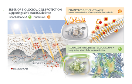
AGE inhibitors
Another potential strategy is the use of topical agents that inhibit the formation or accumulation of AGEs in the skin. One study published in the Journal of Cosmetic Science found that a cream containing carnosine, a peptide that inhibits glycation, improved skin elasticity and reduced the appearance of wrinkles in individuals with ageing skin. Skincare containing NAHP or Acetyl Hydroxyproline inhibits the formation of AGEs significantly (in vitro), most likely through a mechanism where NAHP competes with the proteins for the sugar. Finally, NAHP sacrifices itself in place of the proteins and gets (at least partially) glycated. NAHP also prevents loss of cellular contractile forces in a glycated in vitro dermis model and counteracts the diminished cell-matrix interaction that is caused by glyoxal-induced AGE formation. Take-aways from a study published in the International Journal of Cosmetic Science [2]: 1. NAHP significantly and dose-dependently inhibited the formation of advanced glycation end-products (AGEs) in a protein solution. 2. NAHP dose-dependently inhibited the formation of N-(carboxymethyl)lysine (CML), a specific AGE. 3. In fibroblast-populated collagen lattices, NAHP prevented the glycation-induced disturbance of fibroblast contractile capacity. 4. Ex vivo application of NAHP to skin explants decreased AGE fluorescence compared to glucose-treated samples. Anti-Oxidants I would suggest to combine those ingredients with an ingredient like Licochalcone A. Numerous high ranked publications support that Licochalcone A protects cells from oxidative stress mediated by e.g. UV and HEVIS (blue light) induced reactive oxidative species (ROS). Due to the activation and nuclear translocation of the transcription factor NrF2, the expression of anti-inflammatory, antioxidant and detoxifying enzymes are induced. These enzymes protect the skin cells (like keratinocytes and fibroblasts) from ROS-induced damage, like lipid peroxidation and DNA as well as protein damage. If Licochalcone A is combined with L-Ascorbic Acid, (the most active form of Vitamin C), it supporting skin's own collagen production, provides superior biological cell protection amongst other relevant benefits. Vitamin C (and E) has shown to inhibit protein glycation [4]. SPRAY TAN A study in Redox Biology indicates that sunless tanning with dihydroxyacetone (DHA) causes glycation and potential DNA damage in the epidermis [1].: 1. Glycation: DHA exposure led to the formation of advanced glycation end products (AGEs) in epidermal cells, confirmed by mass spectrometric detection of N-ε-(carboxyethyl)-l-lysine (CEL). 2. DNA damage: DHA induced a cellular stress response, including activation of stress-related genes and phosphoprotein signaling. While not directly measuring DNA damage, these responses are often associated with cellular stress that can lead to DNA damage. 3. Significance: The effects were observed at low millimolar concentrations of DHA, relevant to topical application. The stress response was rapid and pronounced, occurring within minutes of exposure. 4. Location: The effects were primarily observed in epidermal cells and reconstructs, with no mention of dermal effects. This study suggests that sunless tanning with DHA may not be as safe as previously thought, warranting further investigation into its long-term effects on skin health [1], however no need to panic as this negative effect is only seen in the top layer of the skin. DOUBLE TROUBLE: GLYCATION + UVB [3] The combination of GO-AGEs and UVB exposure has a more pronounced effect on skin inflammation and oxidative stress than either factor alone. This suggests a synergistic relationship between glycation and UV exposure in accelerating skin aging processes. 1. GO-AGEs combined with UVB irradiation significantly increased the secretion of pro-inflammatory cytokines (IL-1β, IL-6, IL-8) in skin cells compared to either GO-AGEs or UVB alone. 2. The GO-AGEs + UVB treatment group showed a more than three-fold increase in IL-6 and IL-8 mRNA levels compared to other groups. 3. GO-AGEs + UVB treatment induced significantly higher release of nitric oxide (NO) compared to other groups. 4. The combination of GO-AGEs and UVB enhanced reactive oxygen species (ROS) release, creating about 1.5 times more oxidative stress compared to control and other groups. 5. Cell viability was notably affected in the GO-AGEs, UVB, and GO-AGEs + UVB treatment groups compared to the control group. These findings are significant as they demonstrate that the combination of GO-AGEs and UVB exposure has a more pronounced effect on skin inflammation and oxidative stress than either factor alone. This suggests a synergistic relationship between glycation and UV exposure in accelerating skin aging processes. Use sunscreen daily. . GLYCATION AND SKIN HEALTH Acne In addition to its role in ageing, glycation in the skin has also been linked to a range of skin health problems. One study published in the Journal of Cosmetic Dermatology found that the level of AGEs in the skin was significantly higher in individuals with acne than in those without acne. The study also showed that treating acne with a topical antibiotic significantly reduced the levels of AGEs in the skin. Atopic Dermatitis Another study published in the Journal of Investigative Dermatology found that individuals with atopic dermatitis had higher levels of AGEs in their skin than healthy individuals. This suggests that glycation may play a role in the development of inflammatory skin conditions. Diabetes + Woundhealing The correlation between high sugar levels and skin ageing can be seen in diabetic patients, where one-third of this population has skin complications. A prominent feature of ageing human skin is the fragmentation of collagen fibers, which severely damages the structural integrity and mechanical properties of the skin. Elevated levels of MMP-1 and MMP-2 and higher crosslinked collagen in the dermis of diabetic skin lead to the accumulation of fragmented and crosslinked collagen, thereby impairing the structural integrity and mechanical properties of dermal collagen in diabetes. Collagen crosslinking makes it impossible for them to easily repair, resulting in reduced skin elasticity and wrinkles. Keratinocytes and fibroblasts are the main cells involved in wound healing, but due to the high glucose (HG) microenvironment in diabetics, the functional state of these cells is impaired, thereby accelerating cellular senescence (programmed cell death). Conclusion We can't completely stop the glycation process, therefore it's important that we inhibit it from a young age onwards, hence monitor the sugar intake of our children, use daily SPF and invest in good dermo-cosmetic products containing ingredients like NAHP and powerful anti-oxidants like L-Ascorbid Acid (Vitamin C is needed for the production of collagen) and Licochalcone A (also anti-inflammatory). Preventing signs of ageing, specifically caused by glycation is most effective. If your skin shows (advanced) signs of ageing, you can get visible improvement using skin component (hyaluron, collagen and elastin) bio-stimulating ingredients like Retinol, Bakuchiol, Arctiin, Creatine or Glycine Saponin. Consult your dermatologist if you wish to improve your skin's appearance or skin health issues. Take care Anne-Marie References [1] Perer J, Jandova J, Fimbres J, Jennings EQ, Galligan JJ, Hua A, Wondrak GT. The sunless tanning agent dihydroxyacetone induces stress response gene expression and signaling in cultured human keratinocytes and reconstructed epidermis. Redox Biol. 2020 Sep;36:101594. [2] Knoblich C, et al. N‐acetyl‐L‐hydroxyproline – A potent skin anti‐ageing active preventing advanced glycation end‐product formation in vitro and ex vivo. Int J Cosmet Sci. 2023;1-10. doi:10.1111/ics.12930 [3] Sultana, R., Parveen, A., Kang, MC. et al. Glyoxal-derived advanced glycation end products (GO-AGEs) with UVB critically induce skin inflammaging: in vitro and in silico approaches. Sci Rep 14, 1843 (2024). https://doi.org/10.1038/s41598-024-52037-z [3] Sadowska-Bartosz I, Bartosz G. Prevention of protein glycation by natural compounds. Molecules. 2015 Feb 16;20(2):3309-34. doi: 10.3390/molecules20023309. PMID: 25690291; PMCID: PMC6272653. Special thanks: Ph.D. dr Julia M. Weise Manager Biological Testing & Dorothea Schweiger Lab Manager Facial Skin Biology Beiersdorf HQ Hamburg
There is data suggesting that skin is more prone to sun damage when skin has higher pH levels (>6). The daily use of sunscreen defends the acid mantle by protecting skin cells from sun damage and increasing the skin's ability to protect itself.
Some active ingredients used in sunscreen can increase skin´s optimal pH and are less effective at lowered pH. Ingredients like zinc oxide and titanium oxide (mineral or physical sunscreens) have an optimal “pH window” in which they are more effective. If the pH of zinc oxide or titanium oxide is too low, the oxides will actually dissolve and thus lose efficacy. I understand that some of you might be in favor of only using mineral or physical sunscreen, however with chemical filters or a mix, you most probably maintain a more optimal pH-balance. This is particularly something to consider if you have acne-prone skin, problematic skin, or a sensitive or (very) dry skin type as your skin barrier might already be compromised. Hope you will enjoy the sun well-protected, while maintaining healthy skin. |
CategoriesAll Acne Age Clocks Ageing Aquatic Wrinkles Armpits Autophagy Biostimulators Blue Light & HEVIS Circadian Rhythms Cleansing Collagen CoQ10 Cosmetic Intolerance Syndrome Deodorant Dermaplaning Diabetes DNA Damage DNA Repair Dry Skin Epigenetics Evidence Based Skin Care Exfoliation Exosomes Eyes Face Or Feet? Facial Oils Fibroblast Fingertip Units Gendered Ageism Glycation Growth Factors Gua Sha Hair Hair Removal Hallmark Of Aging Healthy Skin Heat Shock Proteins Hormesis Humidity Hyaluron Hyaluronidase Hypo-allergenic Indulging Jade Roller Keratinocytes Licochalcone A Luxury Skin Care Lymphatic Vessel Ageing Malar Oedema Menopause Mitochondrial Dysfunction Mood Boosting Skin Care Neurocosmetics Ox Inflammageing Peptides PH Balance Skin Photo Biomodulation Polynucleotides Proteasome Psoriasis Regeneration Regenerative Treatments Review Safety Scarring Sensitive Skin Skin Care Regimen Skin Flooding Skin Hydration Skin Senescence Skip-Care Sleep Slugging Sunscreen Tanning Under Eye Bags UV Index Vitamin C Vitamin D Well Ageing Skin Care Wound Healing Wrinkles
Archives
December 2024
|
Anne-Marie van Geloven © 2024 All rights reserved
|